

 PDF
PDF Citation
 Print
Print


INTRODUCTION
The instrumentation of root canals is an important step in root canal treatment, and any failure during the instrumentation process affects the root canal filling procedure [1]. The objective of root canal instrumentation is to perform effective irrigation and root canal filling without changing the original canal configuration and to ensure a conical shape that broadens from the narrowest point of the apical foramen to the canal orifice [2].
Maintaining the original canal configuration and optimal canal form is difficult in curved root canals [3]. Furthermore, procedures carried out using conventional stainless-steel manual files are time-consuming and incapable of achieving, with insufficiently reliable results in terms of achieving the desired root canal shape in narrow and curved canals. These issues affect the success of root canal filling [4]. Several enlargement methods have been developed in order to minimize the risk of failure, which can manifest as ledging, zipping, loss of working length, and apical transportation [5]. Among the various root canal instrumentation methods that have been developed to overcome these problems, rotary nickel-titanium (NiTi) systems are particularly capable of maintaining the original canal shape, thereby achieving better centering. In recent studies, it was reported that rotary NiTi files protected the original canal configuration and reduced the likelihood of iatrogenic errors [67].
Rotary NiTi files are used in endodontic practice due to their higher accuracy and effectiveness than manual files. In comparison with manual files, rotary NiTi files are high-quality materials with a high level of fracture resistance, protecting against the risk of cyclic and torsional fatigue. Duque et al. [8] reported that rotary NiTi files have greater flexibility and cutting abilities in preparing root canals than the manual files. The ProTaper Next (PTN) file (Dentsply Maillefer, Ballaigues, Switzerland) is an off-centered system with a rectangular cross-section and variable taper. This instrument is made of M-wire NiTi material in order to improve its flexibility and fatigue resistance, as the M-wire NiTi material maintains its cutting ability and has a high level of cutting flexibility [910].
The One Shape (OS; Micro Mega, Besancon, France) is a single-file system that is used to prepare root canals by means of its continuous rotary movement. The OS single-file system is made of M-wire material, and it has an asymmetrical cross-sectional geometry throughout the blade [5]. Throughout the length of the file, an uneven cross-sectional pattern of the file produces a drifting wave of motion.
Newly introduced to the market in recent years, the One Curve (OC; Micro Mega) is a single-file system that reduces the root canal instrumentation time, while preserving the original root canal form. The OC system is made of C-wire heat-treated NiTi material with the property of controlled memory (CM); as such, the shape of the file can be flexibly modified before entering the canal to ensure easy access to the root canal. The manufacturer states that the instrument was developed to respond to the need for efficient shaping while respecting the initial root canal anatomy. Therefore, the instrument has a blade structure with a variable cross-section, ensuring the effective removal of debris up to the medium and coronal parts of the root canal. Furthermore, the non-existent ovalization of this instrument secures the apical zone [11].
Three-dimensional (3D) evaluations enable accurate comparisons of the shaping abilities of instruments. Currently, micro-computed tomographic (CT) imaging is accepted as the gold standard in qualitative and quantitative morphologic analyses of root canals, and a major area in which micro-CT imaging is applied is to evaluate the effects of files on the canal anatomy [12]. To our knowledge, no research data on the shaping characteristics of the OC file were yet available when this study was undertaken. For this reason, the aim of the present study was to compare the shaping abilities of the PTN, OS, and OC files in 3D-printed mandibular molars using micro-CT. The null hypothesis of the present study was that there would be no statistically significant differences among these 3 groups.
MATERIALS AND METHODS
Specimen selection and initial micro-CT scanning
In order to ensure standardization, 3D-printed mandibular molars (MM Tooth, Micro Mega) were used in the present study. The entrance cavities of the molar blocks were opened in advance. Standard blocks with a canal length of 19 mm, an apical diameter of 0.15 mm, a taper of 0.02, and a canal slope of 45° were used [13]. Before instrumentation, each of the blocks was scanned using a high-resolution desktop micro-CT system (Skyscan 1275, Bruker, Kontich, Belgium). The scanning conditions were: 100 kVp, 100 mA, a 0.5-mm Al/Cu filter, a pixel size of 10.2 μm, and rotation at 0.2° steps. To minimize ring artifacts, the air calibration of the detector was carried out prior to each scanning. Each sample was rotated 360° within an integration time of 5 minutes. The mean time of scanning was around 2 hours. Other settings included beam hardening correction, and the input of optimal contrast limits according to the manufacturer’s instructions, based on reconstructions of the teeth.
Root canal instrumentation
In mesiobuccal canals, #10 K-type files (Dentsply Maillefer) were operated under ×8 magnification using a dental operating microscope (Opmi-Pico, Karl Zeiss, Jena, Germany) until the file was seen from the apical aspect. Then, the file was taken out from the canal by 1 mm, and this length was determined as the working length. The blocks were randomly divided into 3 groups according to the preparation file used (n = 18).
Root canal instrumentation was performed by a single operator in accordance with the manufacturer’s instructions. For each of the models, instrumentation was performed from the coronal aspect towards the apical aspect until the apical diameter of each tooth's mesiobuccal canal was #25. All the canals were instrumented using a torque-controlled endomotor (X-Smart, Dentsply Tulsa Dental, Tulsa, OK, USA).
In the PTN group, a ProTaper Universal SX was used at 300 rpm with a torque of 4 N·cm to shape the coronal part of the canal. After the SX was used, first the X1 file and then the X2 file were used at the working length. In the OS group, an Endoflare was used at coronal 3 mm to enlarge the coronal section of the canal. Then, the G files (G1 and G2) were used at the working length. The instrumentation was completed in 3 steps with OS (400 rpm, with a torque of 2.5 N·cm) until it reached the working length. If apical resistance was encountered, the file was removed and cleaned. The root canal was irrigated with 2 mL of 5.25% sodium hypochlorite (NaOCl). In the OC group, the One Flare file was used to enlarge the coronal section of the canal. Then, the OC file was pushed forward step-by-step, at 300 rpm and a torque of 2.5 N·cm until the working length was reached. Whenever the file was stuck in the canal, it was irrigated using 2 mL of 5.25% NaOCl.
Irrigation was performed using 2 mL of 5.25% NaOCl after every file replacement and after the completion of root canal instrumentation in each canal. All of the files were used only once to prevent file failure in the root canal.
Micro-CT measurements and evaluations
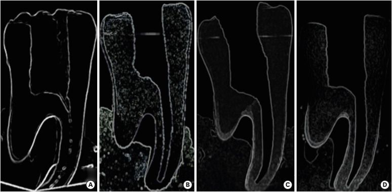
All prepared teeth were scanned with the same micro-CT (Skyscan 1275, Bruker) system, using the same parameters. The NRecon (ver. 1.6.10.4, SkyScan, Kontich, Belgium) and CTAn (ver. 1.16.1.0, SkyScan, Aartselaar, Belgium) software programs were used to reconstruct and measure the samples, following the modified algorithm presented by Feldkamp et al. [14] to obtain 2D axial images. For image reconstruction, ring artifact correction and smoothing were fixed to 0, and the beam hardening artifact correction was set to 40%. The scans were reconstructed to show 2D slices of the roots using NRecon. Several cross-sectional images were reconstructed from the entire micro-CT volume (n = 1,023). CTAn was used for 3D visualization and analysis of the images acquired through micro-CT scanning.
1. Transportation
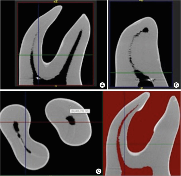
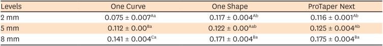
Three cross-sectional planes from the apical end of the root at levels of 2, 5, and 8 mm were used. The pre- and post-instrumentation shortest distances from the edge of the canal to the periphery in all roots were measured in the mesial and distal directions using the DataViewer software (ver. 1.5.6.2, SkyScan). Transportation was calculated following the method presented by Gambill et al. [15].
All constructions and measurements were performed on a 21.3-inch flat-panel color-active matrix TFT medical display (MultiSync MD215MG, NEC, Munich, Germany) with a resolution of 2,048 × 2,560 at 75 Hz and a 0.17-mm dot pitch operated at 11.9 bits (Figure 1).
2. Volume and area measurements
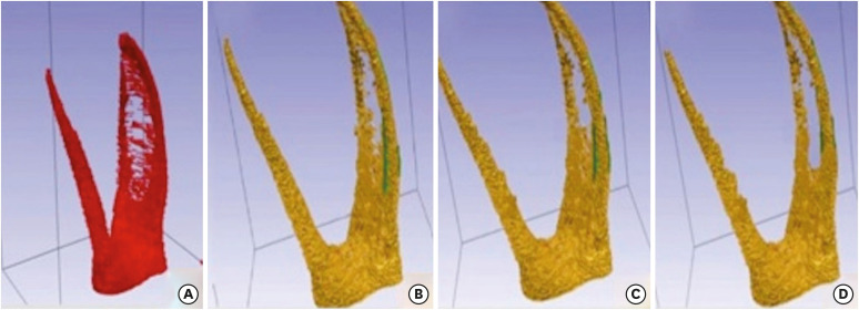
The volume of the mesiobuccal canals was measured before and after instrumentation using CTAn. Additionally, 3D surface representations were prepared from the micro-CT images (Figures 2 and 3). The pulp volume of the mesiobuccal canal of each tooth was calculated using CTAn, which allows the user to “sculpt out” the desired volume from the 3D structure, and by adjusting the brightness and opacity values, to remove unwanted voxels before calculating the final pulp volume. The final canal volume and surface area changes were measured before and after instrumentation.
3. Image evaluation
All images were evaluated and measured by a single observer with 7 years of micro-CT experience (KO). All measurements were taken twice by the same observer, and the mean values of all measurements were included in the statistical analyses. The observer also performed the analyses twice at an interval of 2 months to enable an evaluation of intraobserver variability.
Examiner reliability and statistical analysis
Statistical analyses were performed using SPSS version 20.0.1 (SPSS, Chicago, IL, USA). Intraexaminer validation measures were conducted. To assess intraobserver reliability, the Wilcoxon matched-pairs signed-rank test was used for repeated measurements. The normality of the data distribution was examined using the Kolmogorov-Smirnov test, whereas homogeneity was assessed using the Levene test. It was determined that the data showed a normal distribution. The changes in canal volume and surface area according to the type of canal files used were examined using repeated-measures analysis of variance (ANOVA). Two-way ANOVA was also used to compare the file systems at different canal levels from the apical end of the root. Differences were considered significant at p < 0.05.
RESULTS
Intraobserver consistency
Repeated evaluations and measurements indicated no significant intraobserver differences (p > 0.05). Overall, intraobserver consistency was rated at 91.6% between the 2 sets of evaluations and measurements. All measurements were found to be highly reproducible, and there was no significant difference between the 2 sets of measurements made by a single observer (p > 0.05).
Volume and surface area measurements
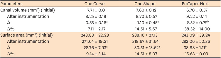
The canal volume increased considerably in all tested systems. No statistically significant difference was found in the change of canal volume and surface area before and after instrumentation between the OC and OS groups (p > 0.05). However, the PTN system showed a significant difference in the change of canal volume and surface area compared to the other 2 groups (Table 1).
Table 1
Mean and standard deviation (SD) values of the baseline parameters among the instrumentation systems
Canal transportation
The statistical analysis demonstrated that location within the canal, the file, and the interaction between those 2 factors were significant. No significant difference was found between the OS and PTN groups. However, a significant difference was found between the apical levels for the systems (p < 0.05). The OC file showed significantly lower transportation values in the apical region than the OS and PTN files. The amount of transportation in the OC group was significantly less than was observed in the other groups. The lowest extent of transportation was observed at 2 mm and the highest at 8 mm (p < 0.05). The average values for the groups are presented in Table 2.
DISCUSSION
The objective of an ideal root canal instrumentation method is to remove the same amount of material from inside and outside the root canals and to maintain the original anatomy of the root canal. Thus, the displacement of the apical foramen should be minimized. This principle is vital during instrumentation in order to prevent iatrogenic complications such as stripping and canal aberrations [16].
In the present study, the curved canals of simulated molar 3D-printed teeth were used, and the extent of canal transportation that occurred using 3 different file systems was evaluated. The root canals of natural teeth vary in size and shape. The use of 3D-printed teeth ensured standardization of the simulated canals' dimensions, including factors such as length, diameter, angle of curvature, and radius of curvature. Furthermore, according to the manufacturer, these 3D-printed teeth are claimed to be perfect reproductions of natural mandibular molars, making each step of the treatment—from coronal flaring to obturation—comparable to real clinical practice [13]. When comparing the shaping abilities of different root canal file systems, it is important to use files with similar apical diameters [17]. In the present study, files with an apical diameter of #25 were used in order to standardize the instrumentation characteristics of files.
Multi-rooted teeth have complex anatomy, and they present greater challenges for successful endodontic therapy than single-rooted teeth. The continuing development of instruments is intended to facilitate this task. In the present study, the canal shaping characteristics of the newly introduced OC system were compared with those of the widely used OS and PTN systems through micro-CT imaging, a nondestructive, reproducible, and well-established method for the 3D assessment of root canal instrumentation [1819202122]. Unfortunately, our results for the OC system cannot be compared with results from other studies because studies with similar experiments have not yet been published.
Canal volume is used to analyze the effects of canal instruments on the removal of dentin. There is no consensus on the amount of dentin that must be removed during root canal instrumentation, but it is recognized that excessive instrumentation of the root canal may result in an excessively thin root [23]. In the present study, an increase was observed in canal volume and surface area in all the file systems during root canal instrumentation, and no significant difference was found between the OC and OS groups. In contrast, in comparison to the other 2 files, the PTN system showed a significant difference in the change in canal volume and surface area. These results might be explained by differences in the design of the instruments. The cross-sections of the OS and OC files are approximately the same [24]. While both the OC and OS files have a triangular cross-sectional geometry, the PTN file has a rectangular shape with an off-centered mass, and the regressive taper of the PTN instrument can be more efficient in the contact area with the canal [25]. These characteristic features of the PTN file produce a mechanical wave (swaggering effect) that goes through the long axis of the instrument. Through these mechanisms, the PTN file can remove more tissue than other instruments of a similar size [26]. Capar et al. [27] also reported that the PTN file with a .06 taper could remove a similar amount of dentin to other instruments with a .08 apical taper because of its asymmetric design.
Numerous studies have compared different file systems [1528]. For instance, Capar et al. [27] compared 6 different file systems (PTN, ProTaper Universal, OS, Reciproc, Twisted File Adaptive, and WaveOne) in terms of canal transportation and surface area based on measurements made using cone-beam CT at 2, 5, and 8 mm from the apex to the crown. Among the 6 groups, they reported no statistically significant differences in terms of transportation, canal curvature, change in surface area, or rate of centering after instrumentation. Celikten et al. [5] examined the shaping ability of PTN and OS files and reported that both file systems showed similar canal transportation and volumetric changes.
These findings are in accordance with the results of the PTN and OS files used in the present study. Since apical transportation exceeding 0.3 mm would cause a remarkable decrease in the impermeability of the root canal filling material, excessive apical transportation could be considered a threat to the success of root canal treatment [29]. Uzunoglu and Turker [30] compared the transportation values obtained for the OS and PTN X2 file systems using cone-beam CT, and they determined that the transportation amounts achieved using these file systems did not exceed this limit and that there was no significant difference between the files. In the present study, in which the PTN, OS, and OC file systems were examined using micro-CT, it was observed that these file systems did not exceed the limit of 3 mm, but the OC file system yielded less transportation in the apical zone than the PTN and OS systems.
Although rotary NiTi instruments are flexible, they may suffer from flexural and/or torsional fatigue [3132]. With technological advancements, however, new generations of instruments with different cross-sections, diameters, tapers, and blades that have various cutting angles have been developed. Interestingly, the most recent advances have been directed towards improvements of the NiTi wire, whereby subtle changes in the proportion of metals and in thermo-mechanical treatments have enabled the development of instruments with a higher level of flexibility and fracture resistance than previous generations [313334]. For instance, CM wires are widely used in rotary instruments because they have more flexibility than conventional NiTi wires [353637]. CM wires are manufactured using a complex heat treatment, with variations in wire composition; specifically, CM wires contain less nickel (52% by weight) than the most commonly used wire, which contains 54.5% to 57% nickel by weight [36]. The OC instrument is produced from a C-wire by using proprietary heat treatment with the CM property and is in the martensitic phase at canal temperature. In the present study, it was determined that the OC file system caused a lesser extent of apical transportation than the other file systems. It is suggested that this difference occurred because the OC file is produced using a C-wire (heat treatment) technical design, whereas the PTN is an M-wire alloy and the OS is manufactured from a conventional austenite 55-NiTi alloy [30].
CONCLUSIONS
No statistically significant differences in the change of canal volume and surface area before and after instrumentation were observed between the OC and OS groups (p > 0.05). Less transportation in the apical zone was achieved when using the OC file system than when using the OS and PTN systems.
 XML Download