

 PDF
PDF Citation
 Print
Print


INTRODUCTION
Post space cleaning and preparation in endodontically treated teeth with oval-shaped canals can be challenging. The circular section of the drills used to remove root canal filling material and prepare the post space often produces untouched walls with residual obturation material [1]. The remaining material reduces the area available for adhesion and affects the bond strength of the cemented fiber post [234]. The use of ultrasonic tips to prepare root canal walls removes the smear layer, leaves the dentin surface free of material residues, and enhances the bond strength of fiber posts, especially in canals with complex anatomies, such as the oval-shaped canals [56].
The introduction of bioceramics in endodontics appears as a viable option in clinical situations where conventional gutta-percha and sealer may not guarantee a good prognosis. The material is a mixture of calcium silicate and calcium phosphate, which form hydroxyapatite on setting, allowing the obturation material to adhere to dentin [7]. The highly alkaline pH improves the bactericidal properties of the material [78]. The nanoparticles of the bioceramic sealers penetrate deeper into the dentinal tubules compared to conventional sealers [8]. While this property is desirable for an endodontic sealer, the cementation of fiber posts requires a clean dentin surface and permeable tubules to achieve a good bond [89].
This study aimed to evaluate the bond strength of fiber posts cemented in oval canals filled with epoxy resin-based or bioceramic sealer and the effect of ultrasonic cleaning of the intracanal post space. The null hypotheses were: 1) The bond strength of cemented fiber posts in oval canals would not differ with the use of bioceramic sealer or epoxy resin-based sealer; 2) The ultrasonic cleaning of the intracanal post space has no effect on the bond strength of fiber posts cemented in oval canals.
Go to : 
MATERIALS AND METHODS
This study was approved by the local ethics committee (CEP: 13.041-545). For this in vitro experimental study, upper and lower human premolars, extracted for various unknown reasons, were preserved in 0.1% thymol immediately after extraction for no more than 4 months until use. A non-probability sample, based on correlated studies of similar methodology published in the literature [91011], was used to select 50 healthy uniradicular premolars, with straight roots, with fully formed apex and oval-shaped root canals. Cone-beam tomography (CB gx 500 fed by I-Cat; Gendex, Chicago, IL, USA) was used to verify the oval shape of the canals (the buccolingual to the mesiodistal ratio of 2 or higher at 5 mm distance from the apex).
Sample preparation
The specimens were cleaned with ultrasonic periodontal tips and polished with prophylaxis brushes to remove traces of adhered hard and soft tissue. Clinical crowns were removed and sectioned, 2 mm above the cementoenamel junction. With a K #10 file (Dentsply Sirona, Ballaigues, Switzerland), the working length (WL) was determined visually with 10× magnification OPMI PICO operating microscope (Carl Zeiss Meditec AG, Oberkochen, Germany). Finally, the samples were mounted on transparent acrylic cylinders.
The root canals were prepared with WaveOne Gold system (Dentsply Sirona) and an X-SMART Plus motor (Dentsply Sirona), using the reciprocating program for the system. After glide path creation with a K #10 instrument, the biomechanical preparation started with the Primary (25/0.07) in a pecking motion up to the WL. The patency of the root canal was verified with a K #10 instrument, irrigated the canal with copious amounts of sodium hypochlorite (NaOCl), and used the Medium (35/0.06) up to the WL.
During canal preparation, it was used 20 mL of NaOCl 2.5%, followed by 5 mL of EDTA 17% and 5 mL of NaOCl 2.5% for a final rinse. The root canals were dried with medium WaveOne Gold paper points (Dentsply Sirona). The teeth were randomly divided with an Excel spreadsheet (Microsoft Office 2016, Seattle, WA, USA) into 2 groups (n = 20), leaving 10 teeth as the control group. The first group (n = 20) was filled with gutta-percha and AH Plus (AHP) (Dentsply DeTrey, Konstanz, Germany), using a 35/0.06 WaveOne Gold gutta-percha point up to WL and the continuous wave of condensation technique with a Calamus Dual (Dentsply Sirona). In the second group (n = 20), the canal walls were moistened with saline carried in a 35/0.06 WaveOne Gold gutta-percha cone. The canal was filled with Bio-C Sealer (BIOC) (Angelus, Londrina, Brazil), and a single gutta-percha point was inserted up to the WL. The gutta-percha cone was cut with the heated plugger of the Calamus Dual in the cervical third and compacted vertically with a cold Machtou plugger (Dentsply Sirona). For the control group, only the last 4 mm of the canal was filled with a WaveOne Gold 35/0.06 gutta-percha cone without a sealer, leaving the cervical and middle thirds free of filling material. The teeth were stored in a culture incubator (Ivoclar Vivadent, Schaan, Liechtenstein) for 7 days to allow the complete set of the sealer.
The post space was prepared, leaving 4 mm of filling material in the apical third of all samples. Samples from each group were randomly subdivided with an Excel spreadsheet, and the 4 study groups were prepared. Group A-I (n = 10): AHP sealer and Gates-Glidden # 2 drill followed by Exacto #1 drill (Angelus) and R2-Flatsonic ultrasonic tip (Helse Ultrasonics, Santa Rosa de Viterbo, Brazil) cleaning, Group A-II (n = 10): BIOC sealer and Gates-Glidden #2 drill followed by Exacto #1 drill and R2-Flatsonic ultrasonic tip cleaning, Group B-I (n = 10): AHP sealer and Gates-Glidden #2 and Exacto #1 drill. Group B-II (n = 10): BIOC sealer and Gates-Glidden # 2 and Exacto #1 drill. Group C (n = 10): control group conformed only by Exacto #1 drill.
The Gates-Glidden #2 drill was used attached to a low-speed handpiece keeping the drill at full speed to ensure the removal of the coronal and middle third gutta-percha. The space created was irrigated with 10 mL of saline, and the Exacto #1 drill was used in the same way that the Gates-Glidden drill to prepare the post space. The R2-Flatsonic ultrasonic tip was used mounted in an ultrasonic device (NSK Brasil Ltda, São Paulo, Brazil) at a frequency of 30 kHz and the cleaning procedures were performed using an OPMI PICO operative microscope at 6x magnification. The post space and the root filling material removal of all samples were verified with the operative microscope and 16x magnification.
After post space preparation, the canals were irrigated with saline, dried with paper points (Dentsply Sirona), and the Exacto #1 fiber posts were cemented with Relyx U200 automix (3M ESPE, Neuss, Germany).
Push-out test
After 7 days, the samples were sectioned perpendicular to the long axis of the root canal with an IsoMet 5000 metallographic cutting machine (Buehler, Lake Bluff, IL, USA) and a 4" Blade Diamond Disc (EXTEC Corp., Enfield, CT, USA) to obtain two 1 mm thickness slices. The slice corresponding to the cervical third was 2 mm from the cementoenamel junction, and the slice classified as the middle third was 5 mm from the cementoenamel junction, resulting in the most central sections of each third.
With a DL 2000 universal testing machine (Instron EMIC, Paraná, Brazil), a 50 kN load was applied from the apical side at a rate of 1.0 mm/min until failure occurred, to determine the fiber post bond strength. The values were obtained in N and converted into MPa by dividing the force by the root canal area of the specimen. The canal area was calculated using the formula: A = P × h, where A is the root canal area of the specimen, P is the perimeter of the root canal walls, and h is the height. P values were calculated by analyzing the specimen images obtained from the BX-43 light microscope (Olympus, Tokyo, Japan), using the Micrometrics SE Premium software (ACCU-SCOPE, New York, NY, USA). The h value was standardized at 1 mm at the time of making the cuts.
Failure analysis
After the push-out test, the specimens were analyzed under a 4× magnification optical microscope to determine the type of failure in the post-cement-dentin interface and classified as: 1) Adhesive failure between the post and luting material or dentin and luting material, 2) Cohesive failure of the post system or luting material or 3) Mixed failure when the samples present a combination of adhesive and cohesive failure.
The values obtained were tabulated and subjected to statistical interpretation using the Stata 11.2 statistical software (StataCorp, College Station, TX, USA) with the Shapiro-Wilk, Kruskal-Wallis, and Mann-Whitney tests (p < 0.05) for the bond strength values between the groups sealed with BIOC and AHP and post space preparation technique and the Shapiro-Wilk and Mann-Whitney tests (p < 0.05) for the area of remaining filling material between the cleaning technique and type of sealer.
Go to :
RESULTS
Bond strength
The results were analyzed according to the post space preparation technique and observed differences in bond strength values between the 2 sealers (Table 1). In samples filled with AHP, those cleaned with ultrasonic tips had higher bond strength values than those prepared only with the post drills, showing significant differences in both areas analyzed. The samples filled with BIOC showed significantly lesser values than those sealed with AHP, regardless of the use of ultrasonic and the root zone analyzed (Figure 1).
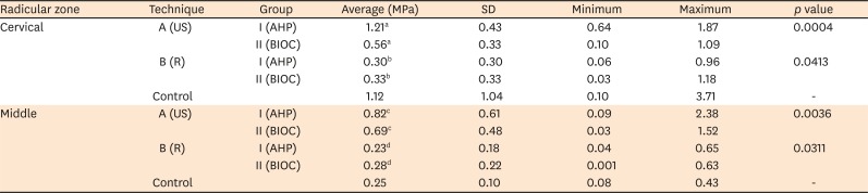
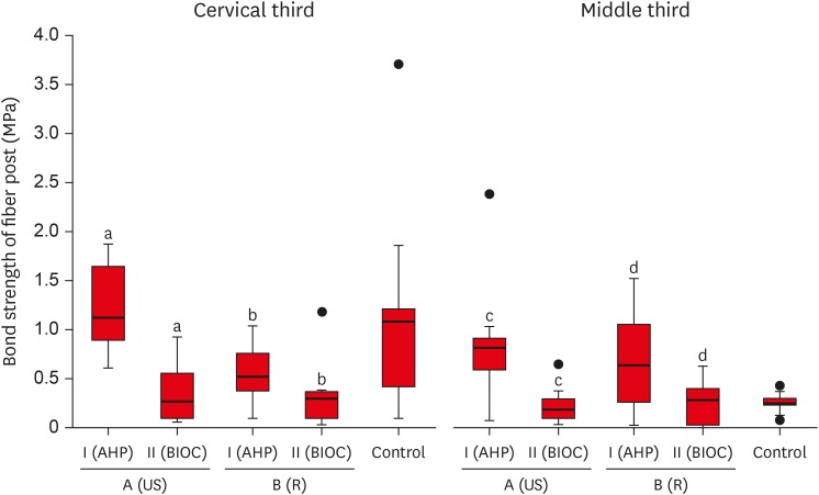 | Figure 1Distribution of all bond strength values in both zones, groups and techniques. It is observed in samples filled with AHP and those prepared with ultrasonic tips obtained higher values of bond strength than those prepared only with the post drills system, with significant differences in both analyzed thirds. The samples sealed with BIOC showed no differences between those prepared with ultrasonic tips or post drills system in the two-thirds analyzed.US, ultrasonic preparation; R, rotatory preparation; AHP, AH Plus; BIOC, Bio-C Sealer.
a,b,c,dSame superscript letters indicate significant diferences between groups (p < 0.05).
|
Table 1
Bond strength values
Using the Mann-Whitney test, statistically significant differences can be observed between the 2 sealers used in all sections analyzed.
US, ultrasonic preparation; R, rotatory preparation; AHP, AH Plus; BIOC, Bio-C Sealer; SD, standard deviation.
a,b,c,dSame superscript letters indicate significant diferences between groups (p < 0.05).
![]()
Area of remaining filling material
The samples prepared and cleaned with ultrasonic tips showed a lesser amount of residual filling material than the samples prepared with the post drill system. On the other hand, samples prepared only with a post drill system showed significant differences depending on the obturation material, being higher in samples filled with BIOC (Figure 2).
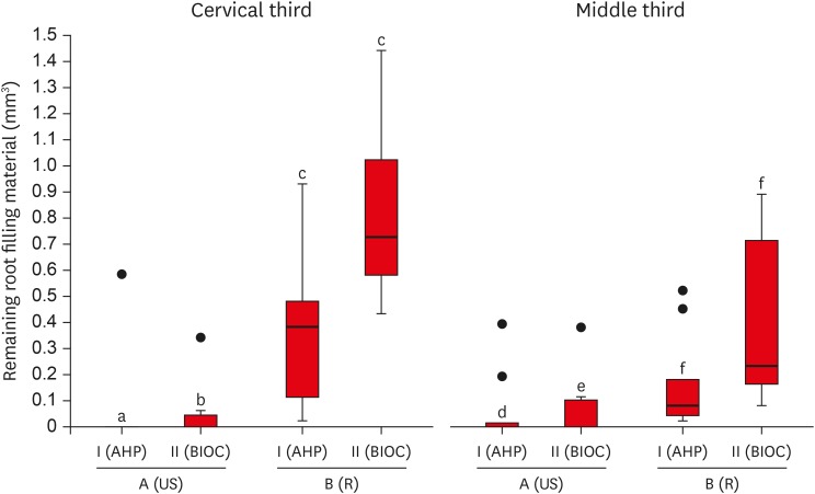 | Figure 2Distribution of the remaining intracanal filling material, in both zones, preparation techniques and sealer. The samples prepared with ultrasonic tips showed no differences and the samples prepared with the post drills system showed significant differences between the 2 sealers. The control group was not considered because the middle and cervical third of the canal were not filled.US, ultrasonic preparation; R, rotatory preparation; AHP, AH Plus; BIOC, Bio-C Sealer.
a,b,c,d,e,fSame superscript letters indicate significant diferences between groups (p < 0.05).
|
Failure analysis
The results obtained from the failure analysis showed similarities between the groups. The most frequent failure, considering all groups studied, was adhesive, which occurred in 84% of the samples analyzed.
Go to :
DISCUSSION
Complete removal of obturation material is critical when fiber post retention in teeth with oval canals is analyzed. Several studies have shown that no instrument can ensure the elimination of all residual filling material in oval-shaped canals in any third [12131415]. Coniglio et al. [15] and Schmage et al. [16] observed that an incomplete debridement of the post space and a thick resin cement layer compromised the bond strength of fiber post retained restorations. The removal of residual obturation material increased the canal space that translated into a thicker layer of adhesive cement, which could have decreased the bond strength [16]. However, Coniglio et al. [15] and Muñoz et al. [17] observed no significant differences in increasing the adhesive cement thickness. The results agree with the findings of the current study that showed significantly higher bond strengths in the samples with complete elimination of the cervical third filling material and a thicker layer of adhesive cement.
An evaluation of the post space preparation protocol showed that the debridement potential of the ultrasonic tips was superior to the rotary system; the technique positively influenced the bond strength of fiber post cemented in AHP sealed samples. The observation can be explained by the use of a new ultrasonic tip developed for the effective removal of filling material in oval canals [5]. The tips allowed for a conservative post space preparation [5] and favored the removal of filling material from the areas that the post drills could not touch [18], resulting in higher values of bond strength. However, the bond strength values of BIOC sealed samples were not influenced by the method of post space preparation. All BIOC sealed samples showed low values of bond strength, regardless of the debridement protocol, demonstrating a distinct influence of the sealer.
An evaluation of the relationship between the sealer and the bond strength of fiber posts showed that when the canal walls were cleaned with an ultrasonic tip, the AHP sealer did not interfere with the adhesion of the resin cement used with the fiber posts. These findings agreed with the results reported by Cecchin et al. [19]. Studies conducted by Chen et al. [7] and Oltra et al. [20] observed that the AHP sealer could be easily removed from the post space when compared to other sealers used in endodontics. Conversely, an analysis of the results obtained with BIOC revealed a decrease in bond strength values, despite complete elimination of the residual filling material and thorough debridement of the post space. A study conducted by Vilas-Boas et al. [4] observed a similar decrease in bond strength and concluded that bioceramic sealers was unsuitable for adhesive cementation of fiber posts. This observation could be due to higher dentinal tubular penetration, salt precipitation, and the effect of the alkaline pH that interfered with the acid conditioning of dentin [20]. Regardless of the obturation technique and post space preparation protocol, BIOC demonstrated low bond strength values than AHP. In the middle third of the root canal, BIOC and AHP had equivalent bond strengths. This observation could be due to the morphological variation between the middle and cervical third of the oval canals [2122]. The medium third needs greater wear for post compared to the cervical third due to its smaller caliber [15]. That wear could eliminate dentin infiltrated by bioceramic sealer eliminating the interference that this kind of materials could generate in the adhesive processes.
An analysis of the type of interface failure in each group showed a higher number of adhesive failures, a finding that coincides with the studies conducted by Kırmalı et al. [9] and Vilas-Boas et al. [4]. This observation could be explained by the lack of techniques and instruments to ensure complete debridement of the canal walls. The result confirms the sensitivity of the bonding technique and the importance of clean walls to maximize adhesive potential.
The results of this study confirm that it is necessary to evaluate the type of endodontic sealer used in a tooth that will be restored with a fiber-reinforced post. To the best of our knowledge, no study has evaluated the cleaning efficiency of the post space of canals filled with bioceramic filling material, making it difficult to compare current results with previous investigations. The literature on bioceramic filling materials is still relatively scarce. Further studies are required for supporting the clinical relevance of this study, especially in the aspects of working mechanism between bioceramic sealers and adhesive cements.
Go to :
CONCLUSIONS
In conclusion, the use of BIOC decreased the bond strength of fiber posts in oval canals, regardless of the cleaning quality of post space preparation. Future investigations comparing debridement protocols and confirming the results obtained in this study would be of paramount importance to the clinician.
Go to :
 XML Download