

 PDF
PDF Citation
 Print
Print


INTRODUCTION
An ideal retrograde filling material should be resistant to dislocating forces [12] to prevent leakage between the root canal system and periradicular tissues [3]. Furthermore, the filling and sealing ability of endodontic cement can affect the long-term outcomes of endodontic surgery [456]. Root-end fillings with zinc oxide and eugenol-based cements, as well as calcium silicate-based materials, have shown a high probability of success [78]. However, the use of biomaterials has been proposed as a way to achieve more predictable outcomes [5679].
The ability of endodontic materials to fill the spaces in the root canal in order to prevent fluid penetration is related to their flow [10]. However, flow and filling ability may not be directly proportional, considering that a material with a better ability to flow linearly will not necessarily provide greater filling capacity [1112].
There are no previously established standards for evaluating the flow of root-end filling materials, and the International Standards Organization (ISO) standard [13] commonly used for the evaluation of root canal sealers does not document a correlation between the flow and fill properties of a material [12]. Thus, more accurate methods are required to evaluate the flow of endodontic materials [10]. For this reason, a novel test model was developed for concomitantly evaluating the flow and fill of endodontic cements using micro-computed tomography (CT) [12]. Micro-CT is a nondestructive, precise, and reproducible imaging tool that provides a 3-dimensional (3D) quantitative evaluation of filling materials [14].
The device developed to evaluate endodontic materials using micro-CT involves the delivery of endodontic cement between 2 glass plates with a metal weight over the top plate, with a similar design to that described in the ISO standards [13]. However, the bottom glass plate is manufactured with a central cavity and 4 grooves extending out horizontally and vertically. In this way, the model enables analysis of flow into the spaces and volumetric analysis, which allows the evaluation of the filling ability of a material in the central and lateral areas [12]. The proposed model using micro-CT assessment showed valid and reproducible results, and has the potential to improve flow analysis. Nevertheless, the influence of the size of the test models on the results has not yet been analyzed.
Since comparing different methods is essential for establishing the most suitable methodologies in endodontic research [15], the aim of this study was to investigate the influence of different sizes of test models on the linear flow and volumetric filling of Biodentine (Septodont, Saint-Maur-des-Fosses, France), intermediate restorative material (IRM; Dentsply DeTrey, Konstanz, Germany), and mineral trioxide aggregate (MTA; MTA-Angelus, Angelus, Londrina, PR, Brazil) using micro-CT. The null hypothesis was that there would be no differences in the flow and filling of the materials according to the test model used.
Go to : 
MATERIALS AND METHODS
Preparation of the test models
The first test model was manufactured according to the technique described by Tanomaru-Filho et al. [12]. A glass plate was fabricated with a central cavity (1 × 1 × 2 mm) (length, width, and height) and grooves extending out horizontally and vertically to the 4 sides. The other 2 dimensions were proposed to facilitate a comparison with the previous study, as follows: 1 × 1 × 1 mm and 1 × 2 × 1 (length, width, and height) (Figure 1). The samples were randomly divided into 3 groups (n = 6 each), according to the root-end filling material used. The complete information regarding the endodontic materials, their manufacturers, composition, and proportions is presented in Table 1. The procedure was performed by a single operator who was previously trained and calibrated. For each material, 0.050 mL was placed in the central cavity of the bottom glass plate, and another glass plate (20 g) and a metal weight (100 g) with a total mass of 120 g were placed on the materials and kept there for 10 minutes (Figure 2), according to the ISO 6876/2012 recommendation [13].
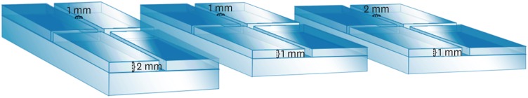 | Figure 1Illustration of the test models with a central cavity and lateral grooves manufactured with different dimensions: 1 × 1 × 2 mm, 1 × 1 × 1 mm, and 1 × 2 × 1 mm (length, width, and height).
|
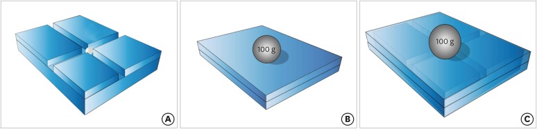 | Figure 2Illustration of the flow and filling ability evaluation process before the assessment using micro-computed tomography. The bottom glass plate with the endodontic cement placed in the central cavity (A). A view representing the assembled device, with the bottom glass plate, the top glass plate, and the metal weight over the cement (B). Another view representing the assembled device, using transparency to show the bottom plate and the metal weight over the material after flow inside the grooves (C).
|
Table 1
Endodontic materials, their manufacturers, their composition, and the proportions used
![]()
Micro-CT scanning and analysis
The glass plates/cement set was scanned with the SkyScan 1176 micro-CT system (SkyScan, Bruker, Kontich, Belgium). The micro-CT parameters were a voxel size of 9 µm, 90 kVp, 278 mA, a 0.1 mm copper filter, and 360° scanning. The linear flow (mm) measurement of the material on each side of the grooves (horizontal and vertical) was analyzed. The mean of the 4 measurements was considered the linear flow for each evaluation. The volume (mm3) filled by the material in the central area was determined as the central cavity filling. The volume (mm3) filled by the materials in the lateral areas was determined up to 2 mm on each side of the central cavity. The mean of the 4 measurements was considered as the lateral cavity filling for each analysis. The data sets were reconstructed using NRecon software (V1.6.10.4, Bruker). The correction parameters for smoothing, beam hardening, and ring artefacts were defined for each material. The flow into the grooves and the filling of the central and lateral cavities were calculated using the CTAn software (V1.15.4.0, Bruker). CTAn was also used to create 3D models of the materials, which were visualized using the CTVol program (V2.3.1.0, Bruker).
Statistical analysis
The normality of the data was tested using the Kolmogorov-Smirnov test. The statistical analysis was performed with 2-way analysis of variance and the Tukey parametric test with a significance level of 5%.
Go to :
RESULTS
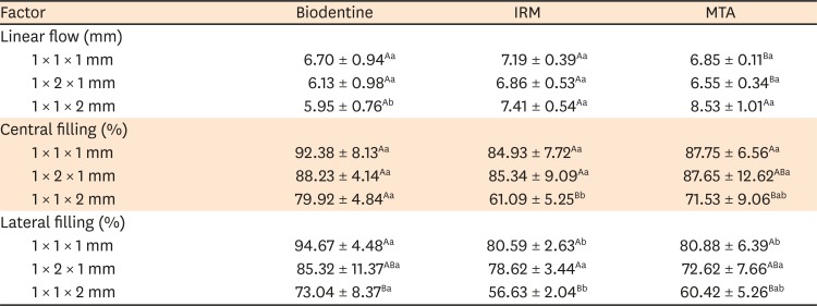
The results are presented in Table 2. MTA and IRM presented similar linear flow and central cavity filling in all test models (p > 0.05). MTA had greater flow when using the test model with a height of 2 mm, with a higher value than Biodentine (p < 0.05). For central cavity filling, IRM and MTA showed worse results in the model with a height of 2 mm than in the model measuring 1 × 1 × 1 mm (p < 0.05). The same occurred for lateral cavity filling in all materials (p < 0.05). Biodentine showed better filling than IRM in the model with a height of 2 mm (p < 0.05). The calculation of the filling of the materials in the central and lateral areas, as well as a 3D model illustrating the flow and filling of the materials into the grooves, can be seen in Figure 3.
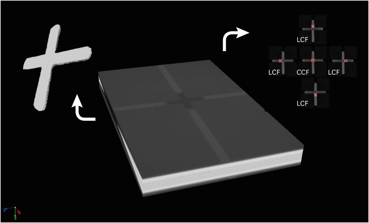 | Figure 3Illustration created in the CTVox software showing the assembled device composed of the bottom glass plate, the cement after flow inside the grooves, and the top glass plate during the scanning process on micro-computed tomography. The flow and filling representation were performed in 3 dimensions using the CTVol software. Central cavity filling (CCF) and lateral cavity filling (LCF) were evaluated using the CTAn software.
|
Table 2
Mean and standard deviation of the results of flow (mm) and filling (%) of endodontic materials evaluated in test models with different sizes (length, width, and height)
The values are mean ± standard deviation. Different lowercase letters on the same line indicate statistically significant differences between the different cements (p < 0.05). Different capital letters in the same column indicate statistically significant differences between the different test models (p < 0.05) (2-way analysis of variance and Tukey test).
IRM, intermediate restorative material; MTA, mineral trioxide aggregate.
![]()
Go to :
DISCUSSION
The flowability of endodontic sealers is evaluated using the ISO 6876/2012 standard [13] because a proper flow may allow filling irregularities [16]. However, this conventional test does not allow a 3D assessment of the filling ability of a material. Thus, it is not possible to determine whether proper flow is associated with adequate filling. Moreover, there is no standard for assessing the flow of root-end filling cements. Therefore, we evaluated the flow and filling of root-end filling materials using micro-CT according to Tanomaru-Filho et al. [12]. According to the authors, volumetric data reflect the ability of a material to fill a space and to flow into lateral spaces, an important property for endodontic materials. Furthermore, the proposed device allowed standardization of the amount of cement and the pressure to be used on the material during the test, similar to the device described in the ISO standard [13].
Numerous studies have proposed using micro-CT to complement conventional evaluations of endodontic materials [12141718192021]. However, since micro-CT is a relatively new methodology, these evaluations have lacked standardization. For this reason, it is necessary to evaluate the influence of the evaluation method on the results obtained.
Based on a new technique already recommended in the literature [12], the current study aimed to assess the influence of the dimensions of the test models on the flow and filling properties of different endodontic cements. Similar to previously observed results [1112], there was no direct correlation between the properties evaluated, since when a material presented a greater flow, a proportional filling of the cavity was not observed. It was also found that the height of the test models had an influence on the results. When a height of 2 mm was used, MTA presented higher flow but less filling capacity. For Biodentine and IRM, lateral cavity filling was also lower when the highest model was used. In contrast, when the smallest dimensions were used, it was possible to observe greater flow and filling. These findings may be directly applied to clinical situations, as root canal systems present small irregularities to be filled by the material. Thus, our null hypothesis was rejected since the test models showed a direct influence on the results.
IRM is a zinc and eugenol-based cement that has been used for several decades. However, its cytotoxicity is an important limitation of this material [7]. Although Biodentine presents lower linear flow in the 2-mm-high model, it showed better filling than IRM. Therefore, it may be suggested that Biodentine has a better filling ability than IRM, which may be supported by previous results indicating that IRM showed high microleakage [22]. However, Tsesis et al. [23] evaluated the apical portion of root canals filled with MTA, IRM, and Biodentine regarding Enterococcus faecalis colonization and observed no differences among the root-end filling materials in the mean and maximal depths of bacterial colonization into the dentinal tubules. Those results suggest that similar success rates for root-end filling can be obtained using calcium silicate-based materials or cements based on zinc oxide and eugenol [8].
However, it is also important to consider that calcium silicate-based materials, such as MTA and Biodentine, are potentially bioactive when placed in direct contact with dentin, and may promote biomineralization [24]. This property enables a better seal for retrograde fillings [25], making them better options for the repair of furcal perforations [1]. Moreover, Akbulut et al. [26] observed no significant difference in the push-out bond strength of MTA-Angelus and Biodentine, in agreement with the similar characteristics observed for both materials in the present study.
Previous studies have evaluated the filling capacity of MTA and Biodentine using micro-CT [272829]. This non-invasive imaging technique provides high-resolution images and enables a 3D volumetric analysis [30], allowing a material's filling ability to be evaluated in terms of the filling percentage [2728]. Furthermore, the methodology employed in the current study allows a concomitant analysis of a material's flow rate and filling of lateral spaces [12], which is more difficult to achieve for root-end filling cements.
Regarding central cavity filling, our results showed that Biodentine and MTA presented similar values in all the test models. This finding corroborates the report of Küçükkaya et al. [27], who observed no significant difference between the obturation quality of MTA and Biodentine in terms of percentage volume of filling materials on micro-CT. The authors used tooth models that simulated perforating internal root resorption in the middle third of the root, and observed that the apical portion of the specimens presented a lower percentage of filling materials than the resorption cavities. Their result agrees with the present study, where the percentage of filling in the central cavity was greater than in the lateral spaces.
It is important to emphasize that when using the test model with the smallest dimensions, Biodentine showed better lateral filling than MTA. This finding may be related to the smaller particle size and higher surface area of Biodentine when compared to MTA. Moreover, the Biodentine radiopacifier is zirconium oxide, which has a smaller particle size and allows less porosity than the bismuth oxide used in MTA-Angelus [31]. Therefore, many studies have stated that a major disadvantage of MTA is its handling properties and its granular consistency. Consequently, additives have been proposed to increase its plasticity. Among these additives, methylcellulose, calcium chloride, calcium lactate gluconate, and propylene glycol improved the handling of MTA [323334]. In Biodentine, a hydro-soluble polymer is incorporated to enhance its handling properties [31].
A recent systematic review was undertaken in order to evaluate the influence of variation in the design of the push-out bond test in endodontic research [35]. The authors concluded that standardization is required for future research, as well as accurate reporting of all test variables, to assess the impact of specific designs on endodontic evaluations. This statement reinforces the importance of conducting studies to establish methods for the reliable analysis of material properties.
Go to :
CONCLUSIONS
The linear flow and filling ability of materials were affected by the size of the evaluated test models. Higher grooves and materials with higher flow showed a lower filling capacity. The test model measuring 1 × 1 × 2 mm showed the best ability to differentiate filling capacity among different materials.
Go to :
 XML Download