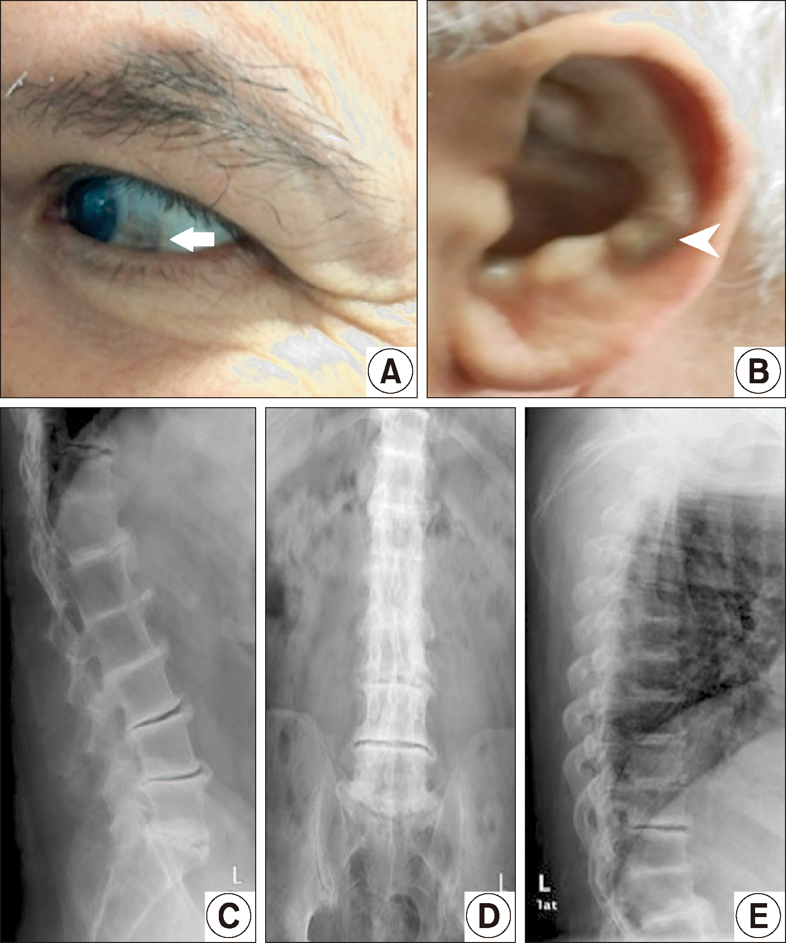
 | Figure 1Photographs show blue-black pigmentation of the sclera (arrow) (A) and ear pinna (arrowhead) (B), X-ray of the thoracolumbar spine, multilevel intervertebral disc space narrowing associated with disc calcifications (C∼E). We received the patient’s consent form about publishing all photographic materials. |
This article has been corrected. See "Erratum: Ochronotic Spondyloarthropathy Mimicking Diffuse Idiopathic Skeletal Hyperostosis" in Volume 27 on page 292.
A 59-year-old man presented with 20 years history of low back pain. The pain was mechanical in nature with early morning stiffness of less than 15 minutes. The patient was followed-up with a diagnosis of diffuse idiopathic skeletal hyperostosis (DISH) by the other physicians. He had partial response to analgesics.
On inquiry, the patient reported that since childhood his urine had turned a dark color on exposure to air. Physical examination revealed blue-black pigmentation of the sclera and ear pinna bilaterally (Figure 1). On laboratory, full blood count, calcium, phosphorus, alkaline phosphatase, erythrocyte sedimentation rate, C-reactive protein and rheumatoid factor were normal. HLA B-27 was also negative. Measurement of 24 hours urinary homogentisic acid levels was 957 μmol/mmol creatinine (reference value, <1 μmol/mmol creatinine). X-ray showed multilevel intervertebral disc space narrowing associated with disc calcifications of the thoracolumbar spine (Figure 1). The diagnosis of ochronosis was established, ascorbic acid and nitisinone were started for the patient. Protein restriction was made in his diet.
Ochronosis is a rare hereditary autosomal recessive disorder characterized by an excess of homogentisic acid deposits in the tissues. The incidence of ochronosis (alkaptonuria) is estimated at 1 case in 250,000 to 1 million live births [1]. Ochronotic arthropathy generally appears in the third decade, commonly affects in which weight bearing joints and the thoracolumbar spine. Narrowing of the intervertebral spaces and widespread, thin, linear, wafer-like disc calcification are diagnostic findings seen on spine radiographs [2]. A potential agent called 2 mg of daily nitisinone has been shown to decrease clinical progression of ochronosis [3]. Disc calcifications must be distinguished from those of the following: ankylosing spondylitis, juvenile idiopathic arthritis, DISH, calcium pyrophosphate dehydrate crystal deposition disease, hyperparathyroidism, Klippel-Feil syndrome, and congenital and acquired fusions of the spine [4]. The diagnosis of ochronosis must be considered for any patient with multilevel vertebral-body disc involvement.
Go to : 
REFERENCES
1. Phornphutkul C, Introne WJ, Perry MB, Bernardini I, Murphey MD, Fitzpatrick DL, et al. 2002; Natural history of alkaptonuria. N Engl J Med. 347:2111–21. DOI: 10.1056/NEJMoa021736. PMID: 12501223.

2. Ventura-Ríos L, Hernández-Díaz C, Gutiérrez-Pérez L, Bernal-González A, Pichardo-Bahena R, Cedeño-Garcidueñas AL, et al. 2016; Ochronotic arthropathy as a paradigm of metabolically induced degenerative joint disease. A case-based review. Clin Rheumatol. 35:1389–95. DOI: 10.1007/s10067-014-2557-7. PMID: 24647979.

3. Ranganath LR, Khedr M, Milan AM, Davison AS, Hughes AT, Usher JL, et al. 2018; Nitisinone arrests ochronosis and decreases rate of progression of Alkaptonuria: evaluation of the effect of nitisinone in the United Kingdom National Alkaptonuria Centre. Mol Genet Metab. 125:127–34. DOI: 10.1016/j.ymgme.2018.07.011. PMID: 30055994.

4. Toth K, Lenart E, Janositz G, Takacs K. 2003; Ochronotic arthropathy. Scand J Rheumatol. 32:315–7. DOI: 10.1080/03009740310003983. PMID: 14690148.

Go to : 


 PDF
PDF Citation
 Print
Print


 XML Download