증례: 21세 여자 환자가 소화불량으로 내원한 외부 병원에서 시행한 복부 CT에서 우연히 발견된 췌장 병변을 주소로 순천향대학교 부속 천안병원에 전원되었다. 환자는 특별한 과거력이나 복용 중인 약물은 없었다. 내원 당시 혈압 122/67 mmHg, 맥박 89회/분으로 신체검진에서 특이 소견은 보이지 않았다. 혈액 검사에서 white blood cell count 6,370/μL; hemoglobin 13.4 g/dL; platelet 320,000/μL; BUN 12 mg/dL; creatinine 0.67 mg/dL; AST 15 IU/L; ALT 9 IU/L; ALP 47 IU/L; γ-GTP 13 IU/L; total bilirubin 0.36 mg/dL; amylase 77 U/L로 특별한 이상 소견은 보이지 않았다.
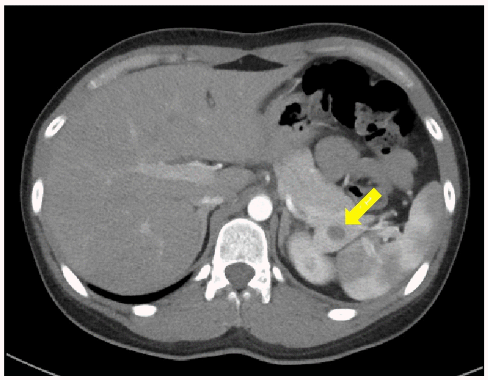
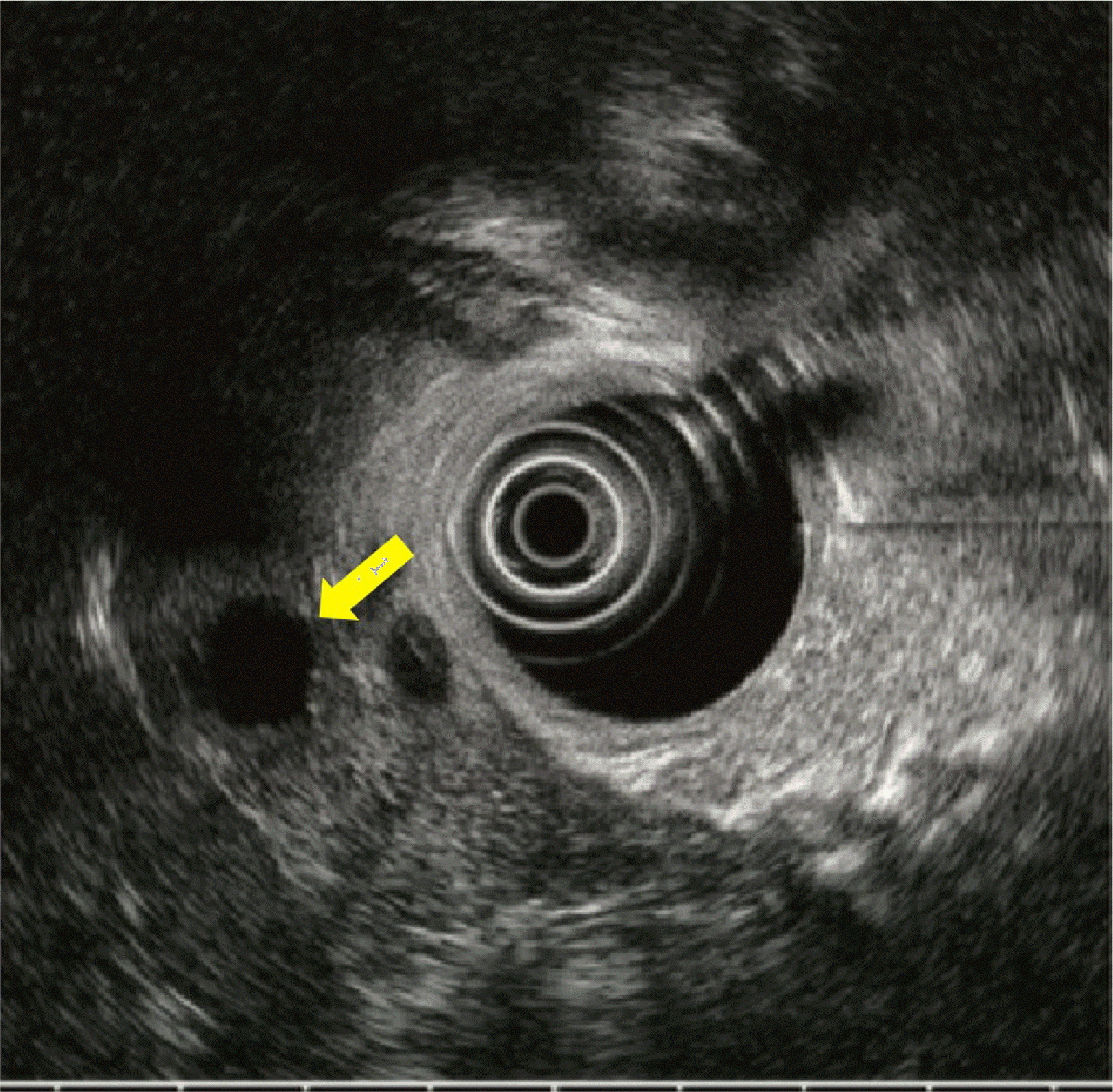
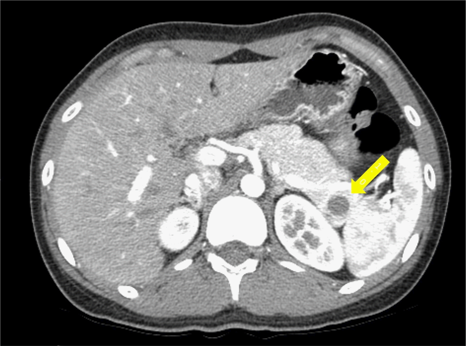
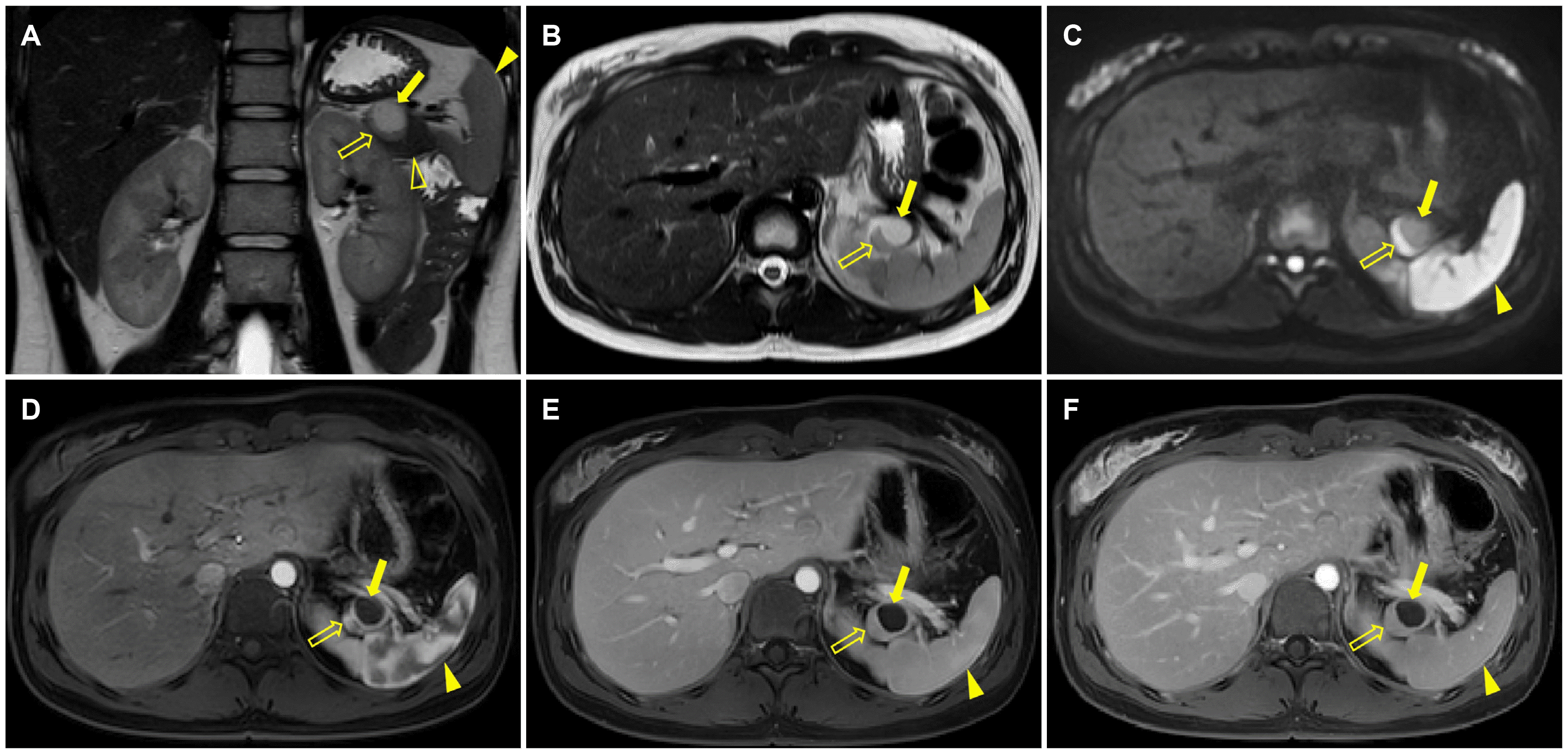
외부 병원에서 시행한 CT에서는 췌장 미부에 약 1.3 cm 크기의 조영증강되지 않는 낭성 병변이 발견되었다(Fig. 1). 순천향대학교 부속 천안병원에서 병변에 대한 추가 검사로 내시경 초음파를 시행하였고, 낭성 병변 내부에 격막이나 고형 결절은 없었다(Fig. 2). 병변은 췌장의 양성 낭성 종양의 가능성이 높을 것으로 생각되었으며, 환자는 이후 6개월마다 추적 관찰을 시행하였다. 처음 6개월 후 시행한 CT에서 해당 병변은 약 1.7 cm로 약간 커졌으나, 여전히 내부에 조영증강되는 격막이나 고형 결절은 없었다(Fig. 3). 12개월 후 시행한 추적 관찰에서는 MRI를 시행하였으며, 낭성 병변은 약 2.5 cm로 크기가 증가되었다(Fig. 4). MRI에서도 낭성 병변 내부에 격막이나 고형 결절은 없었으나, 낭성 병변 주위로 췌장실질과는 다른 신호 강도를 보이는 고형 병변이 관찰되었다. 이 고형 병변은 T2 강조영상과 확산 강조영상에서 고신호 강도를 보였고, 비교적 강한 조영증강을 보였다. 해당 병변은 신경내분비 종양의 낭성 변화 또는 고형 가성유두종양 등으로 생각되었다. 외과 협진 하에 췌장 미부 절제술을 시행하였고, 병리학적 검사에서 췌장 내 부비장에서 발생한 표피 낭종으로 진단되었다. 환자는 수술 후 합병증 없이 퇴원하였으며, 이후 추가적인 외래 경과 관찰은 시행하지 않았다. 후향적으로 MRI 영상을 다시 확인해 보았을 때, 병변의 고형부분은 T2 강조영 상과 확산 강조영상에서 비장과 비슷한 신호강도를 보였고 역동적 조영증강 영상에서도 비장과 비슷한 조영증강 패턴을 보였다.
진단: 췌장 내 부비장에서 발생한 표피 낭종(epidermoid cyst arising from intrapancreatic accessory spleen)
부비장은 영상 검사에서 흔히 보이는 해부학적 변이이다.1,2 부비장은 전체 인구의 약 10%에서 발생하며, 이 중 약 17%는 췌장 미부나 그 주위에 위치한다고 알려져 있다.3-5 CT나 MRI에서 췌장 내 부비장은 신경내분비종양이나 고형 가성유두종양과 같은 췌장의 고형종양과 감별 진단이 필요하다. 부비장은 영상 검사에서 비장과 같은 신호 강도와 조영증강 패턴을 보이며, 특히 동맥기 영상에서 보이는 zebra enhancement pattern은 비장 조직에서 특징적으로 보이는 소견이다. Tc-99m 가열처리 적혈구 스캔(Technetium-99m heat-damaged red blood cell scintigraphy)을 시행하면, 병리학적 진단 없이도 부비장을 확진할 수 있다.4,5
췌장 내 부비장에서 발생하는 표피 낭종은 매우 드문 질환이며, 발생하는 원인에 대해서는 아직 명확히 밝혀진 바가 없다.6 Horibe 등6은 췌장 내 부비장에서 발생한 낭종의 상피 조직이 췌관에서 기인하여 발생하였거나, 부비장 내 이소성 췌장조직에서 만성 염증과 상피 모양 육아종 형성이 반복되며 발생하였을 가능성이 있다고 말하고 있다. 임상적으로 췌장 내 부비장에서 발생하는 표피 낭종을 가진 환자들은 뚜렷한 증상이나 징후가 없는 경우가 대부분이며, 혈액 검사상 CEA와 CA 19-9 같은 종양 표지자는 진단에 큰 도움이 되지 않는다.7,8 부비장 내 표피 낭종은 췌장의 낭성 병변으로 발견되며 낭성 병변 자체의 영상 소견만으로는 점액성 낭성 종양(mucinous cystic neoplasm)이나 장액성 낭선종(serous cystadenoma)과 같은 낭성 종양이나 신경내분비 종양과 같은 고형 종양의 낭성 변화, 고형 가성유두종양과 감별이 쉽지 않다.3,8,9 본 증례에서도 크기 증가가 동반된 췌장 내 낭성 병변을 영상 소견만으로 감별 진단을 하는 데 어려움이 있었다. 하지만 부비장 내 표피 낭종은 낭종 주위 고형 부분이 부비장임을 CT나 MRI에서 인지하는 것이 진단에 도움이 되며, 이는 Tc-99m 가열처리 적혈구 스캔을 통해 확인할 수 있다.3,4,8,9 내시경 초음파를 통한 세침흡인 검사가 진단에 도움을 줄 수 있으나, 고형 부분의 크기가 작은 경우 위음성의 가능성이 높고, 표피낭종 내 액체의 CEA 수치가 높은 경우가 많아 감별에 어려움이 있을 수 있다. 또한 세침흡인 검사는 침습적이고, 낭성 종양이 악성 종양일 경우 검사 중 종양이 퍼질 가능성을 배제할 수 없다는 단점이 있다.3,8
본 증례는 크기가 지속적으로 증가한 췌장 병변을 수술적으로 절제하여 췌장 내 부비장에서 발생한 표피 낭종으로 진단된 증례이다. 일반적으로 부비장 내 표피 낭종은 크기가 작 고 특별한 증상이 없다면 수술적 절제가 필요하지는 않다. 비록 드물지만 비장과 비슷한 영상 소견을 보이는 고형병변 및 이와 동반된 낭성 병변이 췌장 미부에 있다면, 췌장 내 부비장에서 발생한 표피 낭종의 가능성을 고려해야 한다.
REFERENCES
1. Halpert B, Alden ZA. 1964; Accessory spleens in or at the tail of the pancreas. A survey of 2,700 additional necropsies. Arch Pathol. 77:652–654. PMID: 14130052.
2. Freeman JL, Jafri SZ, Roberts JL, Mezwa DG, Shirkhoda A. 1993; CT of congenital and acquired abnormalities of the spleen. Radiographics. 13:597–610. DOI: 10.1148/radiographics.13.3.8316667. PMID: 8316667.

3. Kato S, Mori H, Zakimi M, et al. 2016; Epidermoid cyst in an intrapancreatic accessory spleen: case report and literature review of the preoperative imaging findings. Intern Med. 55:3445–3452. DOI: 10.2169/internalmedicine.55.7140. PMID: 27904107. PMCID: PMC5216141.

4. Kim SH, Lee JM, Han JK, et al. 2008; Intrapancreatic accessory spleen: findings on MR Imaging, CT, US and scintigraphy, and the pathologic analysis. Korean J Radiol. 9:162–174. DOI: 10.3348/kjr.2008.9.2.162. PMID: 18385564. PMCID: PMC2627219.

5. Kim SS, Choi GC, Jou SS. 2018; Pancreas ductal adenocarcinoma and its mimics: review of cross-sectional imaging findings for differential diagnosis. J Belg Soc Radiol. 102:71. DOI: 10.5334/jbsr.1644. PMID: 30386851. PMCID: PMC6208287.

6. Horibe Y, Murakami M, Yamao K, Imaeda Y, Tashiro K, Kasahara M. 2001; Epithelial inclusion cyst (epidermoid cyst) formation with epithelioid cell granuloma in an intrapancreatic accessory spleen. Pathol Int. 51:50–54. DOI: 10.1046/j.1440-1827.2001.01155.x. PMID: 11148465.

7. Higaki K, Jimi A, Watanabe J, Kusaba A, Kojiro M. 1998; Epidermoid cyst of the spleen with CA19-9 or carcinoembryonic antigen productions: report of three cases. Am J Surg Pathol. 22:704–708. DOI: 10.1097/00000478-199806000-00007. PMID: 9630177.
8. Zhang Z, Wang JC. 2009; An epithelial splenic cyst in an intrapancreatic accessory spleen. A case report. JOP. 10:664–666. PMID: 19890189.
9. Sonomura T, Kataoka S, Chikugo T, et al. 2002; Epidermoid cyst originating from an intrapancreatic accessory spleen. Abdom Imaging. 27:560–562. DOI: 10.1007/s00261-001-0145-1. PMID: 12172998.

Fig. 1
Axial computed tomography image obtained at an outside hospital shows an approximately 1.3-cm-size cystic lesion (arrow) in the tail of the pancreas.

Fig. 2
Endoscopic ultrasound image shows an approximately 1.3-cm-size cystic lesion (arrow) in the tail of the pancreas.

Fig. 3
Axial pancreatic phase computed tomography image obtained after 6 months shows that the size of the cystic lesion (arrow) in the tail of the pancreas increased in size from 1.3 cm to 1.7 cm.

Fig. 4
MRI images obtained after 12 months. (A, B) Coronal and axial T2-weighted images show that the cystic lesion (arrow) in the tail of the pancreas increased in size from 1.7 cm to 2.5 cm. There is a solid component (open arrow) around the cystic lesion (arrow). The signal intensity of the solid component (open arrow) is similar to the spleen (arrowhead) but different from the pancreas parenchyma (open arrowhead). (C) Diffusion-weighted image (b-value 800 s/mm2) demonstrates the solid component (open arrow) with similar signal intensity to the spleen (arrowhead). (D) Axial gadolinium-enhanced arterial phase image shows a zebra enhancement pattern of the solid component (open arrow), like the spleen (arrowhead). Note the cystic lesion (arrow) without enhancement in the pancreatic tail. (E, F) On axial gadolinium-enhanced portal venous and 3-minutes delayed phase images, the enhancement of the solid component (open arrow) is similar to that of the spleen (arrowhead).


 PDF
PDF Citation
 Print
Print


 XML Download