

 PDF
PDF Citation
 Print
Print


INTRODUCTION
A gastrointestinal stromal tumor (GIST) is the most common mesenchymal tumor of the gastrointestinal tract, accounting for 1-3% of all gastrointestinal (GI) malignancies.1 The presenting symptoms of GIST include anemia, abdominal pain, and gastrointestinal bleeding. Most patients, however, are asymptomatic and are diagnosed incidentally during an endoscopic examination. Surgery for the complete removal of the tumor is the gold standard of therapy for a resectable GIST and the only chance of cure. On the other hand, recurrence after surgery is common, particularly in high-risk GISTs. Approximately 40-50% of patients have disease recurrence in this subgroup.2 The two main prognostic factors are the mitotic activity and the tumor size. More than five mitoses per 50 high-power fields (HPFs) and a size greater than 10 cm are the factors associated with poor outcomes after surgery.2 Adjuvant imatinib is the key treatment for postoperative high-risk patients with GISTs to decrease the incidence of relapse.3 Nevertheless, imatinib cannot completely prevent the recurrence of GIST in all cases, and side effects, such as hematologic abnormalities, can hinder the maintenance of imatinib therapy. This paper reports a case of gastric GIST with repeated local recurrences, even after a complete resection and adjuvant imatinib therapy, which eventually progressed to metastatic disease.
Go to : 
CASE REPORT
An 88-year-old woman was admitted to Chung-Ang University Hospital due to melena. She had a prior medical history of hypertension and chronic kidney disease. The peripheral blood cell counts showed anemia with a hemoglobin level of 3.8 g/dL. Esophagogastroduodenoscopy (EGD) was performed to determine the source of suspected upper GI bleeding. The endoscopic examination revealed a 2-cm sized, irregular-shaped ulcerofungating mass on the greater curvature, high body of the stomach (Fig. 1A). An endoscopic biopsy was performed to obtain a pathologic diagnosis of GIST. Abdominal CT revealed an 8.9 cm sized exophytic growing, heterogenous enhancing mass adjacent to the gastric high body (Fig. 1B). There was no evidence of a distant metastasis. She had undergone a laparoscopic wedge resection of the stomach for the gastric GIST. A pathology evaluation confirmed that the lesion was GIST with a high risk of progressive disease. The tumor size was 9.0×6.0×5.3 cm, and the mitotic index was a 16/50 HPF. The histologic type was the mixed spindle and epithelioid type (Fig. 2A). Immunohistochemistry confirmed GIST with diffuse positive staining for CD117, CD34, and DOG1, with a low Ki-67 proliferation index (Fig. 2B-E). There was no involvement of the tumor at the resection margin on the microscopic evaluation. After surgery, she received an adjuvant imatinib therapy at an initial dose of 400 mg/day to prevent a recurrence of the disease. On the other hand, recurrent anemia occurred during treatment, and the dose of imatinib was reduced. Despite the dose reduction, however, the severe anemia persisted, and the hemoglobin level decreased to 4.6 g/dL. Imatinib therapy was discontinued after 18 months of maintenance owing to severe drug intolerance. She had undergone EGD 1 year after the surgery and abdominal CT every 6 months, which showed no signs of tumor recurrence. She underwent a follow-up EGD 40 months after surgery, which revealed a 2-cm-sized round-shaped mass at the anastomosis site (Fig. 3A). Subsequent abdominal CT was performed as local recurrence was suspected (Fig. 3B). A laparoscopic wedge resection of the gastric mass was performed because there was no sign of distant metastasis. The histology was a spindle cell type GIST with high risk, showing a tumor size of 2.6×2.3×2.0 cm (Fig. 4A). The mitotic index was more than 10/50 HPF, with a high Ki-67 proliferation index of 30% (Fig. 4B). The resection margin was free from tumor involvement. After 2 months of adjuvant imatinib therapy at the lower dose (300 mg/day), severe anemia occurred again, which led to discontinuation of the drug. Eighteen months after the second surgery, a newly appeared 2.6 cm mass was detected on abdominal CT, and subsequent EGD revealed a mass located at the anastomosis line (Fig. 5A). Considering her very old age and poor performance status, she was followed up without treatment. Six months later, an abdominal CT scan showed progressive disease with invasion to the adjacent organs and multiple intra-abdominal metastases (Fig. 5B). She is now on intermittent imatinib therapy with a reduced dose (200 mg/day) due to her old age and the drug intolerance, while carefully monitoring the hemoglobin count. Follow-up CT taken after 2 months of medication revealed a partial response of the recurred disease, and she is currently being treated in an outpatient clinic setting.
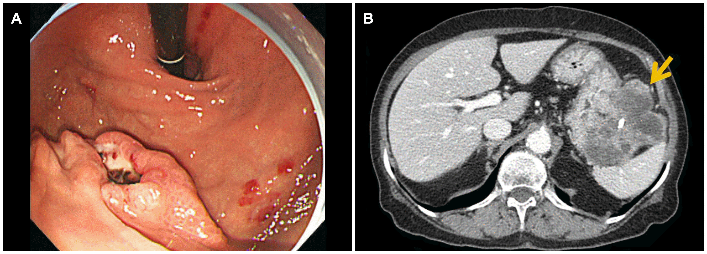 | Fig. 1Endoscopic images of the gastric lesion and abdominal computed tomography findings at the initial presentation. (A) A 2-cm sized, irregular shaped ulcerofungating mass on the greater curvature of the high body of the stomach. (B) An exophytic, heterogeneous enhancing mass with necrosis and central calcification, located adjacent to the gastric high body, closely abutting the splenic hilum (arrow).
|
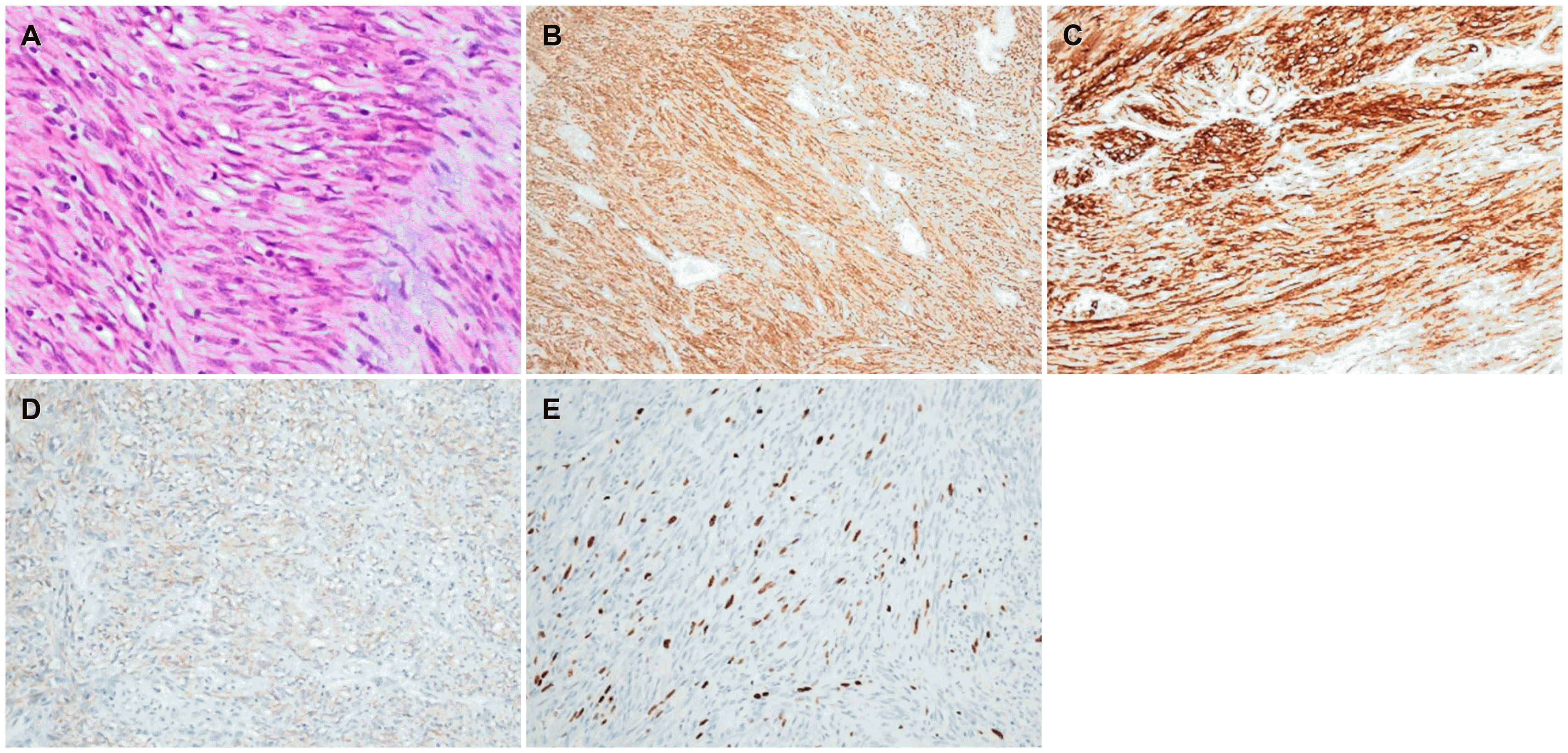 | Fig. 2Histologic features of the gastric gastrointestinal stromal tumor at the first surgery. (A) Histopathological evaluation reveals blandly spindle cells with faintly eosinophilic cytoplasm in a syncytial pattern. Elongated nuclei with inconspicuous nucleoli are noted (H&E, ×400).(B) Diffuse positive cytoplasmic staining for CD117 (Immunohistochemical staining, ×20). (C) Diffuse positive staining for CD34 (Immunohistochemical staining, ×20). (D) Focal positive cytoplasmic staining for DOG-1 (Immunohistochemical staining, ×20). (E) Ki-67 proliferation index is less than 5% (Immunohistochemical staining, ×20).
|
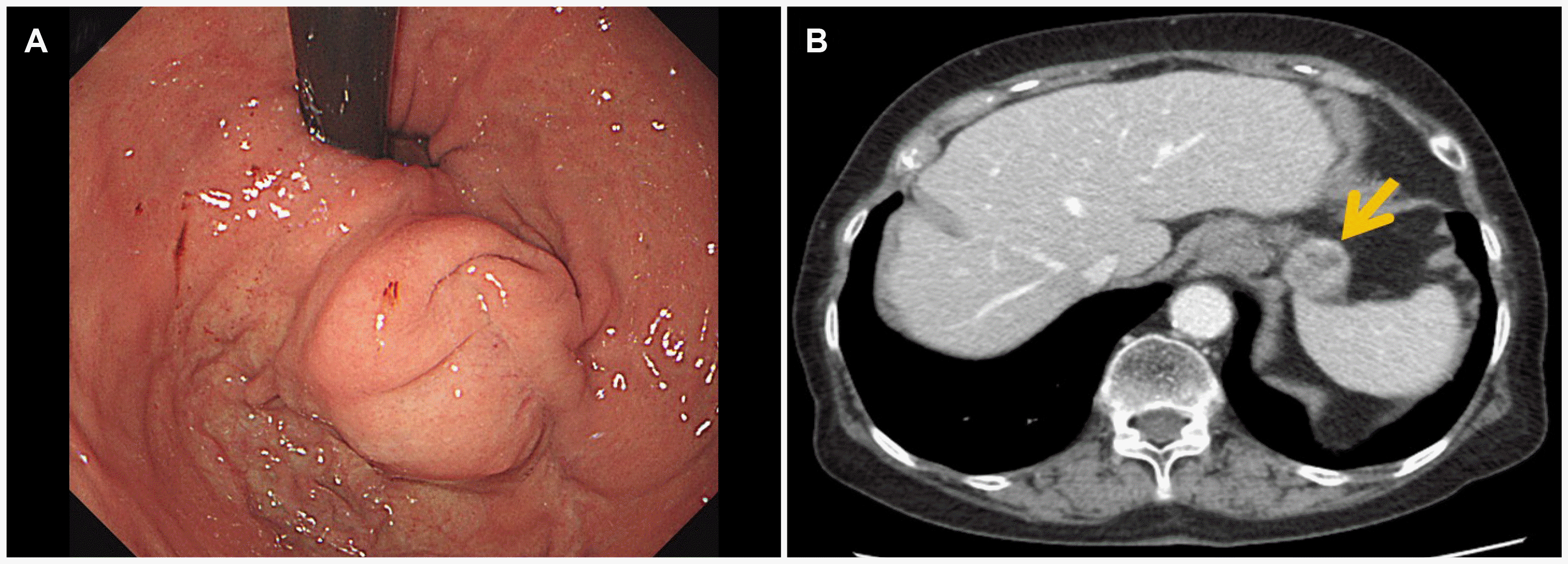 | Fig. 3Endoscopic images of the recurred gastric lesion and abdominal computed tomography findings at the first recurrence. (A) A 2-cm sized spherical shaped subepithelial mass lying on the anastomosis site. (B) An exophytic, heterogeneously enhancing mass at the anastomosis site of the stomach, measuring 2.6 cm, without signs of extragastric metastasis (arrow).
|
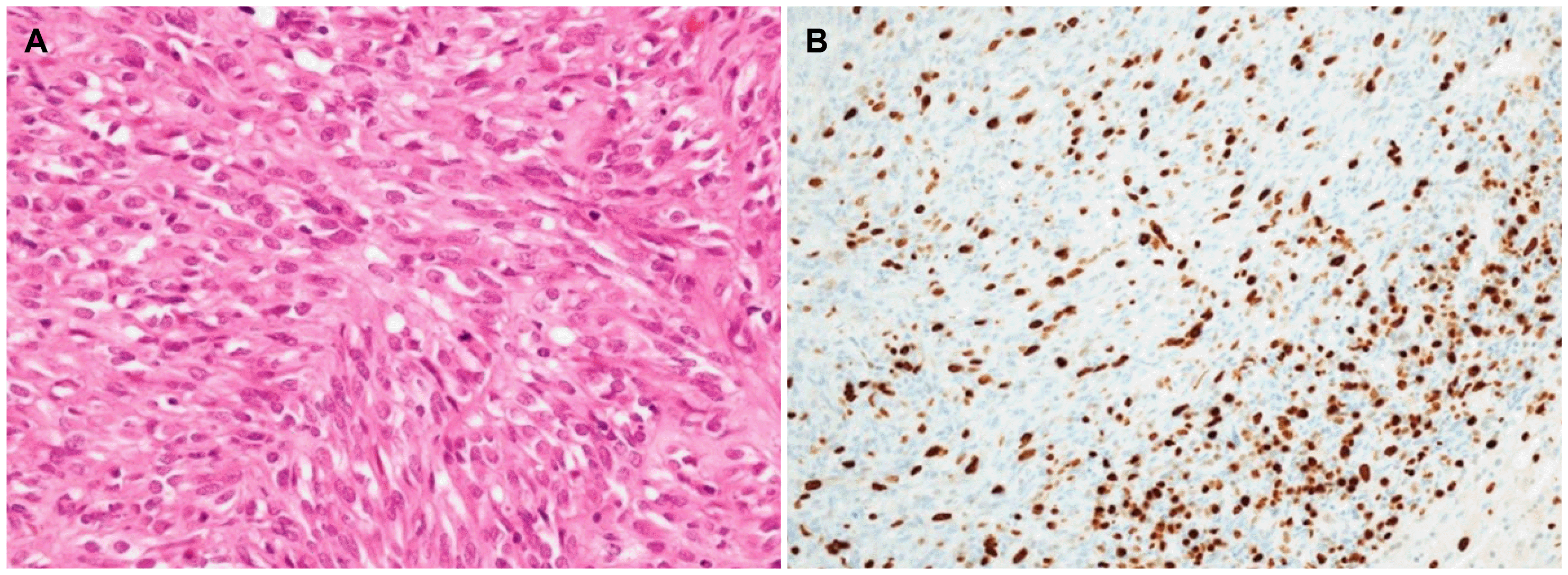 | Fig. 4Histologic features of a gastric gastrointestinal stromal tumor at the second surgery. (A) Histopathological evaluation revealed spindle cell proliferation with frequent mitoses up to 159/50 high-power fields (H&E, ×40) (B) Ki-67 proliferation index is 30% (Immunohistochemical staining, ×20).
|
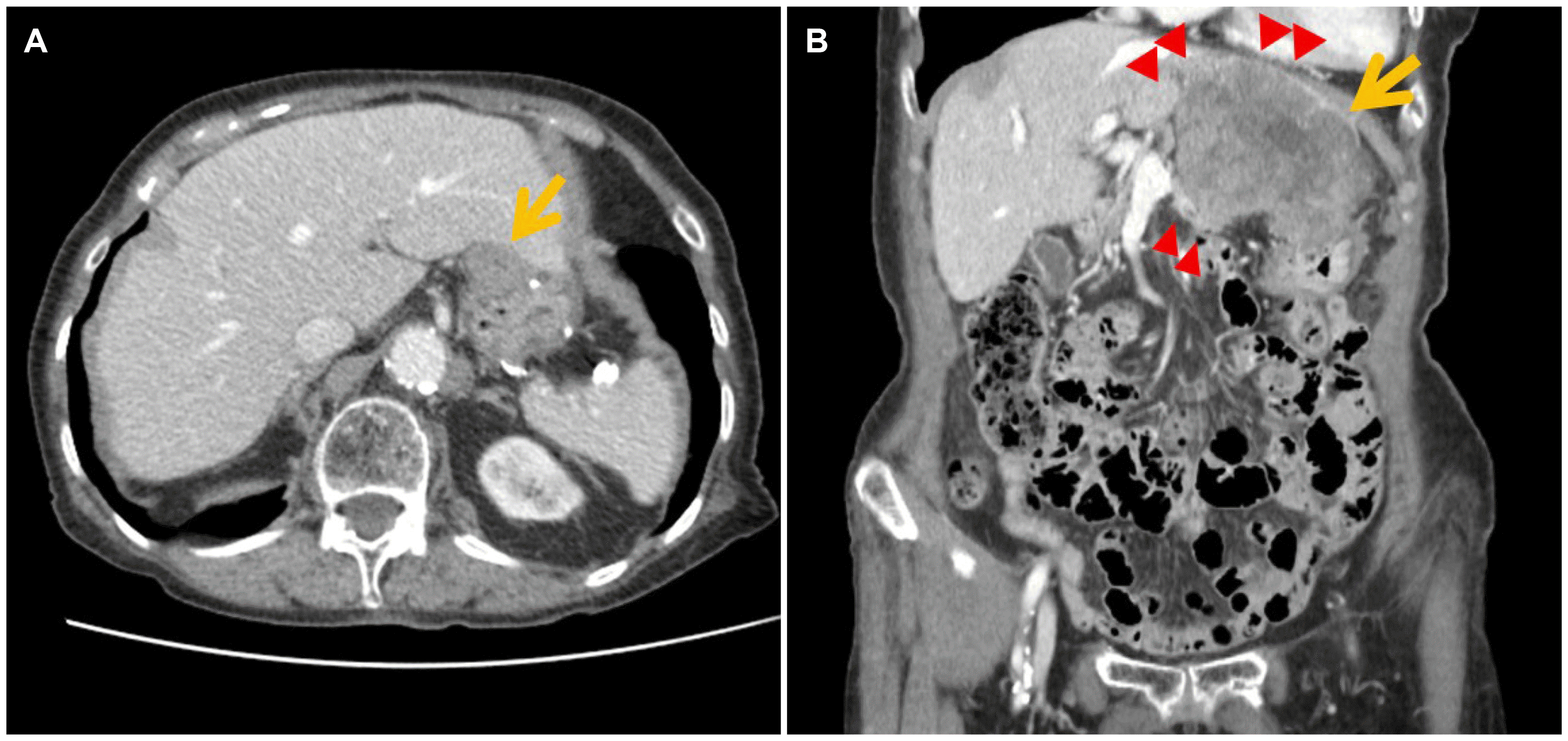 | Fig. 5Follow-up abdominal computed tomography findings after the second surgery. (A) A newly appeared 2.6-cm sized mass was found at the gastric anastomosis site (arrow), 1.5 years after the second surgery. (B) After 6 months of observation, the recurrent gastric lesion increased in size (2.6→9.4 cm, arrow) with invasion to the adjacent liver, pancreas, diaphragm, and pericardial fat (arrowheads).
|
Go to :
DISCUSSION
After complete surgical resection of a localized GIST, approximately half of the patients showed tumor recurrence, and the 5-year disease-specific survival was below 60% in the pre-imatinib era.4 The risk of recurrence varies widely according to the characteristics of the tumor. A high mitotic index and large tumor size are almost uniformly considered factors associated with poor outcomes.2,3,5 Other factors, such as non-gastric primary tumor location, exon 9 mutation, or “wild type” kit, also appear to be deleterious prognostic factors. Intraperitoneal rupture or bleeding while manipulating the tumor during the surgery is another factor associated with a high risk of postoperative recurrence.2
Imatinib has shown its efficacy in treating metastatic and unresectable GISTs, and can also improve the recurrence-free survival after a surgical resection in patients with high-risk GIST. Currently, adjuvant imatinib is a standard therapy in patients who have undergone surgery for GIST with a high risk of recurrence.6 On the other hand, the optimal duration of imatinib therapy after a surgical resection is still controversial. Some studies have shown that adjuvant imatinib for more than 3 years, even 5 years, might decrease tumor recurrence in high-risk GIST and that most recurrences occur after the discontinuation of imatinib.4,5
Two main factors may hinder long-term treatment with imatinib.4 One is the side effects associated with imatinib, and another is the acquired drug resistance. The most common toxic effects of imatinib at 400 mg/day include nausea, vomiting, edema, skin rash, and bone marrow toxicity. Although the drug-related side effects of imatinib are usually mild and resolve with dosage reduction or discontinuation, relatively severe toxicity can occur.7 Previous studies have shown that all patients treated with imatinib experienced one or more adverse events of any grade, and 21×43% experienced at least one Grade 3 or 4 adverse event at a dose of 400 mg/day.4,7,8 These might ultimately alter the efficacy of the imatinib treatment. In the present case, the patient had received adjuvant imatinib therapy only for 18 months. During the maintenance of imatinib, the patient suffered recurrent anemia. The anemia was not resolved completely after a transfusion and supplementation of oral iron. Owing to the severe hematologic adverse effects, she could not continue the imatinib therapy for a sufficient duration. In one report, grade 3-4 hematologic toxicity developed in approximately 20% of elderly patients who underwent imatinib therapy for chronic myelogenous leukemia (CML), which appeared to be related to myelosuppression.9 Nevertheless, imatinib therapy is generally regarded as a safe treatment and is still recommended in elderly patients with CML. Considering that myelosuppression is less common in patients with GIST on imatinib than in patients with CML, a similar treatment strategy can be applied for GIST in elderly patients.7 Close monitoring for toxicity, followed by a temporary dose reduction and treatment continuation can be considered for the maintenance of remission in this patient population.10
In conclusion, this paper reported a case of a completely resected gastric GIST with repeated local recurrence in a very elderly patient. The hematologic side effects of imatinib were controlled with dose reduction and conservative management, and the patient is still alive for 6 years after the initial diagnosis.
Go to :
 XML Download