

 PDF
PDF Citation
 Print
Print


Introduction
The inferior alveolar nerve (IAN) is a large branch of the mandibular nerve and arises from the nerve below the foramen ovale to descend through the infratemporal fossa (ITF) to reach the pterygomandibular space. Before entering the mandibular foramen, the IAN gives rise to the nerve to mylohyoid. Communications between the IAN, lingual nerve (LN), hypoglossal nerve or the auriculotemporal nerve (ATN) have been reported [1-5]. The IAN is clinically important nerve, especially in dentistry so that inferior alveolar nerve blockade (IANB) is better understood [6]. We report a presumably uncommon anatomical variation of the IAN adjacent to the foramen ovale.
Go to : 
Case Report
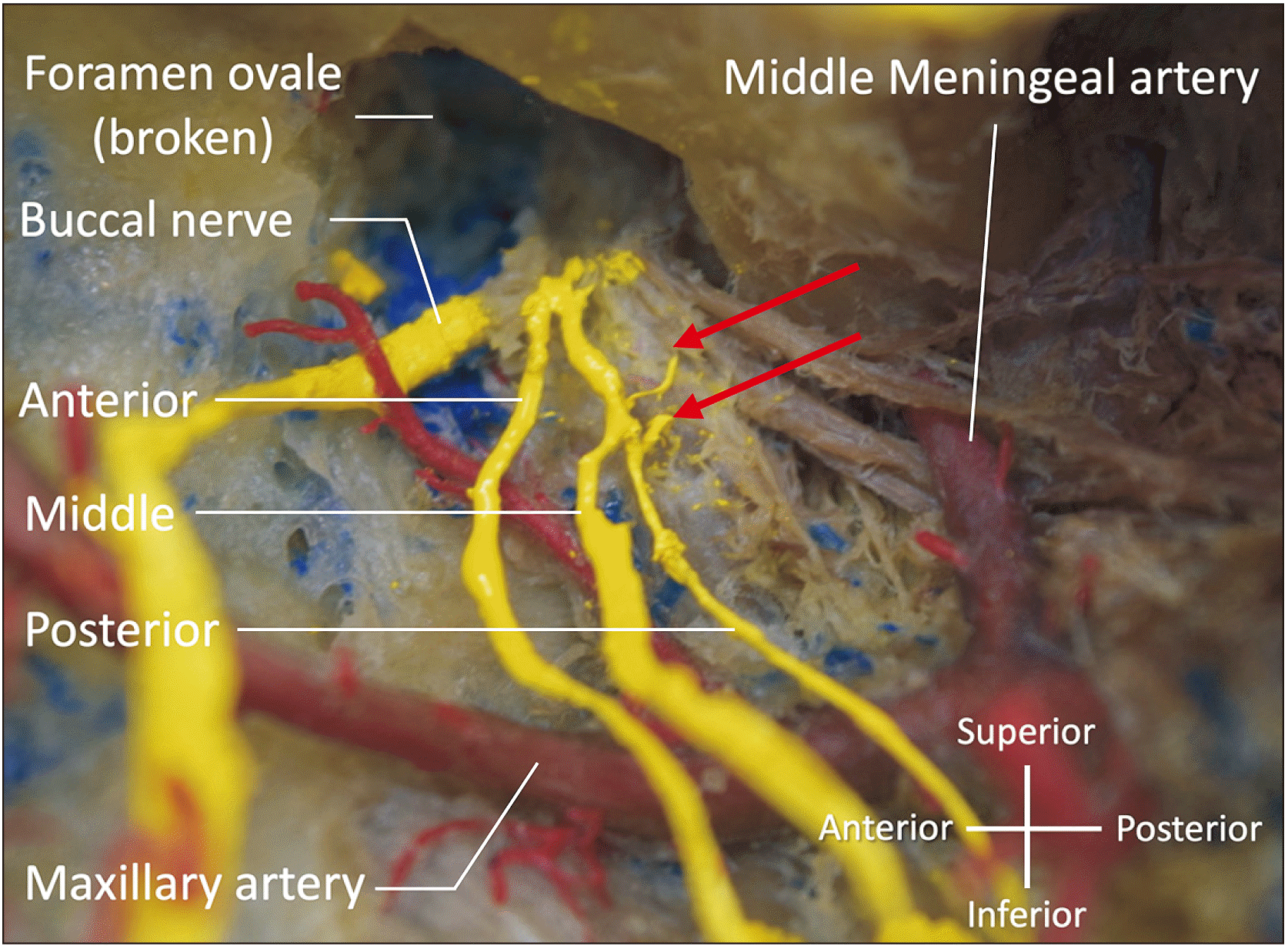
During routine dissection, a variant IAN was found in a formalin-fixed Caucasian male cadaver who was 74-years- old at death. In the left infratemporal fossa, three minor branches (anterior, middle, and posterior branches) arose from the main trunk of the mandibular nerve, passed lateral to the maxillary artery (MA), and joining the IAN (Fig. 1). The anterior and middle branches traveled through the lateral pterygoid muscle (LPM) and the posterior branches traveled between the LPM and tensor/levator veli palatini. The middle and posterior branches arose from the mandibular nerve as a common trunk and then soon bifurcated into two branches. Interestingly, the three branches united and joined the IAN at the root of the lingual branch of the inferior alveolar artery (IAA) and slightly touched the lingual branch. In addition, two, small contributions that originated from a ganglion-like structure, joined the common trunk of the middle and posterior branches, and the posterior branch (Fig. 2). The diameter of the anterior, middle, and posterior branches was 0.68 mm, 1.43 mm, and 0.40 mm, respectively. No other variations were observed in the maxillofacial and neck regions of this cadaver. The IAN on the contralateral side was normal. No scar was found in the head and neck regions. Histological observations were not conducted in this study.
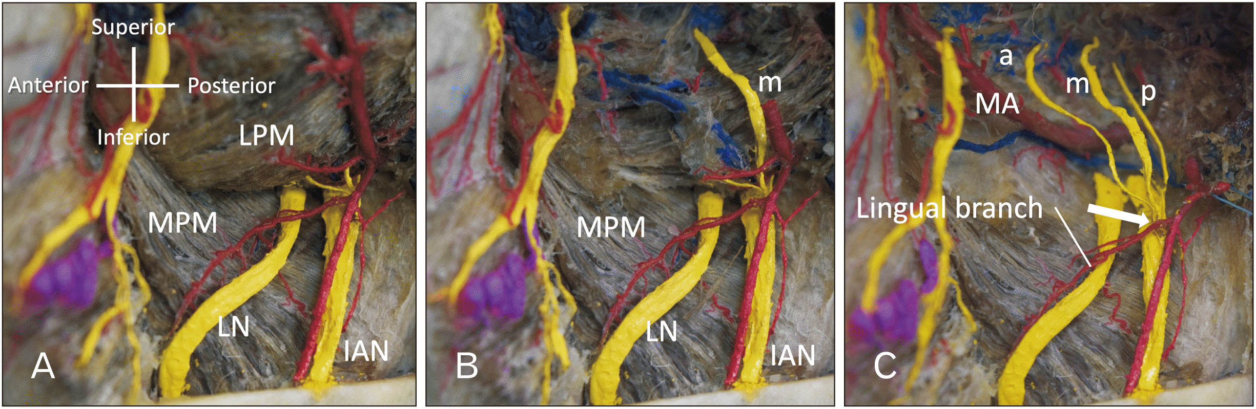 | Fig. 1Lateral view of the three variant branches arising from mandibular nerve in the left infratemporal fossa. The upper half of the mandibular ramus has been removed. (A) The three minor branches are not shown. (B) The middle branch (m) running within the lateral pterygoid muscle is shown. (C) The a, m, and p branches are shown after removal of the lateral pterygoid muscle. Note the three branches forming a common trunk to join the IAN at the root of the lingual branch of the inferior alveolar artery (arrow). a, anterior; IAN, inferior alveolar nerve; LPM, lateral pterygoid muscle; m, middle; MA, maxillary artery; MPM medial pterygoid muscle; p, posterior.
|
Go to :
Discussion
The IAN is one of main trunks of mandibular nerve. The minor three branches we found were oriented from mandibular nerve and connected to IAN. In 2004, Kim et al. [1] classified the branching of the IAN and LN into four types. Type IV was a complex form of branches of the mandibular nerve and its frequency was 6.3%. Iamsaard et al. [2] reported a unique connection between the trunk of the mandibular nerve and the LN communication. Loughner et al. [7] reported three of 52 specimens with three branches of the mandibular nerve that were apparently entrapped in the lateral pterygoid muscle, i.e., anterior and posterior deep temporal nerves and masseteric nerve. These three nerves, in the present case, were resected before dissecting the lateral pterygoid muscle.
Embryologically, the first pharyngeal arch is developed at the 6th week of gestation. Thereafter, branches of the mandibular nerve including the IAN, LN and ATN develop. Anil et al. [8] reported a communicating branch between the ATN and IAN and the MA coursed between the IAN and communicating branch, which appeared to be entrapped. Wolf et al. [9] reported a similar case and described an IAN split by the MA. Moreover, Khan et al. [10] reported three similar cases (out of 50) of a MA splitting the IAN These reported cases have some similar features to the present case. Although three branches found in the present study were very small in diameter, as some of these could distribute mental region via the mental nerve, compression of the nerves by LPM contraction might result in dysfunction of the mental nerve. In addition, the lingual branch of the IAA slightly contacted three branches at its root. This might result in ischemia of the LN as the lingual branch of the IAA supply the LN [11].
In dentistry, IANB is most commonly performed when treating the mandibular molar teeth. In 1973, the Gow-Gates [12] technique, a modified technique of the IANB, was published to target the main trunk of the mandibular nerve. The Vazirani-Akinosi technique is also an alternative option of a modified IANB [13]. Direct injury of variant branches such as the ones found in the present case might occur with either the Gow-Gates or Vazirani-Akinosi techniques due to the higher position of the needle insertion than the regular IANB technique.
A tiny branch between the otic ganglion and common trunk of the middle and posterior branches was also observed. Although postganglionic parasympathetic fibers are not mentioned for the IAN, it might be possible that postganglionic parasympathetic fibers travel with the IAN to be mental nerve and innervate the lower labial glands [14]. Another potential purpose of this fiber might be to join the nerve to mylohyoid to innervate the submandibular gland as the nerve to the mylohyoid runs near the submandibular gland.
In conclusion, we found three contributions from the mandibular nerve connecting to the IAN. These all traveled inside the LPM or between the LPM and tensor/levator veli palatini and all of them were superficial to the MA. Knowledge of such a variation might be helpful to dentists during, for example, anesthetic blockade and various oral surgeries.
Go to :
 XML Download