

 PDF
PDF Citation
 Print
Print


Introduction
Low back pain is one of the most common musculoskeletal disorders that causes a tremendous socio-economic burden for patients due to loss of productivity and increased cost of health care [1]. Although the pathogenesis of low back pain involves numerous and complex causes, the degeneration of the intervertebral disc (IVD) seems to be the main contributing factor [2]. The IVD takes on the role of a spine shock absorber and accommodates compressive loading forces [3]. The disc consists of nucleus pulposus (NP) and an annulus fibrosus (AF) and vertebral end plate (VEP) [4]. Cells in the NP region reside within the extracellular matrix (ECM) rich in proteoglycans, which gives the gelatinous and hydrated properties of the NP matrix rich thereby contributing to the mechanical tensile strength towards the surrounding AF [5].
Intervertebral disc degeneration (IVDD) is a complex and multifactorial process, influenced by genetic, nutritional and mechanical factors [6]. As the cascade of degeneration progresses, production of pro-inflammatory molecules such as the factor of tumor necrosis and interleukins occurs at an alarming rate, simultaneously coinciding with the degeneration process [6]. In addition, endplate calcification impairs the flow of nutrients and exacerbates the environment of hypoxic acid contributing to nutrient insufficiency and inflammatory environment thereby accelerating cell death within NP [7]. As a result of the aforementioned factors, proteoglycan levels and other proteins of the ECM decline leading to disc desiccation and gradual instability [8]. One of the major low back pain underlying causes is IVD degeneration [9]. Furthermore, other pathologies of the disc such as herniation of the disc, spondylolisthesis, stenosis of the spinal canal or facet joint syndrome are also contributed to IVD degeneration [10].
One of the most common surgical approaches for discogenic back pain is Spine fusion [11]. Although existing surgical treatments provide better pain relief than non-surgical interventions [12]. However, they fail to address disc degeneration biology, such as high levels of pro-inflammatory cytokine or the inherent loss of NP cells. In additional, studies have shown that surgery can lead to increased biomechanical stress and the onset of adjacent segment degenerative cascade [12].
Grape is one of the world’s most regularly consumed fruits due to its rich polyphenol content of different biological significance mainly present within the seed (60%–70%) and skin (30%) [13]. Previous research conducted on the phytochemical analysis of grape seed extract (GSE) revealed high amount of phenolic (141.26 mg gallic acid/g), flavonid (68.16 mg quercetin/g) and polymeric procyanidins [14]. The most effective antioxidant is grape seed proanthocyanidin [15]. It was observed that High-performance liquid chromatography analysis showed that Catechin (33.44) and epicatechin (13.03) were the most important phenolic compounds of GSEs. Polyphenol of grape has been reported to have multiple biological activities in grape seeds, including antithrombotic and cardioprotective effects, oxidative stress inhibition, free radical reduction [13]. GSE has numerous biological attributes such as antibacterial, antiviral, antioxidant, anti-inflammatory, anti-allergic, and vasodilatory actions [16].
Grape seeds and their extracts have high amounts of proanthocyanidins that are thought to be powerful antioxidants with anti-inflammatory potential by scavenging reactive oxygen species (ROS) [13]. Previous studies have shown that GSE of proanthocyanidin possesses direct anti-inflammatory effects through immune cell modulation by reducing IL-1β, TNFα [17], osteoarthritis model hyperalgesia and allodynia [18], arthritis model hypersensitivity and TNFα levels [19], streptozotocin-induced diabetic neuropathy model allodynia [19]. In addition, GSE reduced microglia and astrocyte activation in the brain neurons and also cause reduction of Mitogen-Activated Protein Kinases in a model of temporomandibular joint pain through complete Freund’s adjuvant, suggesting an immune-cell-specific mechanism of action [20]. Furthermore, GSE antagonized increased expression of IL-6 due to treatment with IL-17, IL-1β, or TNFα, in human epithelial cells through inhibition of the NF-κB pathway and was also observed in human adipocytes and macrophages [21]. The anti-inflammatory effects of GSE are likely to be mediated through a reduction in cytokine transcription as a result of inhibition of NF-κB activity [21]. This study is therefore aimed at investigating the inhibitory response of GSE on NP cell apoptosis and its anabolic potential in annular puncture induced IVDD in rabbit model.
Go to : 
Materials and Methods
Drugs/chemicals
GSE was obtained from Arab Company for Pharmaceutical and Medicinal plants, Egypt, with 99% purity, dissolved in distilled water and given orally in a dose of 500 mg/kg. All other chemicals used in the study were of good analytical grade.
Surgical technique
All animal handling and surgical procedures were conducted in accordance with the ethical approval from the Health Research Ethics Committee, College of Medicine of the University of Lagos (CM/HREC/07/2019/121).
Twenty eight New Zealand White rabbits (weighing about 1.8–3.0 kg) were used with institutional animal care committee’s approval. The surgical technique was performed using modified Kwon [22] procedure. Briefly, each rabbit was anesthetized with intramuscular injection of xylazine (5 mg/kg) and ketamine (35 mg/kg), and the fur was shaved from the mid back and right flank. After anaesthesia, a lateral plain X-ray was obtained to establish the pre-injection baseline height of the IVDs. The rabbit was then placed in the lateral oblique prone position, and the injection field was sterilized with an alcohol sponge. Initially, the L5–L6 disc was identified through manual palpation of the interspinous space from the mid back and pelvic rim. After confirmation of the exact level, a 21-gauge angiography needle was inserted 3–4 cm ventrally from the midline into the disc space. After brief confirmation of the needle position in the centre of the disc space, the needle was held in the disc space for 30 seconds. Before removal, the needle was rotated 360 degrees. In each rabbit, each of three discs (L3–L4, L4–L5, and L5–L6) was punctured. The L1–L2 and L2–L3 levels were designated as the non-punctured, internal controls. For each level, all procedures for identification and puncture were performed within a calculated time. Special care was taken to minimize contact with the periosteal tissues of the vertebrae because this could cause hypertrophy of the soft tissues and bony structures around the discs. Rabbits were monitored for neurological symptoms. The rabbits were placed in their cages after observation for recovery.
The rabbits were divided into 7 animals per group:
Group A (non-punctured group) received distilled water orally for 4 weeks.
Group B (punctured group) underwent annular puncture and received distilled water orally for 4 weeks.
Group C (punctured treated group) underwent annular puncture and received distilled water orally for 4 weeks and thereafter received 500 mg/kg of GSE orally for another 4 weeks.
Group D underwent annular puncture and received 500 mg/kg of GSE immediately after puncture orally for 4 weeks.
Lateral X-ray of the lumbar spine was taken before and after the experiment to measure IVD height. After the experiments, rabbits were euthanatized and the IVDs were assessed. As an internal control, the non-punctured discs (L1/2 and L2/3) were also assessed.
At the end of the experiment, the animals were sacrificed with intramuscular injection of ketamine (25.0 mg/kg) followed by intravenous injection of sodium pentobarbital (1.2 g/kg).
Determination of percentage disc height index
Lateral plain radiographs of the lumbar spine were scanned and digitally stored with a radiograph machine by a Veterinary Radiologist (collimator-to film distance, 50 cm; exposure, 5 mAs; penetration power, 44 kVp). During the radiographs, special care was taken to minimize axial rotation of the disc space by holding the rabbit in the lateral decubitus position while ensuring the X-ray beam was maintained straight. In addition, each rabbit was treated with a consistent amount of ketamine (35 mg/kg) and intramuscular injection of xylazine (5 mg/kg) in order to provide a similar degree of muscle relaxation to minimize differences in disc height. Vertebral body height and disc height were measured using Scion Imaging Software 4.0 (Scion Corporation, Chicago, IL, USA) and analyzed using image analysis program (Image J; National Institutes of Health, Bethesda, MD, USA).
The IVD height was expressed as the disc height index (DHI) using the method by Masuda et al. [23]. The average IVD height was calculated using measurements obtained from the anterior, middle, and posterior portions of the IVD and was divided by the average of adjacent vertebral body heights. Changes in the DHI were expressed as percentage DHI and normalized to the measured pre-operative IVD height (percentage DHI=post-operation DHI/pre-operation DHI×100).
Histological studies
Histomorphology and organization of collagen and elastic fibres were analysed using Vialle et al. [24] procedure. The intact specimens of the sacrificed rabbits were fixed. The punctured discs and the control discs were harvested for histologic analyses. Tissues were fixed with 10% neutral buffered formalin for 48 hours, decalcified in Ethylinediaminetetraacetic acid solution (National Diagnostics, Atlanta, GA, USA) for 3 days and then processed for paraffin sectioning. Blocks embedded in paraffin were cut into mid-sagittal sections (4 μm in thickness) with a microtome. Sections were stained with hematoxylin and eosin (H&E) to determine the morphology and number of cellular constituents in the AF, NP and EP, Haematoxylin van Gieson stain was used to demonstrate collagen fibres in AF and NP; Verhoeff-van Gieson Stain was used to demonstrate elastic fibres in AF. The slides were analysed under a light microscope. The degree of IDD were assessed by a histological grading scale [23], with scores ranging from grade 4 (normal) to grade 12 (severely degenerated). This grading scale is based on degenerative changes in 4 regions: the AF, the border between the AF and the NP, the parenchyma of the NP, and the matrix of the NP.
Biochemical analysis
Rabbits were randomly chosen for real time quantitative polymerase chain reaction (PCR) using Kwon [22] procedure. The L3–L4 and L4–L5 levels were extracted for the punctured disc while L1–L2 disc was extracted as a non-punctured control. From each disc, the NP was carefully removed from AF and stored separately. The tissues were immediately placed into liquid nitrogen and frozen at –80°C in preparation for PCR analysis. Genes were selected from the representative forms related to the ECM component (collagen type 1 and 2, aggrecan) and catabolic enzymes Matrix Metaloproteinase-13 (MMP-13). The frozen NP samples were homogenized using Mini-Beadbeater; Bio Spec, Bartlesville, OK, USA in 1 ml Trizol reagent (Invitrogen, Carlsbad, CA, USA). RNA was extracted in accordance with the manufacturer’s instructions. The primers for the rabbit-specific genes were designed in accordance with published sequences in Kwon [22], procedure. Gene expression was analysed by RT-PCR using ABI PRISM 9700 (Applied Biosystems, Foster City, CA, USA). Gene expression levels were calibrated using a constitutively expressed housekeeping gene, glyceraldehyde phosphate dehydrogenase (GADPH). A positive standard curve for each primer was obtained using serially diluted cDNA sample mixture. Quantifications of gene expression were calculated (comparative Ct method) using standard curves and normalized to GAPDH in each sample, and then the expression of treated discs was normalized to control discs.
The sequences were as follows: Aggrecan (5’-GCTACGGAGACAAG GATGAGTTC-3’ and 5’-CGTAAAAGACCTCACCCTCCAT-3’), MMP-13 (5’-TGCCCCTCCTCAACAGTAAC-3’ and 5’-GAGCCCGCTGCATTCTTCTT-3’), collagen type II : (5’-TCAGGAATTTGGTGTGGACATA- 3’ and 5’-CCGGACTGTGAGGTTAGGATAG-3’), Type I collagen (5’-GGGCAAGACAGTCATCGAATA-3’ and 5’-GATTGGGATGGAGGGAGTTTA-3’) GAPDH (5’-AAGGCCATCACCATCTTCCA-3’ and 5’-GGATGCGTTGCTGACAATCT-3’).
Determination of expression levels of protein (immunohistochemical analysis)
The expression levels of Bax protein were determined using Le Maitre et al. [25] protocol. Representative IVDD areas were chosen based on the staining of the tissue sections with H&E. Formalin-fixed, paraffin-embedded 3 μm thick sections were de-paraffinized and rehydrated. Endogenous peroxidase activity was blocked by incubating the sections with 3% H2O2 for 10 minutes followed by digestion with 0.01% protease K for 10 minutes. Non-specific binding sites were blocked by incubation with confining liquid for 10 minutes after which the sections were incubated with rat polyclonal antibody to Bax (Cell Signaling Inc., Danvers, MA, USA) at 4°C for 12 hours. After thorough washing, the sections were incubated with biotinylated goat anti-rabbit IgG at 4°C for 60 minutes and then in Streptavidin-HRP for 10 minutes. The final color reaction was developed by incubation with the chromogenic substrate 3, 3’-diaminobenzidine (0.5 mg/ml in Tris). The sections were counterstained with haematoxylin and mounted for examination with an O-max microscope coupled to Image J software.
Statistical analysis
Data were analysed using IBM SPSS Statistics for Windows, Version 24.0 (IBM Co., Armonk, NY, USA). Longitudinal X-ray data were analyzed using two-way analysis of variance (ANOVA) followed by multiple comparison using Bonferroni method. Quantitative real time-PCR data were analyzed using student t-test. Data were presented as mean±standard error of mean. The level of significant was considered at P<0.05.
Go to :
Results
Radiological analysis
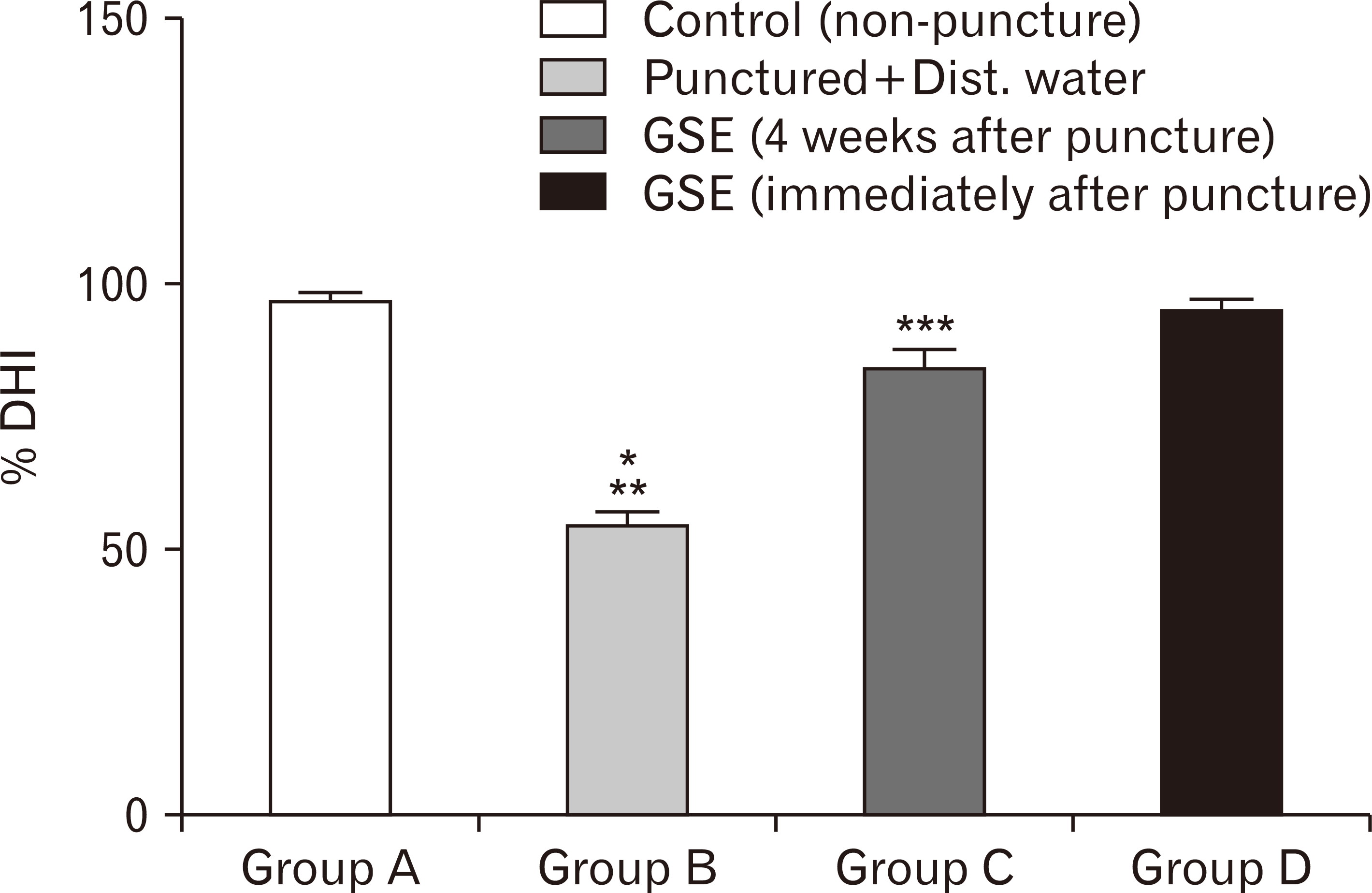
The percentage disc height index (% DHI) of the punctured group showed significant decrease compared to the non-punctured group (group A) (P<0.05) (Fig. 1). The narrowing of the disc height observed revealed the degeneration of the intervertebral disc after 4 weeks of puncture. However, the administration of GSE immediately after puncture (group D) showed no significant difference in the % DHI compared with non-punctured group (group A). Although, there was a significant difference in the % DHI of the group administered with GSE after 4 weeks of puncture (group C) compared with the control and the group that received GSE immediately after puncture (P<0.05) (Fig. 1).
Histological analysis
The histomorphological photomicrograph of the IVD section of the punctured group that received distilled water (group B) showed disorganization of the IVD architecture similar to degeneration such as narrowing of the disc, fissures and collapse of the collagen fibers within the AF, absence of the NP cells. The photomicrograph of the group that received GSE immediately after puncture showed similar morphology with the non-punctured group such as observable numerous chondrocytes like cells (CLCs) in the NP, distinctive alignment of the collagen fibers with the AF, well-organized VEP. However, the photomicrograph of the group administered with GSE 4 weeks after puncture showed regeneration of the NP cells, minimal disorganization of the collage fibers within the AF and restoration of the IVD height compared with group B.
Histological grading scores
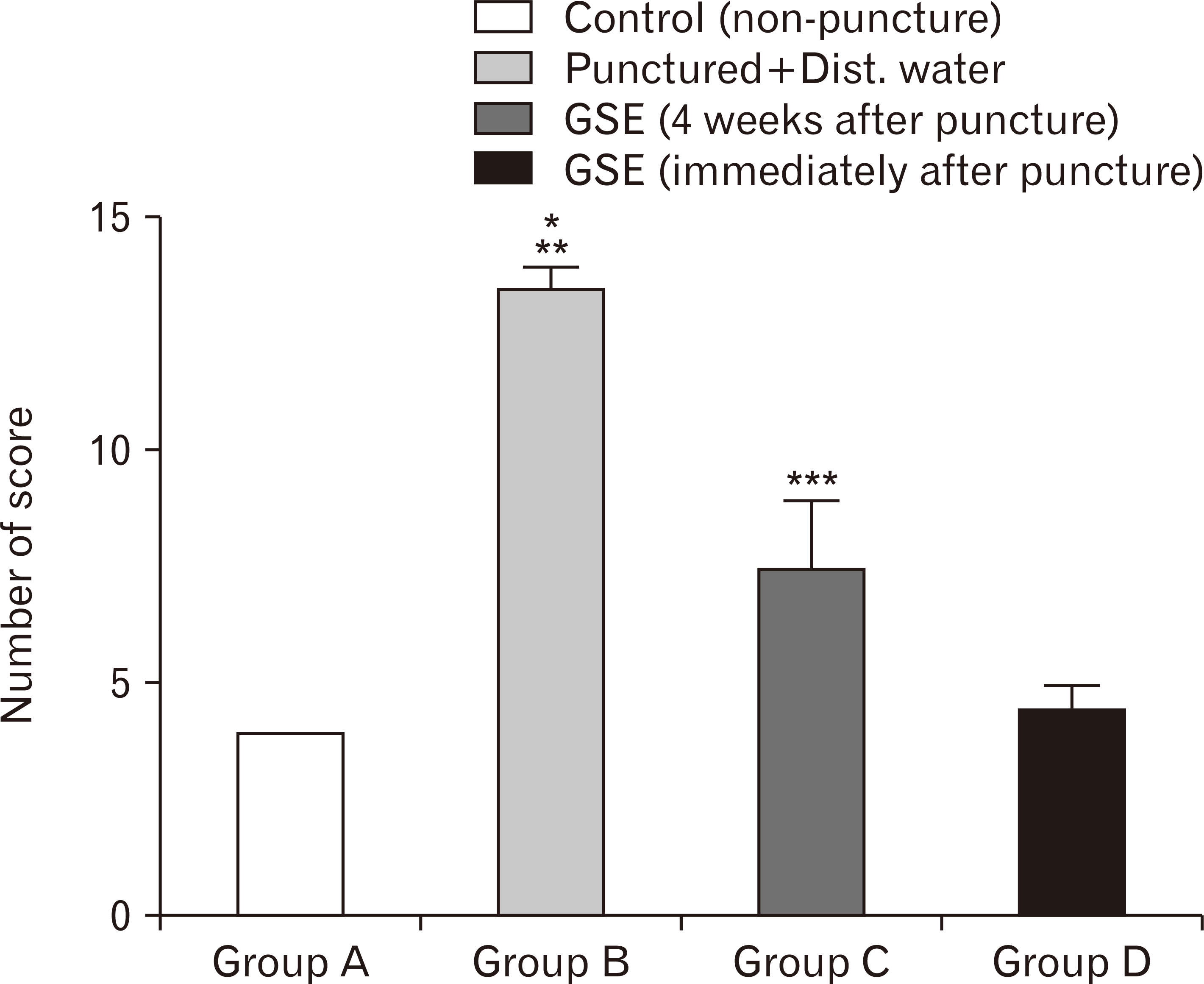
The histological grading score as previously described by Boos et al. [26] was used to ascertain the degree of degeneration of the punctured IVD within the photomicrograph. The level of degeneration varied from 4 (normal) to 14 (severe degeneration). In this present study, the punctured group that received distilled water showed a significant increase in the grading score (13) compared with the control (4) (P<0.05) (Fig. 3). In addition, the punctured group that received GSE immediately after puncture (group D) showed no significant difference in the grading scores (4.5±0.5) compared with the control (4). However, the histological grading score within the group that received GSE 4 weeks after puncture was significantly lower compared with group B (P<0.05) (Fig. 3). Although, the grading score of the group that received GSE 4 weeks after puncture was significantly higher compared with groups A and D respectively (P<0.05) (Fig. 3).
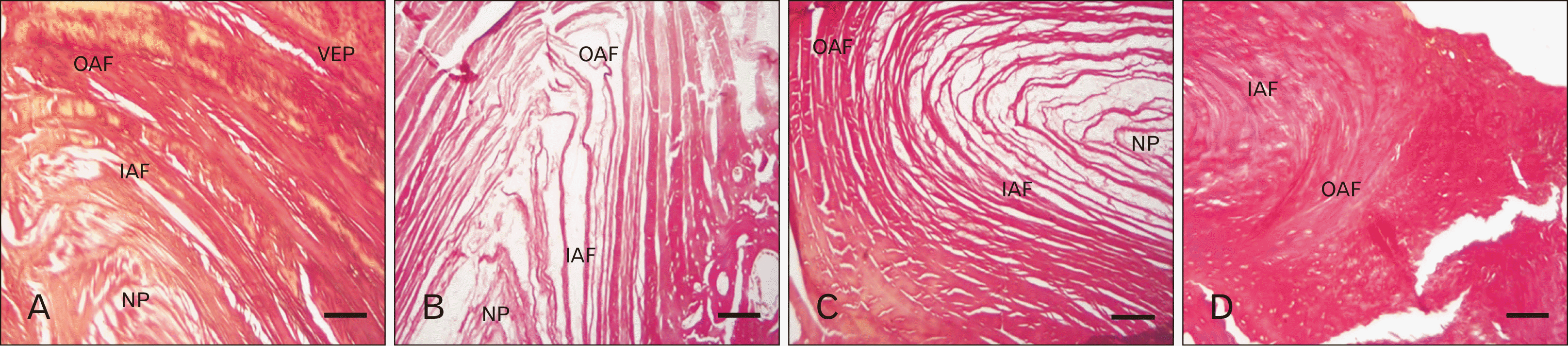
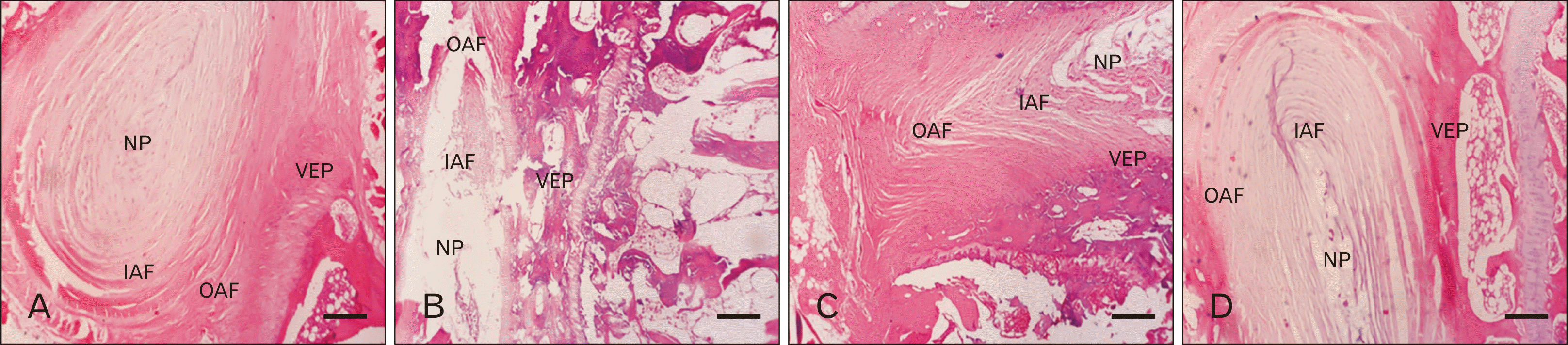 | Fig. 2Photomicrograph of IVD histology in rabbit administered with Grape seed extract in the control and punctured treated groups (H&E, ×100). (A) Group A: showing normal IVD cytoarchitecture with numerous CLCs in the NP. (B) Group B: showing absence of CLCs in the NP and disorganization of collagen fibers in the AF. (C) Group C: showing nearly similar features with group 1. (D) Group D: showing restoration of NP contents. CLCs, chondrocytes like cells; IAF, inner annulus fibrosus; IVD, intervertebral disc; NP, nucleus pulposus; OAF, outer annulus fibrosus; VEP, vertebral end plate. Scale Bars=250 μm (A–D).
|
Morphometry analysis of chondrocyte-like cells in IVD
The number of chondrocyte like cells within the AF (inner and outer) and NP is one of the major predictive features to assess the level of IVD degeneration. In this present study, the punctured group that received distilled water (group B) showed a significant decrease in the CLCs within the NP and inner AF compared to the control (P<0.05) (Fig. 4). The number of CLCs within the
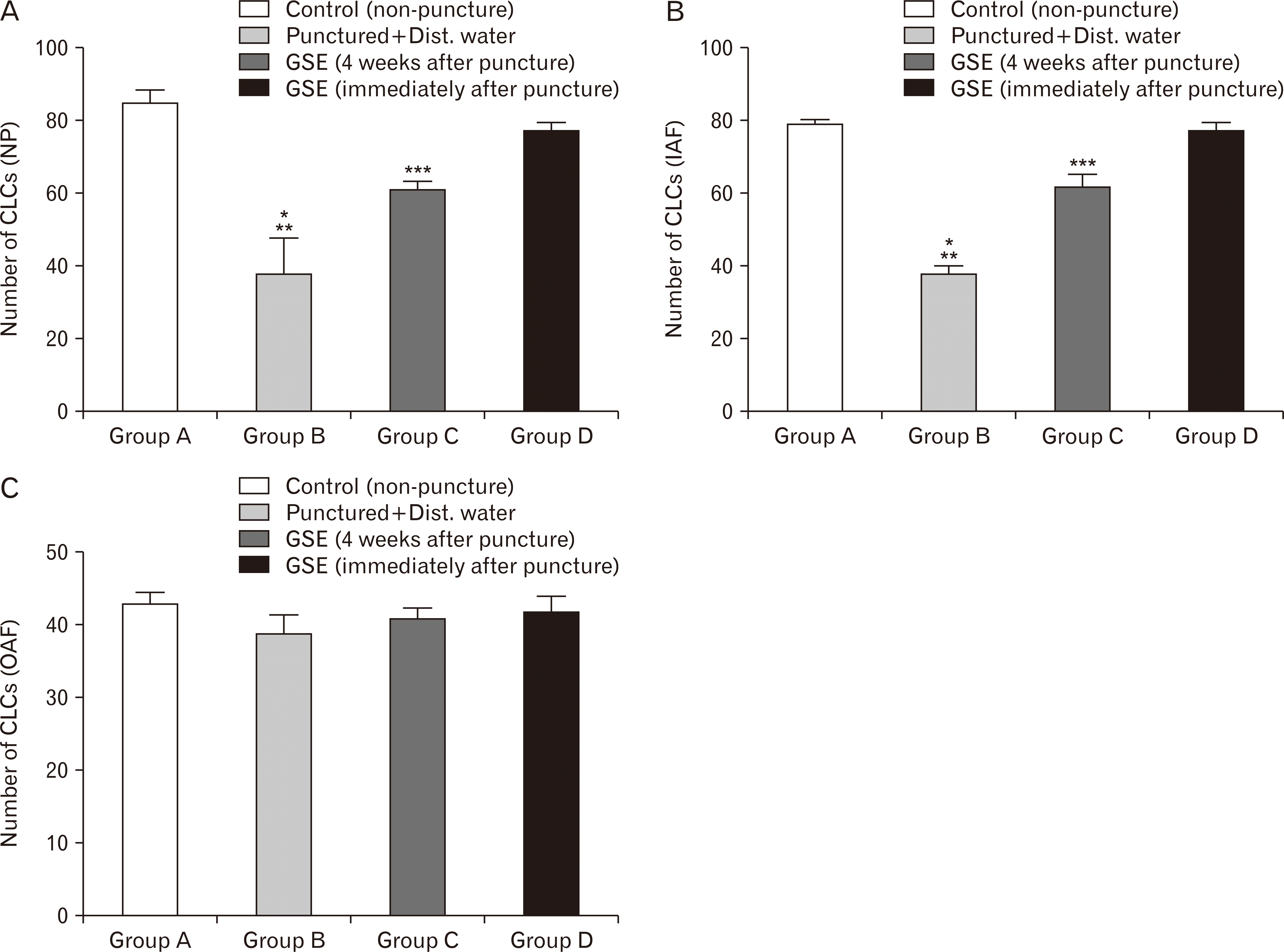 | Fig. 4(A–C) Effect of GSE on morphometric analysis of CLCs in annular punctured induced disc degeneration in rabbit (n=7). Group A, non-punctured group; Group B, punctured non-treated group; Group C, treated group 4 weeks after punctured; Group D, treated group immediately after punctured. CLCs, chondrocytes like cells; Dist., distilled; GSE, grape seed extract; IAF, inner annulus fibrosus; NP, nucleus pulposus; OAF, outer annulus fibrosus. *P<0.05 as compared to group A; **P<0.05 as compared to group C; ***P<0.05 as compared to groups A and D.
|
NP and inner AF in the punctured group that received GSE immediately after puncture showed no significant difference compared to the control. Although, the increase in number of CLCs in the NP and inner AF of group C was significant compared to group B (P<0.05) (Fig. 4). However, there was no significant difference in the number of CLCs in the outer AF across all the groups.
Histological sections of organization of collagen fibres
In the present study, the photomicrograph of the architecture of the collage fibers in the punctured group that received distilled water showed lamellae structure distortion, disorientation of the obliquity of the collagen fibrils and incongruent in the attachment of the lamellae to the VEP compared with the control. However, the group that received GSE immediately after puncture showed similar organization in the alignment of the collagen fibrils and firm attachment of the lamellae to the VEP compared to the control group. Furthermore, the non-punctured group (control) showed normal alternation of lamellae in the AF with longitudinal and oblique arrangements within the outer and inner areas, firm attachment of the lamellae to the VEP thereby forming sharpey’s fibers. Although, the punctured group that received GSE 4 weeks after IVD punctured showed mild disorganization of the collagen fibers compared to the control.
Histological sections of organization of elastic fibres
The photomicrograph of punctured group that received distilled (group B) showed disorganization, distortion and loss of continuity in the alignment of the elastic fibres in AF with longitudinal arrangement of fibre not readily visible compared to the control. However, the photomicrograph of the treated groups (groups C and D) revealed similar morphological organization of elastic fibre after the punctured animals were treated with GSE both immediately after puncture and 4 weeks after puncture respectively showing the attachment of sharpey’s fibers of the AF to the VEP.
Gene expression levels
The present study showed that the gene expression levels of AGG and COL 2 were significantly decline (Fig. 7B, D) with corresponding increase in expression levels of 24. COL 1 and MMP-13 (Fig. 7A, C) within the punctured group that received distilled water compared to the control (P<0.05) (Fig. 7). In addition, the group that received GSE 4 weeks after punctured showed that the gene expression levels of AGG and CoL 2 were significantly increased with corresponding decrease in CoL 1 and MMP-13 compared to group B (P<0.05) (Fig. 7). However, there was no significant difference in the relative gene expression levels among the group that received GSE immediately after puncture compared to the control. Although, the gene expression levels of AGG and CoL 2 were significantly decrease with corresponding increase in the expression levels of CoL 1 and MMP-13 compare to groups A and D respectively (P<0.05) (Fig. 7).
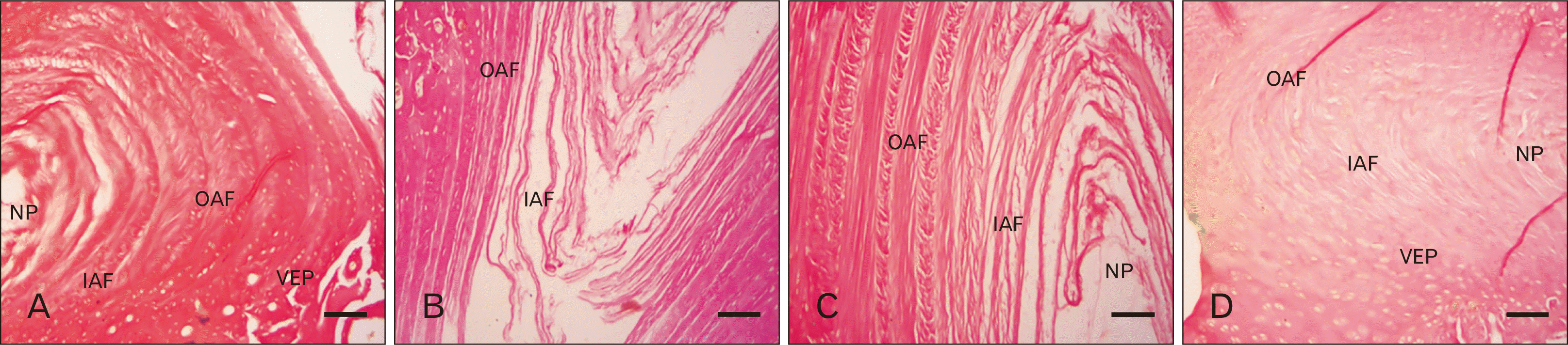
 | Fig. 5(A) Photomicrograph of IVD collagen fibers organization in control rabbit showing parallel arrangements of collagen fibres in AF. (B) Photomicrograph of IVD collagen fibers organization in punctured non-treated rabbit showing disorganization and loss of continuity a of the arrangement of collagen fibres within the outer and IAF. (C) Photomicrograph of IVD collagen fibers organization in treated rabbit after 4 weeks of punctured showing nearly normal re-organization in the distorted arrangements of collagen fibers in both outer and IAF. (D) Photomicrograph of IVD collagen fibers organization in treated rabbit immediately after punctured showing parallel arrangements of collagen fibres in AF (Haematoxylin van Gieson, ×100). AF, annulus fibrosus; IAF, inner annulus fibrosus; IVD, intervertebral disc; OAF, outer annulus fibrosus; VEP, vertebral end plate. Scale Bars=100 μm (A, B, D), 250 μm (C).
|
 | Fig. 6(A) Photomicrograph of IVD elastic fibers organization in control rabbit showing parallel arrangements of elastic fibres in AF. (B) Photomicrograph of IVD elastic fibers organization in punctured non-treated rabbit showing disorganization in the arrangement of elastic fibres within the outer and IAF. (C) Photomicrograph of IVD elastic fibers organization in treated rabbit after 4 weeks of punctured showing nearly normal arrangements of elastic fibers in both outer and IAF. (D) Photomicrograph of IVD elastic fibers organization in treated rabbit immediately after punctured showing parallel arrangements of elastic fibres in AF (Verhoeff van Gieson, ×100). AF, annulus fibrosus; IAF, inner annulus fibrosus; IVD, intervertebral disc; NP, nucleus pulposus; OAF, outer annulus fibrosus; VEP, vertebral end plate. Scale Bars=100 μm (A-D).
|
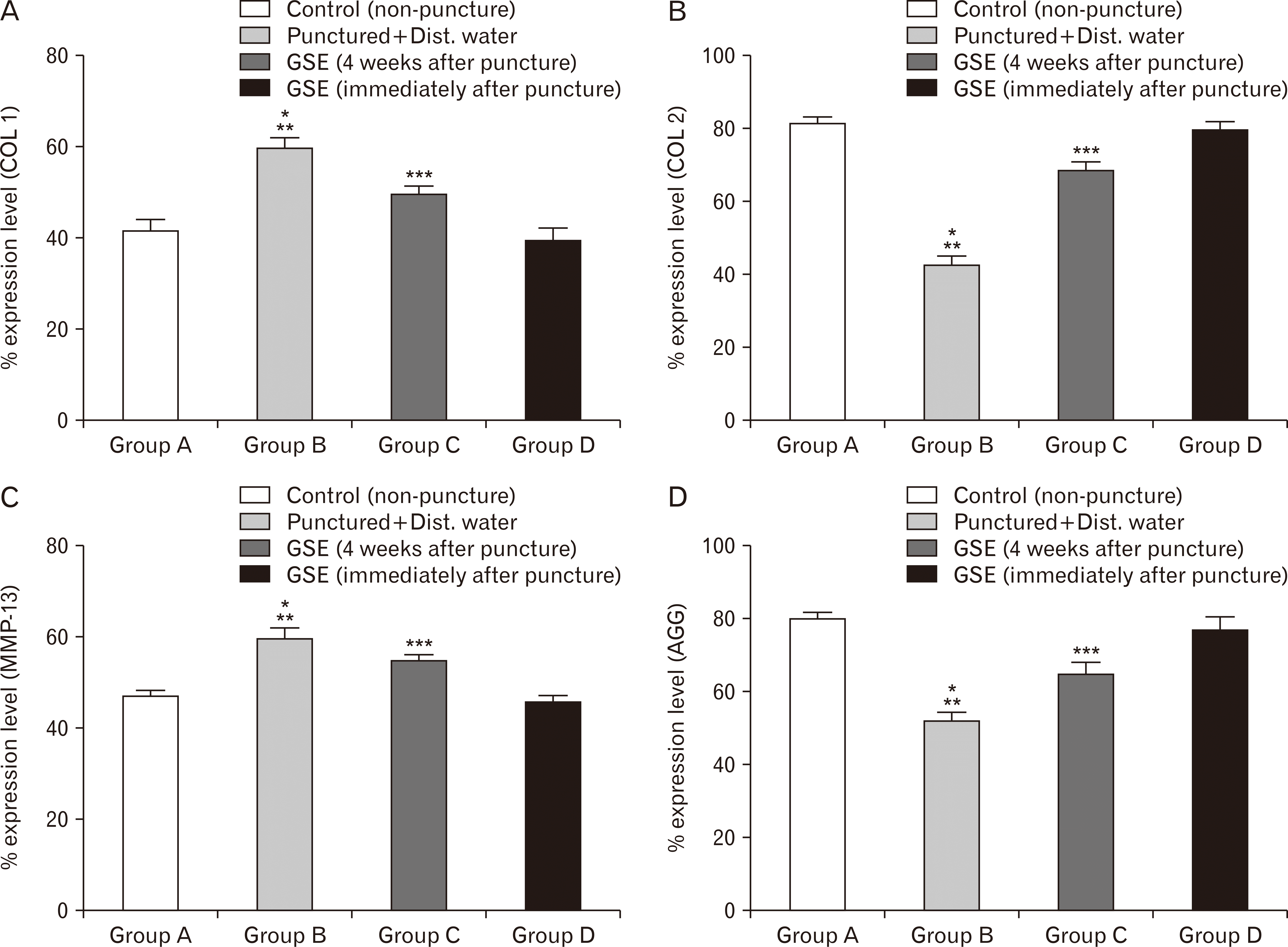 | Fig. 7(A–D) Effect of GSE on levels of gene expression in annular punctured induced disc degeneration in rabbit (n=7). Group A, non-punctured group; Group B, punctured non-treated group; Group C, treated group 4 weeks after punctured; Group D, treated group immediately after punctured. CoL 1, Collage type I; CoL 2, Collagen type II; Dist., distilled; GSE, grape seed extract; MMP-13, Matrix Metaloproteinase-13. *P<0.05 as compared to group A; **P<0.05 as compared to group C; ***P<0.05 as compared to groups A and D.
|
Immunohistochemical analysis
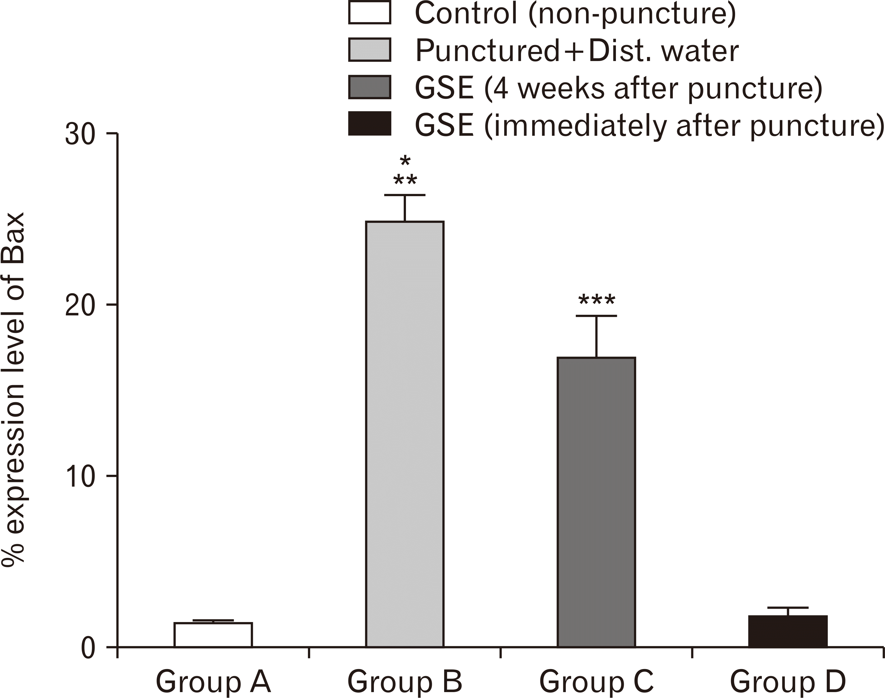
The immunohistochemical analysis in this present study showed that Bax expression level was significantly higher within the punctured group that received distilled water compared to the control (P<0.05) (Fig. 8). However, the treated group that received GSE immediately after puncture showed similar expression level of Bax compared to the control. In addition, the expression level of Bax in the group that received GSE 4 weeks after puncture was significantly higher compared to the control and group D (P<0.05) (Fig. 8). Although, the expression level of Bax among the group that received GSE 4 weeks after puncture was significantly lower compared to the punctured group that received distilled water (P<0.05) (Fig. 8).
Go to :
Discussion
The increased number of people with disability due to low back pain in both developed and developing countries has a major socio-economic impact since the prevention and therapeutic intervention is hampered as a result of non-clarity in the pathophysiology of low back pain [27]. However, the association of low back pain with IVDD has been reported [28]. Although, aetiology of the degeneration of the intervertebral disc implication in low back pain was documented to be a multi-factorial process involving several changes in disc architecture and function, biological and biomechanical alterations, genetic and biochemical composition of the native tissue [28]. Since the pathogenesis of disc degeneration is a multi-factorial process, most treatments modalities are geared towards treating the several pathological conditions arising from degenerated disc rather than addressing the mechanism by which degeneration of the intervertebral disc can be reversed, slowed or prevented. However, recent researches are now focusing on novel therapeutic approaches for the direct management of disc degeneration among which are synthetic peptide or growth factor injections [23], gene therapy [29], phytonutrients and supplements [30], cell therapy and tissue engineering [29].
GSE has anti-inflammatory, antimicrobial, anticarcinogenic, antidiabetic, antioxidant and free radical scavenging properties [31]. In addition, the neuroprotective properties of GSE have been reported in neonatal rat hypoxic-ischemic brain injury model [23].
In this present study, the % DHI of the punctured group showed significant decrease compared to the non-punctured group thereby suggesting degeneration of the intervertebral disc attributed to the use of needle puncture. However, the administration of GSE revealed both preventive and restorative capability of the extracts used immediately after puncture and 4 weeks after puncture. The result corroborates with previous finding after the administration of D-Ribose-L-Cysteine and 1-Isothiocyanato-4-(methylsulfinyl) butane [30]. Radiological and histomorphological observation has been strongly proven to predict the severity of annular puncture induced disc degeneration in animal models. Among the observable characteristics include narrowing of the disc space and height, reduction in water content of the disc, decrease in CLC within the NP, alterations in the morphology of the NP and disorganization of the collagen fibrils within the AF [30].
The present histomorphological findings showed affirmative characteristics of the preventive and restorative response of GSE in annular puncture induced disc degeneration in which the integrity of the NP and AF was improved after GSE administration. In addition, we observed increase cellularity in the NP and the organization of the arrangement of collagen and elastic fibers were preserved and restored after GSE administration immediately after puncture and 4 weeks after puncture respectively. The NP cells are CLCs that are capable of renewal or being prevented to undergo apoptosis due to degeneration processes. Previous observations have showed regenerative properties of several agents on annular puncture induced degeneration of NP cells [22].
The role of proteoglycan in retention capacity of water and swelling of the NP in intervertebral disc has been reported as the main factor acting on the disc for its shock absorbing nature [30, 32]. The decrease in the water content of the NP due to degeneration of the IVD results in morphological disorganization of the disc thereby causing phenotypic changes in the NP cells molecules that were linked to the onset of disc degeneration among which are Collage type I (COL 1), Collagen type II (COL 2), Aggrecan (AGC) and MMP-13 [32]. Our results showed increase in COL 2 and AGC expression with a corresponding decrease in COL 1 and MMP-13 in the GSE administered group compared to the punctured group. Previous studies have expressed several genes that modulated chondrocyte like cells and osteoarthritic chondrocytes [30]. However, research has linked MMP-13 as known proteins that have the capacity of causing degradation of collagens and glycosaminoglycans [32].
Apoptosis of the disc cells has been linked to the occurrence of IVD degeneration thereby leading to compression of the nearby nerve roots which ultimately results in radiculopathy radiculopathy [30]. The complex process in which cells undergo self-destruction without inducing any inflammatory response is referred to as apoptosis. Mitochondria-mediated apoptosis is mainly regulated by pro- and anti-apoptotic proteins. The anti-apoptotic Bcl-2 protein usually form links that maintain mitochondrial membrane thereby prevent the release of mitochondria-derived activator of caspases (cytochrome c). The pro-apoptotic Bax protein has been implicated in forming heterodimers with the anti-apoptotic Bcl-2 thereby causing the formation of pores within the mitochondrial membrane [29]. In this study, Bax expression levels were increased in the punctured group compared to the non-puncture group. However, the expression levels of Bax in GSE administered groups were reduced compared to the punctured group. Previous studies have attributed expression level of Bax to be implicated in the apoptosis of NP cells which ultimately affect the catabolic and anabolic processes involved in cell proliferation thereby leading to disc degeneration [32]. The present study showed that GSE inhibit and reverse cells of the NP, which could be attributed to the flavonoids and phenols present with the GSE. It was reported that several flavonoids showed ameliorative and neuroprotective potential against oxidative stress induce damage by increasing the endogenous antioxidant status thereby protecting the body against the deleterious damage of free radicals [33, 34]. Furthermore, several beneficial importances of polyphenols within the GSE against oxidative stress induced injury include inhibition of inflammation, prevention of LDL oxidation, and preservation of cells and tissues from oxidative stress damage [35, 36].
In addition, GSE was reported to decrease inflammation either by modulation of inflammatory pathways or by reducing ROS levels within the cells and tissue [37]. The procyanidins present in GSE target inflammation through several pathways such as inhibition of release of proinflammatory factors [38] thereby scavenging free radicals by inhibiting formation of pro-inflammatory cytokines. [39]. In addition to the numerous flavonoids and phenolics present in GSE, the inhibition of cell apoptosis observed in this study could also be assisted by the antioxidants presents similar to the antioxidant properties of Vitamin C. Previous study showed that Vitamin C significantly helps in the process of metabolism, collagen synthesis, vasculogenesis, aging and cell proliferation [40].
In conclusion, the present study demonstrated that GSE had preventive and restorative effects on punctured induced intervertebral disc through radiological, biochemical, histological and immunohistochemical studies. GSE enhances the synthesis of matrix proteoglycan and prevent the degradation of collagen fibrils within the disc tissues by increasing aggrecan synthesis and decreased in MMP-13 in the transcriptional level. The present study revealed that GSE has anti-inflammatory, anabolic and antioxidant attributes for treatment of disc degenerative diseases.
Go to :
 XML Download