Introduction
The mandibular foramen (MF) is a superior opening which is found in the medial surface of the ramus of the mandible [
1]. It has an important role to be a passage of mandibular canal which contains neurovascular structures such as inferior alveolar nerve, which is a branch of posterior trunk of trigeminal nerve that is sensory to the mucosa and skin around the lower lip and chin, and inferior alveolar artery, which is a branch of maxillary artery that goes through mental foramen [
1].
MF has an important implication in dental operations, especially in the inferior alveolar nerve block [
1]. So, anesthetic or surgical procedures are required a specific location of the MF, to perform precisely and appropriately, to prevent the operation failure which found mostly because of anatomical variation [
2]. One anatomical variation can be found is accessory MF which is a foramen that usually reported as an unnamed foramen in the body of ramus of the mandible [
3]. The inferior alveolar nerve and artery are also reported to also be seen in this accessory foramen [
4]. There are studies of the prevalence of inferior alveolar nerve block failure is related to the presence of accessory foramen [
4,
5].
Therefore, the anatomical study of the location of MF and prevalence of accessory MF is important for the inferior alveolar nerve block to prevent its failure, especially among the Thai population [
5] because there are no collections of data in Thailand yet. So, knowledge of the location of MF and the prevalence of accessory MF in the Thai population in this study will be helpful for other anatomical and dental studies [
2,
6].
This study aims to study the location of MF from various anatomical landmarks and prevalence of accessory MF from dry mandibles of the Thai population. The study of localization in anteroposterior and superoinferior axes are also noted.
Go to :

Materials and Methods
Samples
This study conducted a cross-sectional descriptive study to examine mandibular bones from the bone bank of the Forensic Osteology Research Center, Anatomy Department, Chiang Mai University. The male samples were 110 bones and the female samples were 110 bones. Collected mandibular samples needed to be from adults (more than 20 years old cadavers) and have at least two teeth in sockets to determine occlusion plane of samples. Exclusively, the damaged bones or bone with pathological diseases such as congenital anomalies, osteoporosis will be uncollected in this study.
Measurement
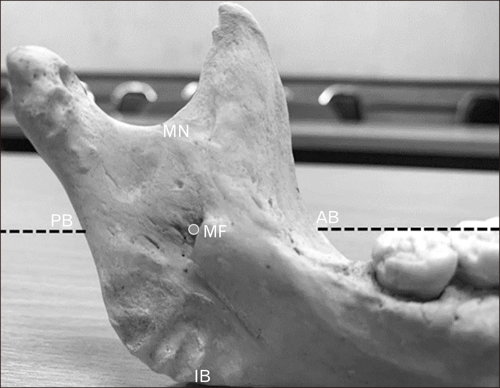
To locate MF, various parameters were measured by digital Vernier calipers of 0.02 mm accuracy on both sides of the mandibles as in
Fig. 1:
 | Fig. 1Parameters measurements from various anatomical landmarks to MF. 1. MF-AB: distance from the midpoint of the MF to the AB of the ramus on occlusion plane. 2. MF-PB: distance from the midpoint of the MF to the PB of the ramus on occlusion plane. 3. AB-PB: distance from the AB of the ramus on occlusion plane to the PB of ramus passing midpoint of the MF or the summation of MF-AB and MF-PB. 4. MF-MN: distance from the midpoint of the MF to the lowest point of the MN. 5. MF-IB: distance from the midpoint of the MF to the IB limited to the base of the mandible. 6. MN-IB: distance from the lowest point of the MN to the IB limited to the base of mandible passing midpoint of the MF or the summation of MF-MN and MF-IB. AB, anterior border; IB, inferior border; MF, mandibular foramen; MN, mandibular notch; PB, posterior border; ‾ ‾ ‾, occlusion plane; ●, midpoint of the MF. 
|
MF-AB: distance from the midpoint of the MF to the anterior border (AB) of the ramus on occlusion plane
MF-PB: distance from the midpoint of the MF to the posterior border (PB) of the ramus on occlusion plane
AB-PB: distance from the AB of the ramus on occlusion plane to the PB of ramus passing midpoint of the MF or the summation of MF-AB and MF-PB
MF-MN: distance from the midpoint of the MF to the lowest point of the mandibular notch (MN)
MF-IB: distance from the midpoint of the MF to the inferior border (IB) limited to the base of the mandible
MN-IB: distance from the lowest point of the MN to the IB limited to the base of mandible passing midpoint of the MF or the summation of MF-MN and MF-IB
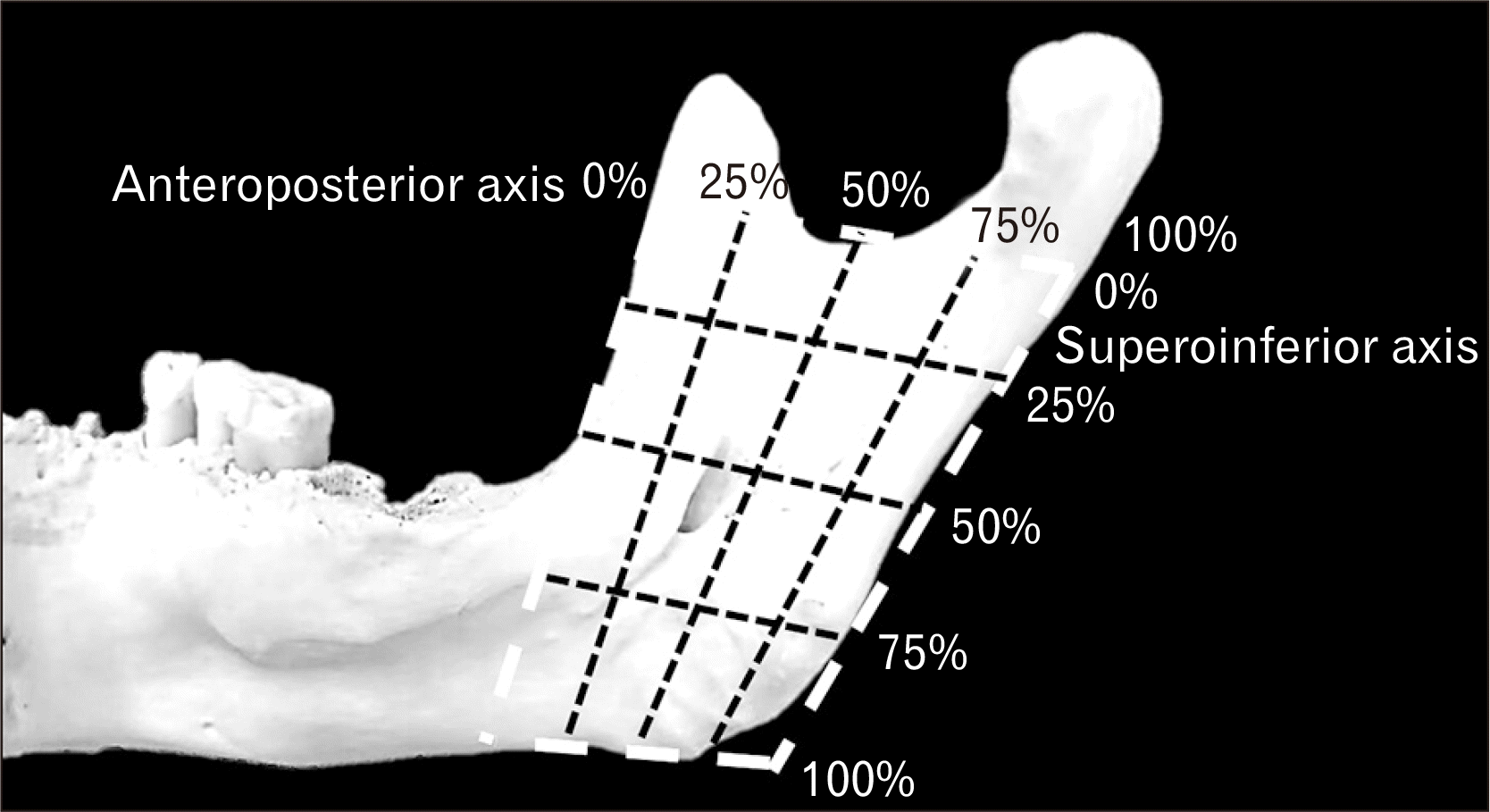
To be more precise, the location of MF was identified by the calculation of the prior parameters. Anteroposterior localization was calculated by percentile relation of MF-AB to the AB-PB and superoinferior localization was calculated by percentile relation of MF-MN to the MN-IB. The quartiles of MF both anteroposteriorly and superoinferiorly were indicated by the value from 0% to 25%, the first quartile; from 26% to 50%, the second; from 51% to 75%, the third and from 76% to 100%, the fourth as in
Fig. 2.
 | Fig. 2Mandibular foramen localization in anteroposterior and superoinferior axes. 
|
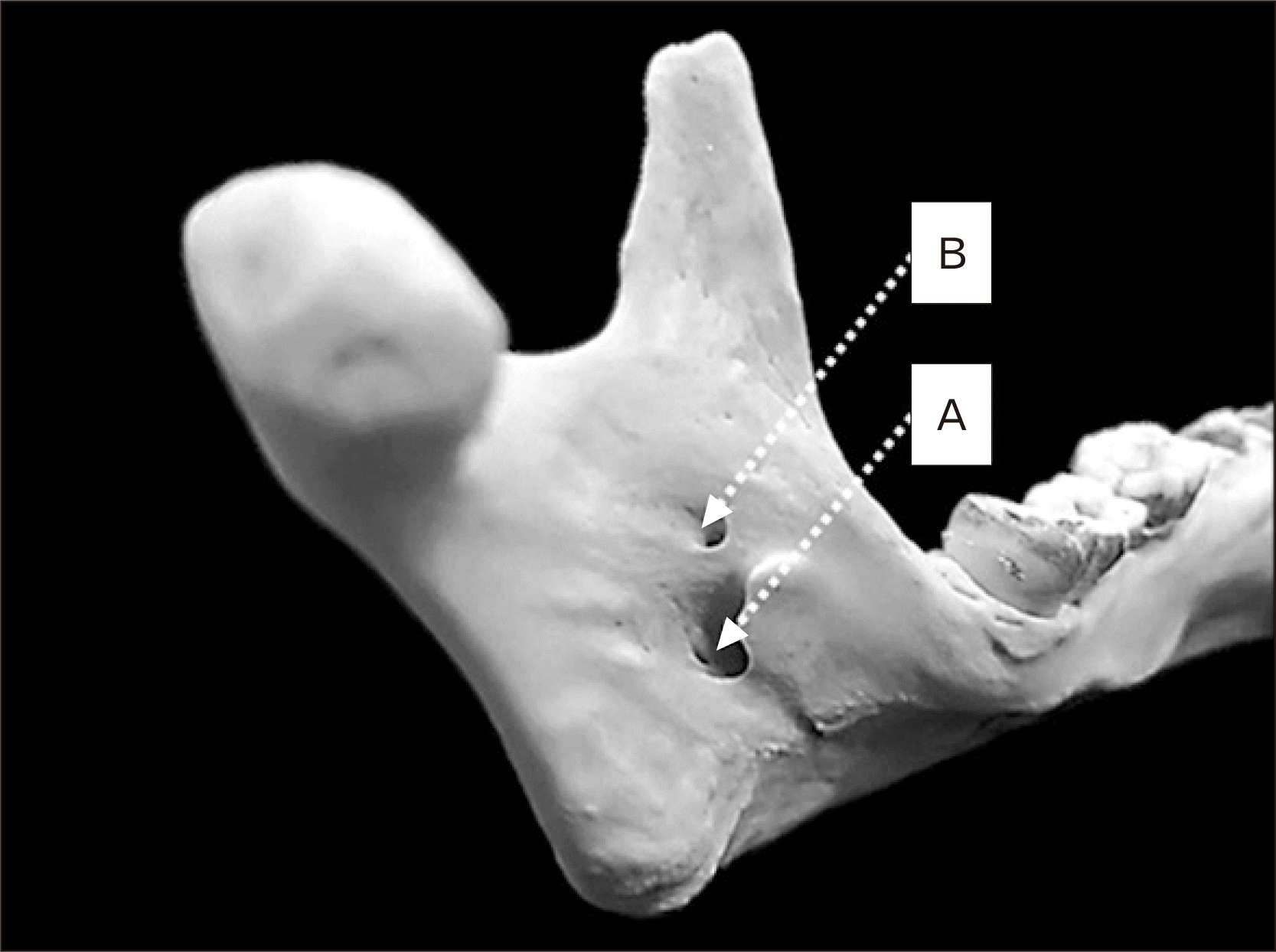
Further study, observation of the presence of accessory MF on both sides of mandibles by normal visual observation as
Fig. 3.
 | Fig. 3Accessory mandibular foramen observation. A, mandibular foramen; B, accessory mandibular foramen. 
|
The intra-observation was collected by the repetition of measurement similar parameter by one person three times from 20 samples, and inter-observation also was collected by another person measured parameter compared to mean of data from intra-observation from similar 20 samples. This process will give more reliability to the measurement procedure.
Statistics analysis
The various parameters indicated the location of MF was calculated to figure out mean and standard deviation. The parameters were compared between mandibular foramina in different sex. Also, the parameters were compared between both sides of mandibles among total samples, male samples and female samples.
The percentile of anteroposterior and superoinferior localization were calculated mean and standard deviation to find quartiles of MF in each mandible. The modest quartile of foramen would obtain a representative quartile of localization.
The accessory MF observations were calculated frequency and percentage classified by the absence of accessory MF, single accessory MF either right or left side, double accessory foramina either right or left side, bilateral accessory foramen, and single accessory foramen on one side and double accessory foramina on another side.
All the prior parameters were carefully statistic analyzed by IBM SPSS Statistics for Windows, Version 26.0 (IBM Co., Armonk, NY, USA) and Microsoft Excel 2016 (Microsoft Corp., Redmond, WA, USA). The descriptive analysis was employed for describing the central tendency and dispersion of data and an independent sample t-test was used as a test of significance under P-value<0.05 was considered as statistical significance.
Go to :

Discussion
This study of the location of MF in the Thai population has difference MF location from other international studies in means, standard deviations and statistically significant differences between the left and right sides of mandibles. Sella Tunis et al. studied that anatomical structure of mandible is significantly correlated to masticatory muscles cross-sectional area, which are temporalis muscle and masseter muscle [
7]. In other words, if muscle mass increases, the mandible structure will be e.g. widening ramus, larger coronoid of the mandible. Increasing muscle mass can result in some pathologic conditions such as TMJ disorders, psychological disorders or physiological conditions such as chewing [
8]. Simione et al. [
9] reported structural differences in foods especially the hardness of food is affecting chewing performance. Therefore, a variety of food which is mostly related to masseter muscle increasing in size, affecting the mandible structure, leading to different MF location in different ethnicity as
Table 4 [
5,
10,
11,
12].
Table 4
Differences of means, standard deviations and MF localization of between previous studies and this study
|
Author |
Pop. |
Distance (mm) |
|
Localization |
|
|
Right
MF-AB |
Left
MF-AB |
Right
MF-PB |
Left
MF-PB |
Right MF-MN |
Left
MF-MN |
Side |
Anteroposterior
axis (%) |
Quartile |
Superoinferior
axis (%) |
Quartile |
Ennes and
Medeiros [10] |
Brazil |
9.40±2.03 |
6.90±2.06 |
8.60±1.20 |
8.40±1.77 |
18.30±3.25 |
17.50±3.37 |
|
Right |
56.43 |
Third |
53.27 |
Third |
|
|
|
|
|
|
|
|
Left |
56.33 |
Third |
52.43 |
Third |
|
Shalini et al. [5] |
South India |
17.11±2.74 |
17.41±3.05 |
10.47±2.11 |
9.68±2.03 |
21.74±2.74 |
21.92±3.33 |
|
Right |
56.73±3.44 |
Third |
49.68±3.46 |
Junction of second and third |
|
|
|
|
|
|
|
|
|
Left |
62.2±2.32 |
Third |
46.51±5.10 |
Junction of second and third |
|
Padmavathi et al. [12] |
South India |
- |
- |
- |
- |
- |
- |
|
Right |
- |
Third |
- |
Junction of second and third |
|
|
|
|
|
|
|
|
|
Left |
- |
Third |
- |
Junction of second and third |
Oguz and
Bozkir [11] |
Turkey |
16.9 |
16.78 |
14.09 |
14.37 |
22.37 |
22.17 |
|
Right |
- |
Right |
- |
- |
|
|
|
|
|
|
|
|
Left |
- |
Left |
- |
- |
|
Present study |
Thailand |
20.00±2.16 |
18.66±2.31 |
19.17±3.14 |
18.91±2.79 |
20.89±3.14 |
20.97±2.84 |
|
Right |
51.25±5.20 |
Third |
42.54±5.40 |
Second |
|
|
|
|
|
|
|
|
Left |
49.75±4.77 |
Second |
42.92±5.27 |
Second |

Ennes and Mediros [
10] studied MF location in Brazil population found the means and standard variations from various anatomical landmarks: Right MF-AB is 9.40±2.03 mm, left MF-AB is 6.90±2.06 mm, right MF-PB is 8.60±1.20 mm, left MF-PB is 8.40±1.77 mm, right MF-MN is 18.30±3.25 mm, left MF-MN is 17.50±3.37 mm, and means from left and right sides have no statistically significant difference. Ennes and Mediros [
10] studied MF localization in Brazil population found the right and left anteroposterior localizations are at 56.43% and 56.33% respectively. They are in the third quartile similarly. Moreover, right and left superoinferior localization are at 53.27% and 52.43% respectively which are also at the third quartile [
9]. In addition, Oguz and Bozkir [
11] have investigated to localize the MF in adult dry bone of Turkish population. Right MF-AB is 16.9 mm, left MF-AB is 16.78 mm, right MF-PB is 14.09 mm, left MF-PB is 14.37 mm, right MF-MN is 22.37 mm, left MF-MN is 22.17 mm with no SD value.
Padmavathi et al. [
12] reported MF localization from the South Indian population found right and left anteroposterior localizations are at the third quartile. Differently, right and left superoinferior localization are at the junction between the second and third quartile.
Shalini et al. [
5] studied MF location in South Indian population found the means and standard variations from various anatomical landmarks: Right MF-AB is 17.11±2.74 mm, left MF-AB is 17.41±3.05 mm, right MF-PB is 10.47±2.11 mm, left MF-PB is 9.68±2.03 mm, right MF-MN is 21.74±2.74 mm, left MF-MN is 21.92±3.33 mm, and means from left and right sides have no statistically significant difference. This present study has differences from previous studies which are Right MF-AB is 20.00±2.16 mm, left MF-AB is 18.66±2.31 mm, right MF-PB is 19.17±3.14 mm, left MF-PB is 18.91±2.79 mm, right MF-MN is 20.89±3.14 mm, left MF-MN is 20.97±2.84 mm and there are some statistically significant differences between left and right sides in AB-PB, MF-IB, MN-IB parameters which is differed from previous studies showed no statistically significant difference in these parameters. Nonetheless, between male and female AB-PB, MF-IB and MN-IB parameters also have a statistically significant difference. Besides of location of the MF, the localization of MF is also noted finding the difference between this study and previous studies as
Table 4 [
5,
10,
11,
12]. Shalini et al. [
5] also reported MF localization from South Indians found right and left anteroposterior localizations are at 56.73%±3.44% and 62.2%±2.32% respectively. They are in the third quartile similarly. Moreover, right and left superoinferior localization are at 49.68%±3.46% and 46.51%±5.10% respectively which are at the junction between the second and third quartile. This present study has differences from previous studies that are right and left anteroposterior localizations are at 51.25%±5.20% and 49.75%±4.77% which are at the third and second quartile respectively. Moreover, right and left superoinferior localization are at 42.54%±5.40% and 42.92%±5.27% respectively which are at the third quartile. Hence, there is a statistically significant difference in anteroposterior localization between the left and right sides. On another side, there is no statistically significant difference in localizations between males and females.
Accessory MF that contains accessory inferior alveolar nerve is due to embryogenesis, initially, the inferior alveolar nerve has 3 branches. Later, the branches of the nerve will be fused together as one inferior alveolar nerve but in a variation of the accessory foramen, the branches can be separated and passed through the normal MF and accessory MF [
12]. The studies of the prevalence of accessory MF are shown in
Table 5 [
5,
13,
14].
Table 5
Differences of the prevalence of accessory MF between previous studies and this study
|
Author |
Population |
Unilateral (%) |
Bilateral (%) |
One-sided unilateral-one sided bilateral (%) |
Total (%) |
|
Shalini et al. [5] |
South India |
22.05 |
10.30 |
- |
32.36 |
|
Galdames et al. [13] |
Brazil |
23.40 |
19.10 |
- |
42.60 |
|
Lima et al. [14] |
Brazil |
26.60 |
23.30 |
- |
50.00 |
|
Present study |
Thailand |
22.72 |
6.81 |
1.36 |
30.89 |

Shalini et al. [
5] found out that the prevalence of accessory MF in the South Indian population has unilateral foramen 22.05%, bilateral foramen 10.30%, so, in total 32.36%. While Galdames et al. [
13] found prevalence in Brazil differently which is unilateral foramen 23.40%, bilateral foramen 19.10%, in total 42.60%. Additionally, Lima et al. [
14] also found prevalence in Brazil population which is unilateral foramen 26.60%, bilateral foramen 13.30%, in total 50.00%. This study yet conducted prevalence of accessory MF classified as unilateral foramen 22.72%, bilateral foramen 6.81%, right single accessory MF with left doubled accessory MF 0.91%, left single accessory MF and right doubled accessory MF 0.45%. which concluding in total 30.89%
The implication of location and localization of MF in the Thai population can be done in many dental surgical and anesthetic procedures [
14]. The location of MF may be reached from anatomical landmark especially IB of the ramus of mandibles due to physical examination of jawline before the procedure. The statistical analysis showed the statistical significance of the MF-IB parameter between males and females. So, there can be some estimations of the location of mandible foramen differently in males and females before any procedure. As well as localization, there is a statistically significant difference between the right and left side of anteroposterior localization. The right anteroposterior localization is at the third quartile, while left anteroposterior localization is at the second quartile. Accordingly, the alveolar nerve block depth of injection should be done differently which right side may be deeper than the left side. The prevalence of accessory MF is still important to the inferior alveolar nerve block. Even though the location of the accessory MF is not significantly affecting the injection but the presence of accessory MF may affect the dosage of anesthetic injection which infers as the presence of accessory MF may need more doses of anesthetic injection. Among Thai population found around one-third of the population has accessory MF who may need a higher dosage of anesthetic injection in inferior alveolar nerve block.
In this study, the measurement of parameters is also finding the reliability of measurement by compare intra-observer error and inter-observer error. The intra-observer error is tested the hypothesis by one-way ANOVA test finding there is no statistically significant difference within each observer (accepting H0 hypothesis).
In conclusion, according to the study of location and localization of MF and prevalence of accessory MF in Thai population found differences between ethnicities. This knowledge can be applied in clinically dental surgical and anesthetic procedures especially inferior alveolar nerve block.
Go to :


 PDF
PDF Citation
 Print
Print


 XML Download