

 PDF
PDF Citation
 Print
Print


Introduction
It is essential for the maxillofacial surgeons, radiologists and dentists to have a sound knowledge about anatomical variations at the infratemporal fossa as this region is hard to access surgically [1]. It was reported that the variations of mandibular nerve and arteria maxillaris (MA) may lead to compression of neurovascular contents causing symptoms of altered sensorium and pain in the craniofacial region [2]. The lingual and inferior alveolar nerve (IAN) are the chief branches of the posterior division of mandibular nerve in the infratemporal fossa. The MA and pterygoid venous plexus are the important vessels in this region. The face receives sensory innervation from the branches of fifth cranial nerve, mandibular nerve being one among them. Mandibular nerve gives the IAN, which supplies the mandibular teeth. Morphological deviations in the infratemporal region and difficulty in identifying the anatomical landmarks have been reported to have resulted in ineffective nerve blocks [3-5]. There are few reports of accessory mandibular foramina and accessory IANs available [6], the same about double roots forming the IAN are rarely reported. Here we report an unusual case of two roots which are forming the IAN in a female cadaver.
Go to : 
Case Report
We report a variation of right sided infratemporal region in an embalmed female cadaver aged approximately 70 years (Fig. 1). The IAN was found to emerge as double roots from the posterior division of mandibular nerve. The duo formed a loop around the pterygoid part of MA. One of the pair was superficial (root 1), while the other (root 2) coursed deep to the MA (Fig. 1). The superficial root of IAN measured 14 mm and the deep root was 20 mm in length. Later these two roots joined to form the IAN. The IAN accompanied its associated vessel to enter the mandibular foramen as usual. The nerve to mylohyoid was seen arising 10 mm below the formation of IAN. The artery to medial pterygoid (AMPt) was passing deep to both the roots of IAN and it measured 15 mm before it reached medial to lateral pterygoid plate and passed deep to it. This AMPt was coursing between the deep root of IAN and lingual nerve (LN) (Fig. 1). The MA measured 69 mm at the infratemporal region. The auriculotemporal nerve (ATN) arose as a solitary root and was found to be unusually thick (Fig. 1). The ATN was running superficial to the arteria meningea media. The LN was originating deeper to the pterygoid part of internal MA. The neurovascular variations observed in this case are schematically represented in Fig. 2.
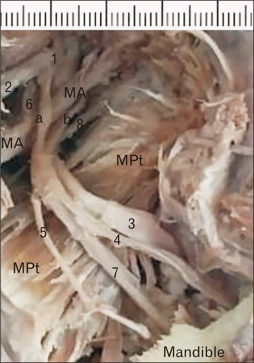 | Fig. 1Cadaveric specimen showing the variations observed in the right infra-temporal fossa of a female cadaver. 1, posterior division of mandibular nerve; 2, ATN; 3, lingual nerve; 4, IAN; 5, nerve to mylohyoid; 6, middle meningeal artery; 7, inferior alveolar artery; 8, artery to medial pterygoid; a, superficial root of IAN; b, deep root of IAN; ATN, auriculotemporal nerve; IAN, inferior alveolar nerve; MA, arteria maxillaris; MPt, medial pterygoid.
|
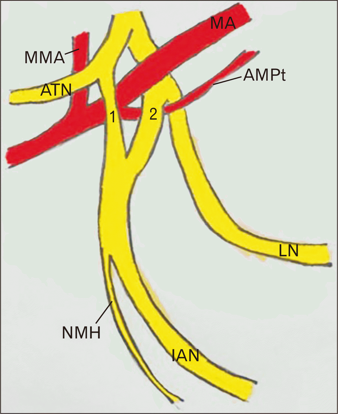 | Fig. 2Schematic diagram showing the variations observed in the present case. 1, superficial root of IAN; 2, deep root of IAN; AMPt, artery to medial pterygoid; ATN, auriculotemporal nerve; IAN, inferior alveolar nerve; LN, lingual nerve; MA, arteria maxillaris; MMA, middle meningeal artery; NMH, nerve to mylohyoid.
|
Go to :
Discussion
According to Wolf et al. [6], if the MA is enclosed between the nerves, it can lead to complications. It is also possible that pulsating MA can compress the surrounding nerves leading to symptoms like pain, tingling and numbness in their area of distribution [6, 7]. Nerve compression has been reported to trigger recurrent attacks of facial pain and is among the causes of trigeminal neuralgia [6]. The MA can also be injured during the nerve block procedures, if they are looped by the nerve roots. This may cause severe bleeding and hematoma formation. Either of the roots of IAN as observed in the present case can be compressed by MA. It can cause tic douloureux and altered sensation of gingival mucosa [8, 9]. Chen et al. [10] opined that the surgical decompression is the best alleviate for the trigeminal neuralgia due to arterial compression.
Double formative roots of the IAN was reported previously by Wolf et al. [6] and Khan et al. [11]. These variations are analogous to the present observation in the present study. On the other hand, Sumalatha et al. [7], Pai et al. [12] and Quadros et al. [13] reported cases of multiple roots forming the IAN. If there are multiple nerve roots, the chances and effects of nerve compression are doubled. Dodd et al. [14] reported that perineural spread of tumours are often overlooked by operating surgeons. Primary and secondary metastatic carcinomas can occur in the infratemporal fossa. Pretterklieber et al. [15] reported that, in 4.9% of their cases, the MA was piercing the IAN. They also observed that in one case, IAN and LN, both had two roots because of perforation by the MA. A unique case was documented by Kalra et al. [16] where there was anomalous coexistence of binary roots of ATN, which passed superficial to the arteria meningea media. Gülekon et al. [17] observed that 50% of their specimens had single root of ATN. Here also a similar occurrence of solitary root was noted. Ortug and Moriggl [18] observed a condition in which MA was perforating the LN.
Babu amd Thomas [19] reported an interesting finding of the MA which was passing inside the nervus auriculotemporalis formation. Anatomical relationships amid the neurovascular structures, muscles and their associated bony articulations in the infratemporal fossa favours high occurrence of various entrapment syndromes. Since it is not uncommon to find variations in the infratemporal fossa, the clinicians should be aware of the possible variations along with the normal anatomy. This will help them to avoid misinterpretations and subsequent complications. Also, Pai et al. [12] speculate that these types of neurovascular association variations should be considered in the future radiological and clinical studies.
During the development, mandibular nerve and its branches are derived from the migration of neural crest cells at the rostral end of the embryo. This migration occurs anteriorly through the mesoderm of the first pharyngeal arch. This happens due to interactions of multiple cell matrices [20] along with chemo-repulsions [21, 22]. Sometimes, this neural crest cell migration is prevented by factors which are released by the caudal somites [23, 24]. Since there are separate pathways for the development of motor and sensory fibres, there is a possibility of occurrence of two nerve bundles, explaining the developmental basis of the two roots of IAN, which is observed in this cadaver.
The myoblastic cells of the pterygoid mass will form a vascular arcade, which later forms the MA. Initially the stapedial artery supplies this network, but later taken over by the external carotid artery. During this taking over, most of the vascular network regresses except for few vessels, which forms MA. If there are variant nerve loops, these persistent vessels may remain in these loops. This leads to the development of MA inside the nerve loop [25, 26]. This could be the reason for the observed variation of the MA encircled by the IAN-superficial and deep roots.
It was reported that, incomplete fusion of nerves may lead to accessory foramina in the mandible [27]. Lee et al. [28] from their histoloical observations reported that, the intra-mandibular course of IAN has a larger trunk, which consists of two separate nerve bundles. These bundles are of mental and dental nerves, and covered with the same epineurium. The mental nerve gets separated from it near the premolar region [28], because the mandibular canal splits into the mental and incisive canals near the premolar area [29]. Here the present study observed that there were two roots of IAN being observed in the infratemporal fossa. This could be hypothesized that these two roots are of dental and mental nerves, which are formed separately from the posterior division of mandibular nerve. These roots have joined near the mandibular foramen to have a solitary intra-mandibular course and might have split later again near the mental foramen.
In conclusion, variations in the branching pattern of mandibular nerve and their aberrent communications have an upper hold in understanding the effectiveness of nerve blocks and complications in dental surgery and regional anesthesia. It must be taken into account that the failure of anesthetic effect upon its administration during regional surgeries can also be due to the variations in the structures pertaining to the infratemporal fossa. This necessitates adopting alternative methods of regional aneasthesia. The complex anatomical relationships between the neurovascular structures in the infratemporal fossa favours increased incidence of nerve entrapments as well. We opine that such relevant variations should be included in the list of causative factors of disorders like trigeminal neuralgia.
Go to :
 XML Download