

 PDF
PDF Citation
 Print
Print


Introduction
Cancer is a life-threatening illness affecting millions of individuals worldwide. During the last century, the only available treatment for cancer patients was its surgical removal. Lately, chemotherapy use led to better survival rates. Thus, it has been broadly used for treating different neoplasms either alone or accompanying surgery and/or radiotherapy [1] regardless of its cytotoxicity to healthy tissues [2].
A 5-fluorouracil (5-FU), thymidylate synthetase inhibitor, is a widely consumed chemotherapeutic agent that acts via inhibition of DNA synthesis. It affects not only the malignant cells but also the rapidly dividing cells e.g. epithelium lining the intestinal mucosa [3]. Accordingly, its effectiveness, like other chemotherapeutic agents, is often constrained by its serious side-effects [4].
One of the major incapacitating side-effects of chemotherapy is mucositis that occurred as early as 24–48 hours after its administration. It has been stated that 60% of patients on chemotherapy experience intestinal mucositis [5]. Such intestinal mucositis aggravates patients’ sicknesses ranging from nausea and vomiting to inflammation, ulceration and sepsis [6]. Consequently, it corrupts the patient’s quality of life during and after treatment and maybe a life-threatening condition. Besides, it delays treatment completion and results in chemotherapeutic dose reduction [4]. These injurious adverse effects are associated with distorted membrane permeability and compromised water movement across the membranes of the intestinal cells [7]. The available treatment for intestinal mucositis strives for reducing the symptoms severity instead of curing or preventing the cause [8]. Therefore, patients with cancer pursue unusual solutions to balance the devastating manifestations of both disease and its treatment.
Ancient herbal medicine has been used for years to treat different diseases and enhance better health. Rhubarb (Rh) is a herb with a long, fleshy stalk, frequently used in cooking. Its dried rhizomes were habitually used in China as a medicine for gastrointestinal complications such as diarrhoea, constipation and inflammation [9]. This medicinal effect has been assigned to its stalk extract which may be water or ethanol extract [10].
Aquaporins (AQPs) are 13 different integral membrane proteins channels (AQP 0–12) acting as bidirectional water channels. As they increase the membrane permeability to water by 10–100 times [11], they are copious in tissues dependent on water permeability to achieve proper metabolic functions such as kidney, lung, brain and gastrointestinal tract [12,13].
In the digestive tract, a large volume of water is secreted in its upper part in addition to 2 l of water ingested with food each day to provide quick balancing for the osmotic pressure of the contents in the intestine [11]. One litre is secreted by the salivary glands [14], 2 l by the stomach [15], 2.5 l by pancreas [16], 0.6 l by the liver [17] and 1 l by small intestine [18]. Then, water is absorbed by the intestines with nutrients, where about 9 l of water is absorbed every day [19].
In the gasstrointestinal tract, AQPs allow transcellular transport of water in two directions. However, each one permits mainly unidirectional water movement depending on the organ in which it is present and the osmolarity across the cell membrane [11]. In the intestines, AQP-4 in the basolateral membrane of the absorptive cells is responsible for the control of the bidirectional water movement, mainly water absorption. Thus, AQP-4 guarantees sufficient hydration and optimal stool consistency [13].
Matrix metalloproteinases (MMPs) are zinc-dependent endopeptidases that interpose inflammation and tissue remodelling. MMP-9 is unnoticeable in normal tissue but is highly expressed in inflammatory and cancerous conditions via several active signalling molecules and target pathways [20]. It is usually secreted by the epithelial and the immune cells as neutrophils and sometimes macrophages [21].
The tumor necrosis factor-α/nuclear factor-kappa B (TNF-α/NF-κB) signalling pathway was postulated to regulate various aspects of innate and adaptive immunity and to modulate the inflammatory responses. When the canonical NF-κB pathway is triggered by TNF-α, a vast range of genes involved in the immune and inflammatory cascades are upregulated [22]. Previous studies demonstrated that amelioration of the TNF-α/NF-κB signalling pathway would improve the intestinal inflammatory conditions and protect the intestinal epithelium [23].
This study aimed at detecting the role of AQP-4, TNF-α, NF-κB, and MMP-9 in the histological and biochemical changes occurred in the ileum secondary to 5-FU, a chemotherapy model, and the potential therapeutic effect of Rh water extract on these changes in adult male albino rats.
Go to : 
Materials and Methods
Animals
Forty-five adult male Wistar albino rats ( 200 g) were used in this study. They were treated according to the guidelines approved by the Animal Use Committee of Cairo University and the principles of laboratory animal care (CU-III-F-41-19). The rats were housed in the laboratory animal house unit of Kasr Al-Aini, Faculty of Medicine, Cairo University. The rats were kept under the same environmental conditions for 48 hours before starting the experiment to adapt to the new environmental conditions. They were provided with ordinary rat chow and water ad libitum and housed at 24°C±1°C in normal light/dark cycle. Each rat was kept separately in a cage to monitor the occurrence of diarrhoea.
Materials
5-fluorouracil: purchased as vials for injection (EBEWE Pharma Ges.mbH. Nfg.KG, Unterach, Austria). Each contains 250 mg 5-FU/5 ml.
Rhubarb: purchased from Harraz for Food Industry & Natural Products Company, Cairo, Egypt and was authenticated by the Pharmacognosy Department, Faculty of Pharmacy, Egyptian Russian University, Cairo, Egypt. A herbarium specimen is stored for further reference.
Anti-matrix metallopeptidase-9 antibody: a metalloproteinase mouse monoclonal antibody (cat # MA5-14228; Labvision, Thermo Fisher scientific, Cambridge, MA, USA).
Anti-nuclear factor-kappa-ß (NF-kB p65) antibody: rabbit polyclonal antibody (cat # PA5-16545; Labvision, ThermoFisher Scientific).
Anti-aquaporin-4 (AQP-4) antibody: rabbit polyclonal antibody (cat # YPA1171; Chongqing, Biospes, China).
Experimental design
The rats were equally divided into three groups (n=15):
Group І (control group)
The animals were equally subdivided into three subgroups:
‒ Subgroup IA: received no treatment.
‒ Subgroup IB: each rat received a single intraperitoneal (IP) injection of 1ml saline on day 6 of the experiment.
‒ Subgroup IC: each rat received distilled water (1 ml/day) via a gastric tube on a daily basis for the whole experimental period (8 days). Additionally, on day 6, it received a single IP injection of 1 ml saline.
Group ІI (5-FU group)
On the 6th day of the experiment, each rat received single IP injection of 150 mg/kg 5-FU freshly dissolved in 1 ml saline.
Group ІII (Rh-treated)
Each rat was given Rh water extract (20 mg/kg/day) dissolved in 1 ml distilled water via a gastric tube for the whole experimental duration (8 days) [6]. Also, each animal was treated as in group II on day 6 of the experiment.
Experimental procedure
Rh water extract preparation
About 2.5 kg of Rh stems were used and cut into sections (1 cm length). The alcohol-soluble components in these sections were extracted by boiling the sections in absolute alcohol (ethanol) then, getting rid of the extract after its cooling. The marc left after ethanol extraction was subjected to extraction with boiling water to yield the aqueous extract. Such extract was dehydrated and crystalized by lyophilization at –40ºC to get a lyophilized powder.
This method of preparation provided 4 g Rh powder/500 g fresh Rh. Fractionation of the Rh water extract indicated that the active glycopeptide constituent was water-soluble and alcohol insoluble [28].
Calculation of the mean incidence of diarrhoea
The calculation of diarrhoea incidence depends on the stools form and consistency, not frequency. At day 7 and day 8 of the study, a score for each animal in each group was recorded as follows; 0: no diarrhoea; 1: minor diarrhoea (somewhat wet and soft stool); 2: moderate diarrhoea (wet and shapeless stool with moderate perianal staining of the fur); and 3: severe diarrhoea (watery stool with severe perianal staining of the fur) [24]. For each day, the mean score for each group was calculated as follows:
Mean score for each group in each day=
(n: number of rats in each group, n=15)
Then, calculation of the mean score for the two days was estimated and approximated to a whole figure.
Biological samples collection
After eight days (day nine i.e. three days after 5-FU administration), the animals of the three groups were weighed then euthanized using anaesthesia overdose with IP injection of ketamine (90 mg/kg)/xylazine (15 mg/kg) in the animal house, Faculty of Medicine, Cairo University. The gastrointestinal tract of each rat was dissected and emptied. 2–4 cm from the last 90% of the small intestine (ileum) were obtained for histological and biochemical investigations, respectively.
Biochemical investigations
Ileal segments (4 cm) of each group were homogenized in 10% iced phosphate buffer saline (PBS, pH 7.4) for 10 minutes in a glass manual homogenizer. The homogenate was centrifuged at 10,000×g for 20 minutes at 4°C. The resulting supernatant was used for measuring the value of TNF-α using an ELISA kit supplied by (cat # ELR-TNF-α-1; Ray Biotech, Norcross, GA, USA) and the value of AQP-4 by an ELISA kit provided by (cat # MBS3808306; MyBioSource, San Diego, CA, USA), according to the manufacturer’s instructions.
Histological studies
The ileal segments for histological examination (2 cm) of all animals of all groups were fixed in 10% formol saline, kept for 24 hours then processed to paraffin blocks. Six micrometres-thick sections were cut and stained with:
‒ Hematoxylin and eosin stain (H&E)
‒ Periodic acid Schiff (PAS) reaction
‒ Immunohistochemical staining for:
MMP-9: a marker for extracellular matrix degradation [25]. It appears as a cytoplasmic reaction in the inflammatory, epithelial and endothelial cells.
NF-κB: an inflammatory marker expressed in the cytoplasm and the nuclei of both inflammatory and intestinal mucosal cells [8].
AQP-4: It is detected in the cytoplasm and the basolateral membrane of the enterocytes [13].
Immunostaining required pretreatment by boiling for 10 minutes in 10 mM citrate buffer (cat # 005000) pH 6 for antigen retrieval and leaving the sections to cool in room temperature for 20 minutes. Then, the sections were incubated for 1 hour with the primary antibodies. Immunostaining was completed by the use of Ultravision One Detection System (cat # TL-060-HLJ). Counterstaining was done using Lab Vision Mayer's hematoxylin (cat # TA-060-MH). Negative control sections were prepared using the same procedure after omitting the primary antibodies. Citrate buffer, Ultravision One Detection System and Ultravision Mayer's hematoxylin were purchased from Labvision, ThermoFisher Scientific, USA.
‒ Morphometric studies
At a magnification of ×400, ten non-overlapping fields/section were obtained to measure:
Mean value of mucosal thickness (sum value of villus height and crypt depth) in H&E stained sections.
Mean area percent (%) of PAS-positive (+ve) reaction.
Mean area % of MMP-9, NF-κB, and AQP-4 immunoreaction.
Image analysis was done using Leica Qwin-500 LTD-software image analysis computer system (Cambridge, England) at Histology Department, Faculty of Medicine, Cairo University.
‒ Statistical analysis
The data were expressed as mean±standard deviation (SD) and were analyzed statistically using IBM SPSS Statistics for Windows, Version 21.0 (IBM Co., Armonk, NY, USA). This was done using one-way analysis of variance ANOVA followed by “Tuckey” post hoc test. The results were considered statistically significant when the P-value was <0.05.
Go to :
Results
Changes in the incidence of diarrhoea and the body weight
Group II showed a significant increase in the mean incidence of diarrhoea and a significant decrease in body weight versus group I & III. In addition, group III revealed a significant increase in the mean incidence of diarrhoea compared to group I (Table 1).
Table 1
The effect of aqueous rhubarb extract administration on the incidence of diarrhea, body weight (gm) and TNF-α and AQP-4 values (pg/ml) in ileal homogenates
| Group | Group I | Group II | Group III |
|---|---|---|---|
| Incidence of diarrhea | 0 | 3 | 2 |
| Mean body weight | 195.00±7.20 | 130.00±8.70a) | 167.00±12.90a,b) |
| Mean value of TNF-α | 24.00±2.40 | 116.70±7.20a) | 63.50±10.50a,b) |
| Mean value of AQP-4 | 1.10±0.26 | 0.23±0.01a) | 0.95±0.07b) |
Values are presented as number only or mean±SD. TNF-α, tumour necrosis factor-α; AQP-4, aquaporin-4. For incidence of diarrhea; 0=no diarrhea, 1=minor, 2=moderate, 3=severe. a)Significantly different from respective GpI (control) group at P<0.05, b)Significantly different from respective Gp II (5-fluorouracil) group.
![]()
Biochemical results
In the 5-FU group, the ileal homogenates demonstrated a significant increase in the mean value of TNF-α and a significant decrease in that of AQP-4 in comparison to control and treated groups. However, group III revealed a significant decrease in the mean value of AQP-4 versus group I (Table 1).
Histological analysis
H&E stain
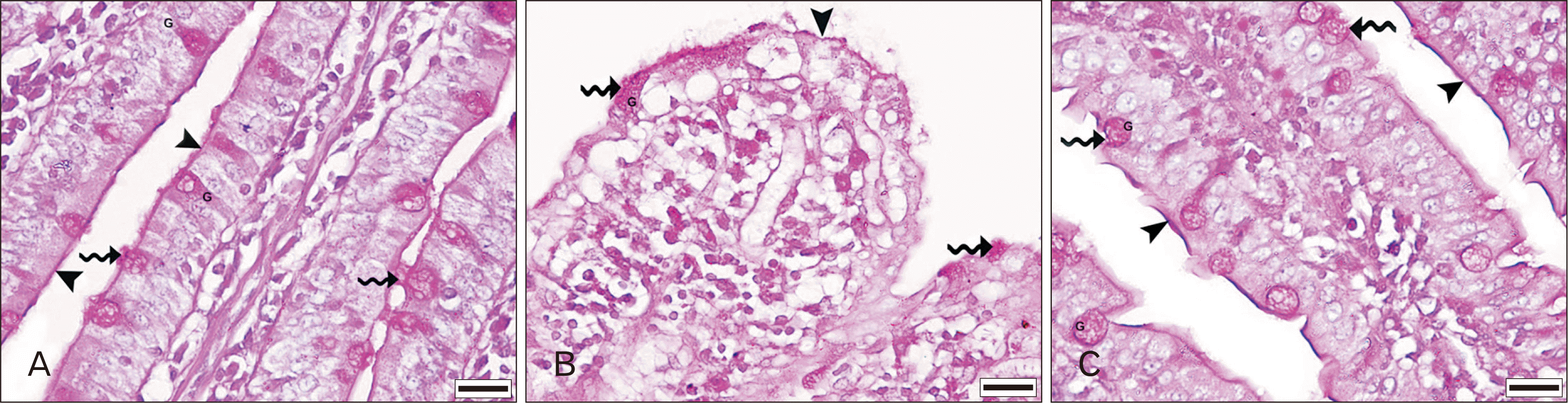
Ileal sections of the control group revealed normal histological architecture. The wall was formed of mucosa, submucosa (loose connective tissue [C.T.] with Peyer’s patches lymphatic nodules in the anti-mesenteric border) and muscularis externa. The mucosal layer showed the presence of intestinal villi and intestinal glands (Fig. 1A–D).
 | Fig. 1Photomicrograph of H&E stained ileal sections showing: (A–D) control group: normal tissue architecture demonstrating M (Vi, c), muscularis M (curved arrow), submucosa (*) containing PP and ME (ic, ol). The villi are displaying a core of LP and EP formed of simple columnar cells, E, with brush border and G. The intestinal c are separated by LP and lined with E, G, and basally located P. Intra-epithelial L are noted in the epithelial lining of the Vi and the c. (E, F) A 5-FU group: obvious mucosal damage with most of the epithelial cells showing multiple V, some of them seemed with deeply eosinophilic cytoplasm and shrunken darkly stained nuclei (arrow heads), and some appearing swollen and dissolute while others are nearly absent (wavy arrow). Some of the Vi appeared F while others showed areas of mucosal U with complete loss of the epithelial lining. Disorganization of the c and marked mononuclear inflammatory cell In within the mucosal connective tissue are noted. Some of the BV are dilated and/or disrupted. (G, H) Rh-treated group: nearly normal tissue architecture, yet few epithelial cells with cytoplasmic V and few pyknotic nuclei, some inflammatory cellular In and slightly disorganized c are noted. Scale bars=200 μm (A), 100 μm (B, E, G), and 20 μm (C, D, F, H). BV, blood vessels; c, crypts; E, enterocytes; EP, epithelium; F, fused; G, goblet cells; ic, inner circular; In, infiltration; L, lymphocytes; LP, lamina propria; M, mucosa; ME, muscularis externa; ol, outer longitudinal; P, Paneth cells ; PP, Peyer’s patches; Rh, rhubarb; U, ulceration; V, vacuolations; Vi, villi; 5-FU, 5-fluorouracil.
|
In the 5-FU group, the ileal sections revealed mucosal damage. Most of the epithelial cells showed multiple vacuolations and some of them seemed with deeply eosinophilic cytoplasm. Most of the nuclei were shrunken and darkly stained, some appeared swollen and dissolute while others were nearly absent. Additionally, some of the villi appeared fused while others showed areas of mucosal ulceration with complete loss of the epithelial lining. Moreover, there was disorganization of the crypts. Furthermore, marked mononuclear inflammatory cell infiltration within the mucosal C.T. corium was detected. Additionally, some of the blood vessels were dilated and/or disrupted (Fig. 1E, F).
However, Rh-treated group demonstrated nearly normal ileal sections except for the presence of few epithelial cells cytoplasmic vacuolations, few pyknotic nuclei, some inflammatory cellular infiltration and slightly disorganized crypts (Fig. 1G, H).
PAS reaction
There was abundant+ve PAS reaction in the sections of group I filling the cytoplasm of goblet cells. Additionally, there was+ve reaction in the brush border of the enterocytes (Fig. 2A). This reaction was drastically diminished in the sections of group II (Fig. 2B). Then, it became abundant again in group III with Rh treatment (Fig. 2C).
 | Fig. 2Photomicrograph picture of PAS reaction in the ileal sections of the experimental groups: (A) control group: abundant+ve PAS reaction filling the cytoplasm of the G (wavy arrow), in addition to+ve reaction in the brush border (arrowhead) of the enterocytes. (B) A 5-FU group: minimal+ve PAS reaction in a few G (wavy arrow) and the brush border (arrowhead). (C) Rh-treated group: abundant+ve PAS reaction in the cytoplasm of many G (wavy arrow) and the brush border (arrowhead). Scale bars=20 μm. G, goblet cells; PAS, periodic acid Schiff; 5-FU, 5-fluorouracil.
|
Immunohistochemical Stain for
‒ MMP-9: Minimal+ve MMP-9 immunoreaction in some cells of the mucosal LP of group I (Fig. 3A). However, in group II, there was abundant+ve cytoplasmic reaction in the inflammatory cells within the mucosal C.T. and in the epithelial cells covering the villi (Fig. 3B). Such+ve reaction was markedly minimized in group III (Fig. 3C).
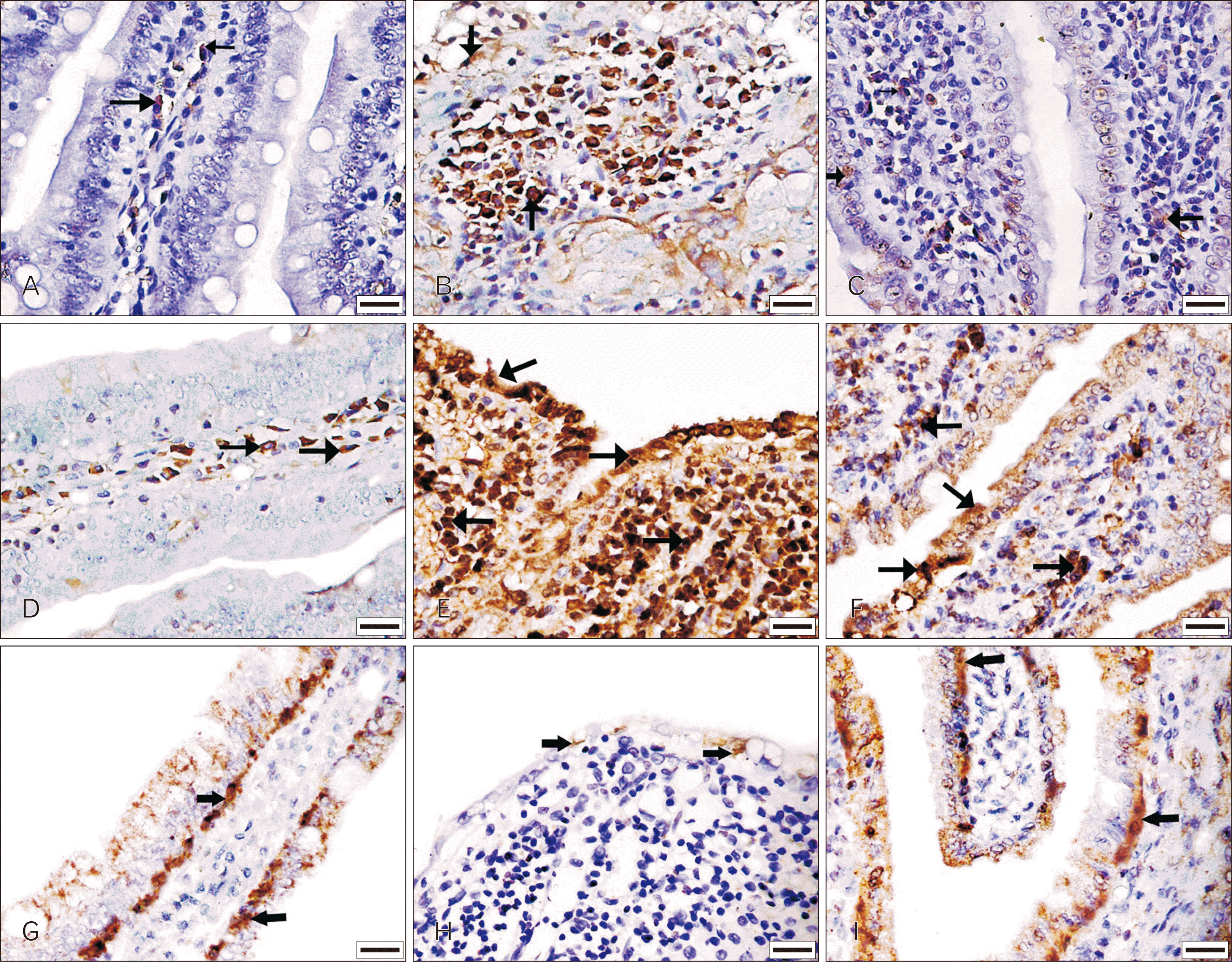 | Fig. 3Photomicrograph of immunohistochemistry in the ileal sections for MMP-9, NF-κB and AQP-4. Anti-MMP-9 immunohistochemistry showing: (A) control group: minimal+ve immunoreaction (arrows) in some cells of the mucosal LP (B) 5-FU group: abundant+ve cytoplasmic immunoreaction (arrows) in the inflammatory cells within the mucosal C.T. and the epithelial cells covering the Vi. (C) Rh-treated group: minimal+ve immunoreaction (arrows) in few inflammatory and epithelial cells. Anti-NF-κB immunohistochemistry showing: (D) control group: few+ve cytoplasmic immunoreactions (arrows) in some cells of the mucosal C.T. (E) A 5-FU group: obvious increased+ve immunoreaction (arrows) involving both the cytoplasm and the nuclei of most of the epithelial and inflammatory cells (F) Rh-treated group: some epithelial and inflammatory cells showing+ve immunoreaction (arrows) which is mainly cytoplasmic with few nuclear immunoreactions. Anti-AQP-4 immunohistochemistry showing: (G) control group: obvious+ve AQP-4 immunoreaction (arrows) mostly in the basolateral regions in addition to the cytoplasm of the enterocytes. (H) A 5-FU group: minimal+ve AQP-4 immunoreaction (arrows) in the epithelial cells (I) Rh-treated group: increased+ve AQP-4 immunoreaction (arrows) mainly in the basolateral regions in addition to the cytoplasm of the enterocytes. Scale bars= 20 μm. AQP-4, aquaporin-4; C.T., connective tissue; LP, lamina propria; MMP-9, matrix metalloproteinase-9; NF-κB, nuclear factor-kappa B; Rh, rhubarb; Vi, villi; 5-FU, 5-fluorouracil.
|
‒ NF-κB: Control group showed few+ve cytoplasmic immunoreactions in some cells of the mucosal C.T. (Fig. 3D). A radical increase in this+ve reaction was seen in sections of group II where it extended to involve both the cytoplasm and the nuclei of most of the epithelial and inflammatory cells (Fig. 3E). However, in group III, some cells showed+ve immunoreaction which was mainly cytoplasmic with few nuclear reactions (Fig. 3F).
Morphometric and statistical analysis
In the 5-FU group, there was a significant decrease in the mucosal thickness and the mean area % of the PAS+ve reaction as compared to control and Rh-treated groups. Also, there was a significant increase in the mean area % of MMP-9 and NF-κB+ve immunoreaction versus group I & III. Meanwhile, the mean area % of AQP-4+ve immunoreaction was significantly decreased in group II when compared to group I & III (Table 2).
Table 2
Quantification of the mean thickness of the mucosa (μm), the mean area % of PAS+ve reaction and the mean area % of+ve immunoreaction of MMP-9, NF-κB, and AQP-4
| Group | Group I | Group II | Group III |
|---|---|---|---|
| Mucosal thickness | 580.9±46.1 | 352.5±22.0a) | 553.0±24.5b) |
| PAS+ve reaction | 25.7±1.7 | 5.4±1.2a) | 24.2±1.5b) |
| MMP-9+ve immuoreaction | 2.9±0.7 | 22.9±1.1a) | 3.1±1.0b) |
| NF-κB +ve immuoreaction | 7.6±1.1 | 36.3±2.3a) | 8.5±2.0b) |
| AQP-4+ve immuoreaction | 17.8±1.0 | 3.4±0.7a) | 16.0±1.2b) |
![]()
Go to :
Discussion
This work aimed at detecting the role of AQP-4, TNF-α, NF-κB, and MMP-9 in the histological and biochemical changes occurred in the ileum of adult male albino rats secondary to 5-FU administration, a chemotherapy model. Additionally, it highlighted the potential therapeutic effect of Rh water extract on these changes.
Ileum was chosen in this study to demonstrate 5-FU induced intestinal mucositis and diarrhoea as it is responsible for absorption of 6.5 l/day while colon absorbs only 1.3 l/day of water [26]. So, the ileum is much more critical than colon in AQP-4 dependent water absorption [27].
Enterocytes apoptosis revealed following 5-FU administration could be explained by its direct toxic effects including; inhibition of DNA synthesis and reactive oxygen species (ROS) production with subsequent oxidative stress (OS) and inflammatory response [28]. Increased ROS was proved to be due to the over-production of TNF-α in the injured tissues [10] by the tissue fixed macrophages (the major source), fibroblasts, smooth muscle, epithelial and endothelial cells [29]. Both OS and TNF-α increase the recruitment of inflammatory cells and induce tissue inflammatory response [30]. Such tissue inflammation together with OS led to necrotic and apoptotic cell death [31]. These effects were enforced in the current study by the significant increase in the mean value of TNF-α in the ileal homogenates of the 5-FU group compared to group I. Further support was achieved by the marked inflammatory cell infiltration observed in the C.T. of the ileal mucosa.
Furtherly, the inflammatory cells produce more proinflammatory cytokines especially, TNF-α, and interleukin-Ib (IL-1b) [30]. TNF-α triggers the up-regulation of NF-κB and its translocation into the nucleus [32,33]. In the nucleus, NF-κB increases the gene expression and the translation of the proinflammatory cytokines that previously activated it such as TNF-α, IL-1b, and IL-6 [34]. This was enforced in the current study by the significant increase in the mean area % of NF-κB immunoreaction and its appearance in the nuclei of the epithelial and inflammatory cells in group II versus control. Similar results were detected in a former study [35] where NF-κB and TNF-α were increased two days following 5-FU treatment.
Mucosal ulceration, a fusion of the villi and disorganization of the crypts detected in this work and formerly described [36] could be clarified by apoptotic and necrotic cell death that affected not only the enterocytes but also all other epithelial cells [37]. In this study, affection of goblet cells was supported by the significant decrease in the mean area % of PAS reaction in group II compared to group I. This result coincides with the previously reported reduction in goblet cells with subsequent disruption of the mucin secretion and intestinal flora function followed by intestinal mucositis [28]. Additionally, endothelial cell death in the current work was manifested by the disruption of some blood vessels. Such affection might result in ischemia of the intestinal mucosa and consequently epithelial cell death [38].
Apoptosis of intestinal stem cells was suggested as both small intestine and hair follicles have the same stem cells (leucine-rich repeat-containing G protein-coupled receptor 5) [39] and the hair follicles’ stem cells underwent apoptosis with consequent alopecia following chemotherapy [40]. Thus, the proposed stem cell affection and the resultant decreased cellular proliferation might be added to the cell death to explain the significant reduction in the ileal mucosal thickness in the 5-FU group versus the control group. This explanation is in line with what was stated previously [24].
Interestingly, the 5-FU group showed a significant increase in the mean area % of MMP-9 immunoreaction versus the control group. MMP-9 is one of the MMPs, produced by different cells to preface tissue remodeling through degradation of the extracellular matrix. Its increased production was proved to occur in response to pro-inflammatory cytokines as TNF-α [20] whose high level was stated succeeding 5-FU injection in this work. Accordingly, the increase in MMP-9 could result in lysis of the extra-cellular components that might lead to the recorded ulceration and inflammation [25]. Additionally, MMP-9 is concerned with the initiation and augmentation of the chemotherapy-induced inflammatory reaction. This occurred via stimulating the production of the inflammatory mediators [41] which in turn could activate MMP-9 action and vice versa.
Moreover, increased MMP-9 could lead to disruption of the tight junctions between the epithelial cells through degradation of their cytoplasmic proteins [42]. Dysfunctional tight junctions between epithelial cells and endothelial cells of the intestinal barrier result in increased intestinal permeability and development of intestinal mucositis, oedema and diarrhoea [43]. This explanation is similar to that reported previously in cases of brain oedema [44].
Bearing in mind that diarrhoea induced by chemotherapy is one of the harshest sequelae of the intestinal mucositis as it might lead to nutritional disturbance, electrolyte imbalance and immune system dysfunction [45]. It was of great importance to explain the possible mechanisms of the significant increase in the mean incidence of diarrhoea observed in the 5-FU group of the current study. First, it could be enlightened by the increased intestinal permeability due to tight junction disruption by MMP-9 mentioned before.
Second, it could be based on the significant decrease in the mean level of AQP-4 in ileal homogenates and the mean area % of its immunoreactivity in the 5-FU group compared to the control group. Decreased AQP-4 expression is followed by a substantial decrease in water absorption from the small intestinal lumen to the blood with consequent diarrhoea. The decrease in AQP-4 expression was similarly documented in previous works where stool-water content was elevated in AQP-4 knockout mice [46], its colonic mRNA expression was decreased in mice having diarrhoea following 5-FU administration [3] and its mRNA and protein were reduced in cases of entero-toxigenic Escherichia Coli-induced diarrhoea [13].
Reduced AQP-4 was presumed to result from either death of the enterocytes and/or chemotherapy-induced inflammatory response. As it was documented that reduced AQP-4 following 5-FU treatment was prevented by neutrophil elastase inhibitor [47] where neutrophils are the first cells to reach the inflamed intestine following 5-FU treatment [48]. In addition, reduced colonic expression of AQP1, 3, 4, 8 in diarrhea induced by irinotecan (an anti-cancer agent) was illuminated by increased NF-κB, TNF-α and prostaglandin E2 (one of the inflammatory mediators) [49].
All the previous effects of 5-FU (direct toxicity, OS, inflammation, cell death, and reduced cellular proliferation) result into marked mucosal damage [50] that lead to reduced food intake, severe pain on eating and marked diarrhea [49]. Consequently, there was detection of a significant reduction in the body weight following chemotherapy administration when compared to the control group.
In the current study, Rh was proved to ameliorate 5-FU induced intestinal mucositis. This is suggested to occur through the anti-inflammatory effect of Rh water extract components (e.g., tannins and rhein). They could reduce the pro-inflammatory cytokines where IL-4 and interferon-γ were reduced by tannins [51] and TNF-α, IL-6 and IL-1b were decreased by rhein [52]. Moreover, rhein was documented to prevent NF-κB activation with the consequent decrease in the production of proinflammatory cytokines [52]. Another component of Rh water extract is polysaccharides which also have an anti-inflammatory effect [51]. Further support to this assumption came from the significant decrease in the mean level of TNF-α in the ileal homogenates and the mean area % of NF-κB positive immunoreaction in Rh-treated group in comparison with 5-FU group and its non-significant increase versus control group.
Furtherly, the Rh-treated group revealed minimal inflammatory cell infiltration in the ileal sections. This could be explained by the Rh capability to decrease the recruitment of the inflammatory cells [52]. Additionally, it can decrease the intestinal expression of myeloperoxidase (the neutrophil marker) following 5-FU [24] inhibiting neutrophils migration to it.
Moreover, Rh has an anti-oxidative effect on cases of 5-FU induced mucositis [24]. As rhein ability to decrease ROS production such as nitric oxide and cyclooxygenase-2 was documented [52]. This, in turn, reduces OS-induced apoptotic cell death.
Decreased oxidative and inflammatory induced cell death together with increased cell proliferation effect of Rh [52] were presumed to be the direct causes of the demonstrated normal villi and crypts structure with few pyknosis and vacuolations. This was enforced by the significant increase in the PAS area % and ileal mucosal thickness in Rh-treated group compared to 5-FU group.
The most profound influence of Rh-water extract in this study was its anti-diarrhoeal effect documented by the significant decrease in its incidence in group III. This could be explained by the ability of tannins to control water influx and efflux in and form the enterocytes, respectively [54] through inhibition of AQP-3 [51]. AQP-3 is known to cause water secretion in oesophagus, stomach, small intestine [55]. Moreover, tannins have protein denaturing effect that forms a protective layer of protein tannates on the surface of the intestinal epithelium increasing its resistance to local irritations and hence, decreasing intestinal secretion [54].
Also, Rh appeared to significantly decrease MMP-9 area % in the treated group compared to the 5-FU group which was non-significantly increased than the control group. This was followed by decreased epithelial and endothelial tight junctions degradation reducing intestinal and vascular permeability and consequently diarrhoea. Cui et al. [53] documented that Rh significantly decreased the extracellular soluble vascular endothelial cadherin concentration (the main component of cellular adhesion released after their damage). They added that Rh protected the capillary endothelium from the injurious agents and increased the formation of new capillaries.
A further explanation for the anti-diarrhoeal effect of Rh came from its influence on AQP-4. As the treated group showed a significant increase in AQP-4 mean value in the ileal homogenates and its immunopositive area % versus group II. This could be elucidated by the anti-inflammatory, anti-oxidative, anti-apoptotic and proliferative effects of Rh [52,53]. Furtherly, extracellular Rh was recorded to block water secretion direction of AQP-4 in the enterocytes, keeping only the water absorption direction. This occurs via its ability to bind to the interstitial AQP-4 pores due to its similarity to AQP-4 in shape and size. This blocks the influx of water into the enterocytes and decreases water secretion to the intestinal lumen [24].
The anti-diarrheal influence of Rh together with the preservation of the intestinal mucosa detected in this study and the normalization of food intake proved previously [24] could explain the significant increase in the body weight of group III than group II.
Despite the documented anti-diarrheal effect of Rh, the mean incidence of diarrhoea in Rh-treated group showed a significant increase when compared to the control group. This finding might be clarified by the significant decrease in AQP-4 mean level and mean area % of its immunoreaction in this group versus the control group. Such AQP-4 decrease was presumed to be due to the imperfect amelioration of the toxic effect of 5-FU by Rh which was enforced by the presence of few cells with cytoplasmic vacuolations and/or nuclear condensation and mild inflammatory cell infiltration in the Rh-treated group.
This study concluded that 5-FU, a chemotherapy model, elicited intestinal mucositis via increased TNF-α and NF-κB induced inflammatory reaction and/or OS. Such intestinal mucositis is characterized by marked diarrhoea caused by increased MMP-9 and decreased AQP-4. Rh water extract could partially ameliorate these effects through its anti-inflammatory, antiapoptotic, anti-diarrhoeal and proliferative effects.
Go to :
 XML Download