

 PDF
PDF Citation
 Print
Print


Introduction
Environmental exposure to heavy metals is a public health hazard that has been linked to cognitive and neurological deficits [1]. Exposure to heavy metals can occur through contaminated food, water, air, or in industrial work settings [2]. To date, the majority of research investigating the deleterious effects of environmental toxins on neurocognition has focused on heavy metals such as lead and mercury [2]. Of increasing interest in recent years are the health risks and/or benefits of exposure to the transition metal known as vanadium.
Vanadium (NaVO3) as a potentially toxic environmental pollutant induces oxidative damage in the central nervous system [3, 4]. Vanadium deposition in the brain might be associated with the pathogenesis of certain neurological disorders, and this, after long-term exposure can result in more severe pathology. Most research on vanadium neurotoxicity has been conducted after acute exposure; however, some populations are exposed to this Vanadium for a lifetime [5]. Exposure to neurotoxic metals such as vanadium occurs through various sources including heavy metals mining [6], combustion products of vanadium bearing fuel oils [7], forest fires and volcanic emissions. Large quantities of vanadium compounds have also been reported to be released into the environment mainly through the burning of fossil fuels having vanadium contaminated crude as seen in oil-producing communities such as Venezuela, the Arabian Gulf, the Gulf of Mexico and the Nigerian Niger Delta [8, 9].
Medicinal herbs have been known to be effective following their roles in the treatment of various diseases in humans and animals [10]. Sandpaper leaf, also known as Ficus exasperate Vahl leaf (FEVL) is a type of medicinal plant with various medicinal properties [11]. It is widespread in tropical Africa from Mozambique, Zambia, and northern Angola to Senegal, Ethiopia and also in the southern part of the Arabian Peninsula and India [12].
The toxicology analysis and phytochemical constituents of Ficus exasperata revealed the presence of flavonoids, tannins, saponin, alkaloids, and glycosides [4, 13]. Fafure et al. [4] demonstrated that F. exasperata Vahl improved motor activities in mice exposed to manganese chloride. Available reports in western Nigeria indicate that leaves of F. exasperata Vahl exhibit antiulcer, hypotensive, hypoglycemic, hypolipidemic, anti-inflammatory, anxiolytic, oxytocin inhibiting, anticonvulsant, antinociceptive, antimicrobial, anticandidal, insecticidal and pesticidal activities [14, 15], and the decoctions and infusions of F. exasperata leaf have been used traditionally in the management and treatment of different human diseases including diabetes mellitus, hypertension, and certain cardiovascular dysfunctions [16]. Although sandpaper leaf has been shown to possess many properties, there is a dearth of information on the effect of saponin fraction on the anti-inflammatory properties of sandpaper leaf. Hence, this study sought to investigate in mice whether saponin fraction of F.exasparata Vahl leaves could counteract the noxious effects induced by a subchronic treatment of vanadium which can serve as a readily accessible and inexpensive alternative for treating parkinsonism/Parkinson-like diseases. The animals were tested for motor coordination using the rotarod test (RT) and parallel bar test (PBT), and the integrity of dopaminergic (DA) neurons of the substantia nigra (SNc) was evaluated with histological/immunohistochemical approaches.
Go to : 
Materials and Methods
Animal procurement and care
Forty male adult Balb/c mice weighing between 25-35 g were used for this study. The mice were collected from the animal handling facility of the department of anatomy from the University of Delta State, Nigeria. The mice were allowed to acclimatized for two weeks and had free access to rodent chow (purchased from the ABUAD feed mill, Ado Ekiti, Nigeria) and water; they were also exposed to 12 hours by 12 hours dark and light period.
Collection and identification of plant
F. exasperata Vahl leaves were collected during its blossoming stage in February from farmland in Ikole Ekiti South-Western Nigeria. The plant was identified at the University of Lagos Herbarium as F. exasperata Vahl leaves with herbarium number 7786.
Preparation of plant materials
The F. exasperata Vahl leaves were dried for five days and then pulverized into a fine powder using an electric blender. Saponin was extracted from the fine powder (250 g) in the Abuad chemistry Laboratory. A 250 g of the fine powder was weighed into a big beaker while 100 ml of 20% ethanol was added and mixed properly; it was placed in a water bath at 55ºC for 4 hours. It was continuously stirred for 15 minutes each for the four hours until it became concentrated. Di ethyl ether was added to it after the 4 hours (not in the water bath) and stirred vigorously to get pure saponin, after which N-butanol was also added and stirred. A total of 5% sodium chloride was also put and allowed to decant; it was then filtered to get the saponin extract, which was later left in the water bath at 60ºC until it was properly dried.
Research ethical approval
Ethical clearance for this study was obtained from the Health Research Ethics Committee (HREC) of the Afe Babalola University, Ado Ekiti, Ekiti State, Nigeria (AB/EC/19/02/001).
Experimental design and administration
The mice were randomly divided into four groups (n=10); Group A (control group) received normal saline for 21 days, Group B with subgroup B1 (Vanadium) and B2 (Withdrawal). Group B1 received normal saline for 14 days and NaVO3 for seven days. Group B2 received normal saline for seven days, NaVO3 intraperitoneally and withdrew for seven days. Group C received 10 mg/kg of NaVO3 intraperitoneally for 7 days followed by 50 mg/kg of Saponin fraction of F. exasperata Vahl leave extract orally for 14 days; Group D received 10 mg/kg of NaVO3 intraperitoneally for seven days followed by 100 mg/kg of Saponin fraction of F. exasperata Vahl leave extract orally for 14 days.
After administration, five mice from each group were sacrificed by cervical dislocation. Their brains were harvested and transferred into 30% sucrose in a frozen environment to preserve the chemicals for biochemical analysis (malondialdehyde [MDA], catalase [CAT], and glutathione [GSH]). Also, another five mice from each group were sacrificed by cervical dislocation and their brains were perfused with 10% formal saline and quickly transferred to a specimen bottle containing 10% formal saline.
Neurobehavioral study
Motor activity and coordination were measured in the animals using rotarod (IITC Life Science, Woodland Hills, CA, USA), and PBT. All tests were done on mice using a digital video recorder and were analyzed later for motor activity and coordination.
Rotarod test
Motor coordination was assessed on the mice using rotarod following treatment with Vanadium. Each animal was placed on the rotarod, and the time spent on the rotating bar was recorded as the latency of fall. This is a test of motor learning and coordination. Mice are placed on a rod made of PVC doweling (~5-cm in diameter) that has an etched surface. The rod is then activated so that it accelerates at a defined rate. The mice will begin to walk/run until they cannot stay on any longer, at which point they fall from the rotating rod (approximately 30-cm). With repeated exposure, mice show increased latencies, which is a measure of motor coordination and cerebellar learning. The latency of fall was calculated, which is the time the animals will fall off of the rotating bar and the number of times the animals cling to the rotating bar and rotate with it is termed passive rotation.
Parallel bar test
Two metal bars, each 1meter long and 2 mm thick were placed on an elevated wooden platform, 50 cm high above the floor. Subsequently, the animals were placed perpendicular to the axis of the metal bars at the midpoint of their length (i.e. 0.5 m mark). With the use of a timer, the duration for each animal to complete a 90-degree turn on the double bar was recorded (as the latency of turn [LOT]) in seconds for 3 minutes trial. Also, the time taken for an animal to cover the 0.5 m to one end of the bars was determined.
Immunohistochemistry
The experimental animals were sacrificed 48 hours after the last injection, and neurobehavioural studies were done, based on previous studies that revealed significant activation of astroglia. Mice were sacrificed under ketamine anesthesia and transcardially perfused with paraformaldehyde (4% in 0.1 M phosphate buffer, pH 7.4). For immunohistochemistry studies, sections from the SNc (10 µm thick) were coronally cut on a microtome and immunoreacted with primary antibodies directed against glial fibrillary acidic protein (GFAP)- to mark the astrocytes, tyrosine hydroxylase (TH 1:400, Bio-Techne, Oxford, UK) to mark the DA neurons, and dopamine transporter (DAT) to mark the dopaminergic transporter. For diaminobenzidine visualization of GFAP, the proper biotinylated secondary antibody (goat anti-mouse IgG for GFAP from Vector, United Kingdom) was used, and the avidin-biotin-peroxidase protocol (ABC; Vector Laboratories, Inc., Peterborough, UK) was applied. The 5 µm-sections were mounted on gelatin-coated slides, dehydrated and cover-slipped.
Microscopy analysis
All sections were studied under a Leica DM 750 microscope (Leica Microsystems Heidelberg, Mannheim, Germany) connected to a digital camera (Leica ICC 50) and a computer. Photomicrographs of stained sections were obtained, reported and imported into Image J image analysis software (National Institute of Health, Bethesda, MD, USA) for analysis.
Statistical analysis
Relative brain weight and motor coordination performance of the treated group were compared with those of control using the One-way ANOVA followed by Newman-Keuls for post hoc in case of significance. All results presented here are mean±SEM and are considered statistically significant at P<0.05.
Go to :
Results
Phytochemical analysis of gutenbergia nigritana leaves
Methanol and aqueous F. exasperata Vahl leaves extracts contained: phenol, tannins, steroids, flavonoids, saponins, alkaloids and glycosides which have been identified by other researchers in various plants and in different parts of plants [17, 18] but the activities of phenol, tannin alkaloids, saponin and glycosides were more pronounced with the methanolic extract as seen in Table 1. Saponins are group of steroid or triterpenoid glycosides, also found to be effective antimicrobial substances in vitro against a wide array of microorganisms by inhibiting the membrane bound enzymes [19]. They have been reported to possess substantial anti-carcinogenic activities due to their anti-oxidant and inflammatory properties [20].
Assessment of motor coordination performance
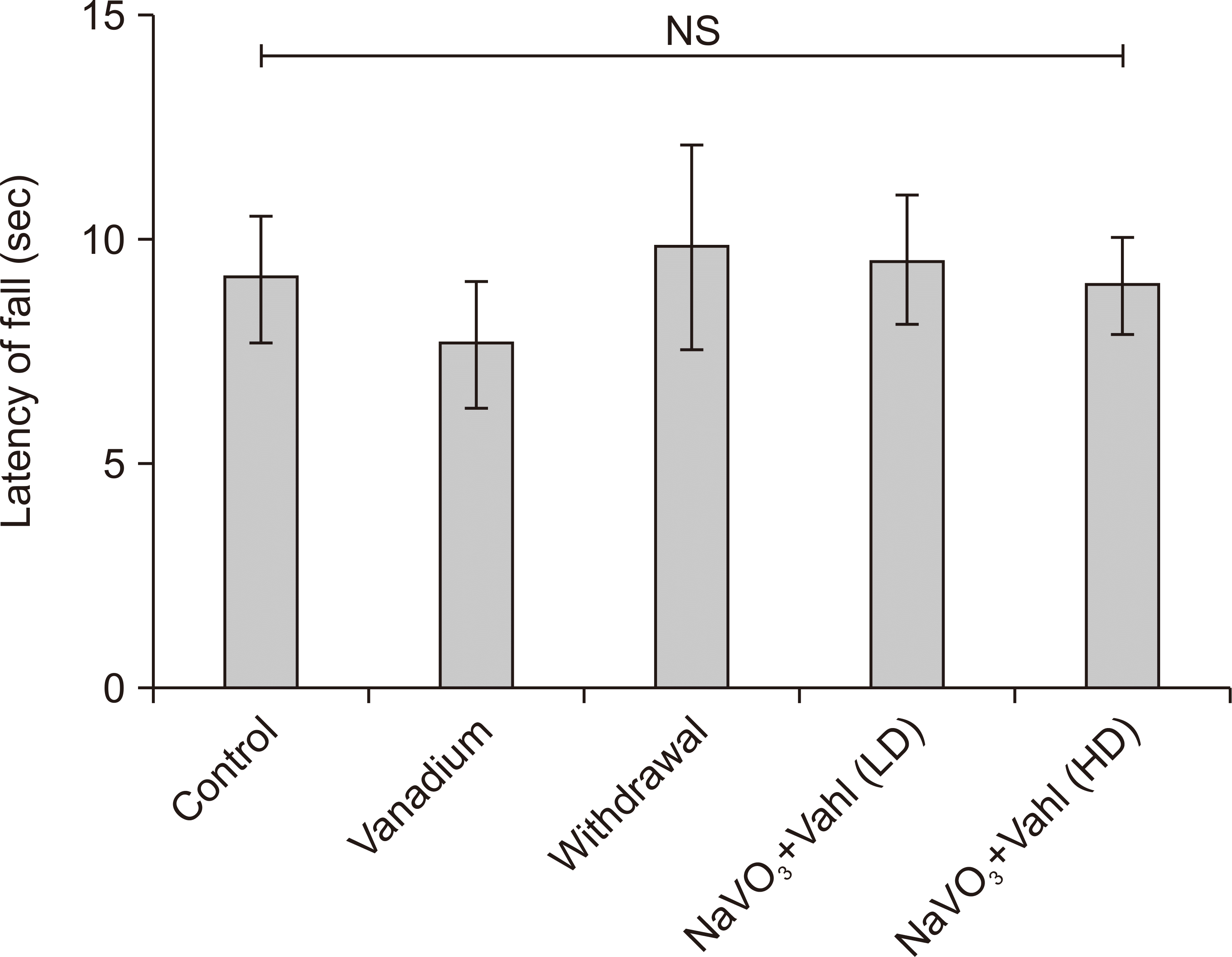
Animals with good motor coordination are expected to spend more time on the rotating bar [4, 21]. Vanadium group shows significantly decrease motor activity when compared to the control group, while the F. exasperata Vahl group and post-treated groups (NaVO3+Vahl) show a significant increase in the retention time as they spent more time on the rotating bar compared to vanadium exposed group (**P<0.01). However, the withdrawal group showed a significant increase in the retention time on the rotating bar compared to the vanadium exposed group (Fig. 1).
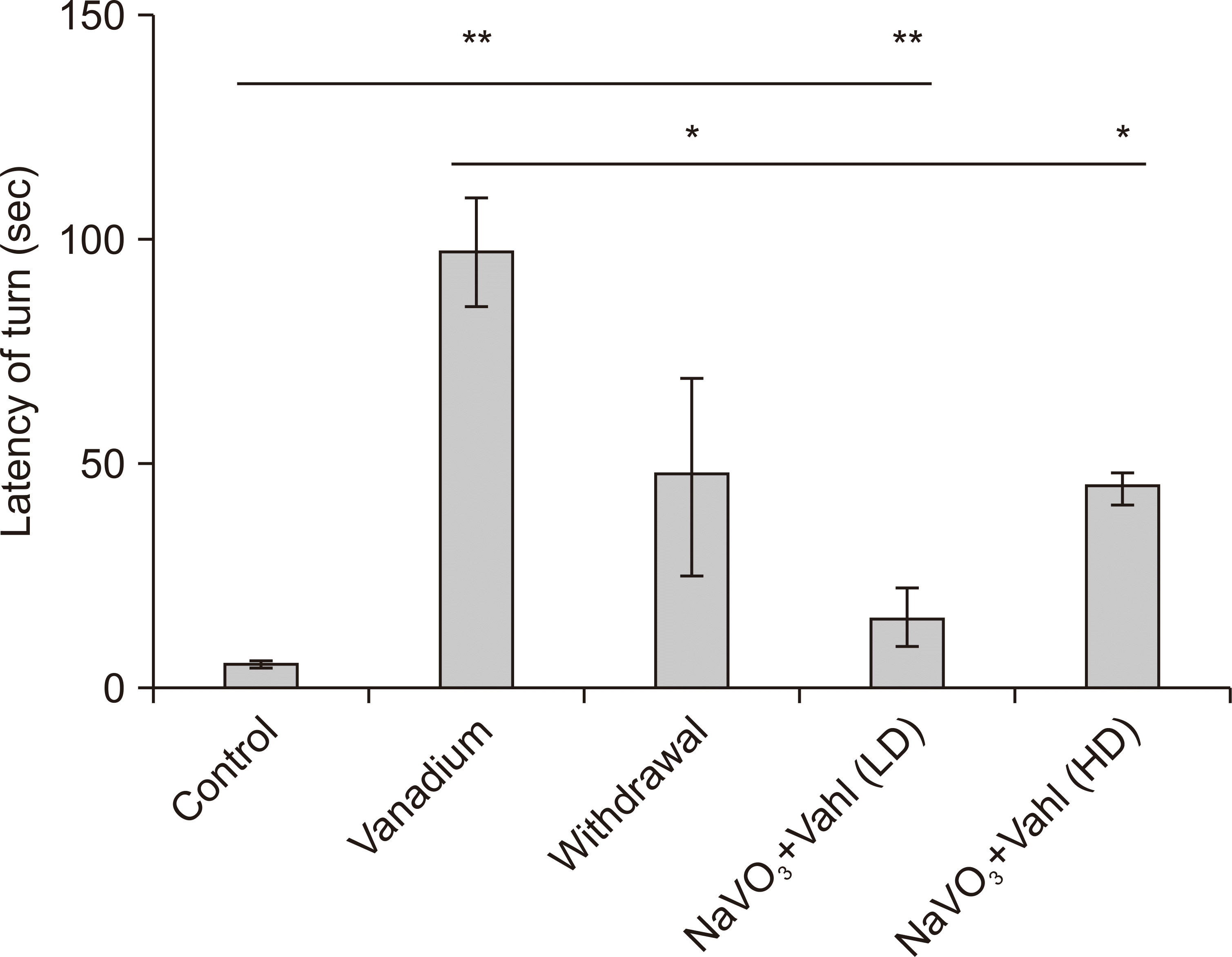
Significant increases in LOT scores were considered as abnormal motor coordination when the treatment groups were compared against control, as shown in Fig. 2 below. Vanadium exposed mice exhibited increased LOT (time to turn) and total time spent during the PBT when compared to the control group. There was a significant decrease in the LOT of post-treated groups and the withdrawal group when compared with the vanadium exposed group. However, control (saline) group exhibited a decrease in time of turn compared to the vanadium exposed group (*P<0.05, **P<0.01).
Analysis of biochemical assay
Lipid peroxidation
Fig. 3 showed no significant difference of MDA level when comparing control group with the vanadium, withdrawal, NaVO3+Vahl (low dose) and NaVO3+Vahl (high dose) group. However, the group exposed to vanadium showed increased level of MDA, while a reduction in the level of MDA was seen in NaVO3+Vahl (high dose) group when they were compared with the control group.
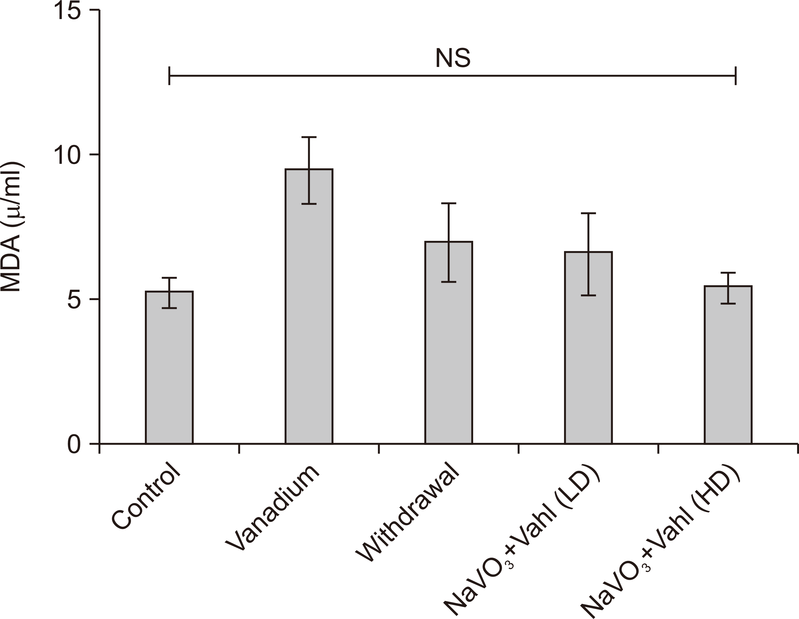 | Fig. 3Graph representing the concentration of MDA (units) per mg in brain tissue. There is no statistical significant different in the MDA between the control and vanadium treated groups. Sodium metavanadate and low dose of Ficus exasperata Vahl (NaVO3+Vahl LD), Sodium metavanadate and low dose of F. exasperata Vahl (NaVO3+Vahl HD). HD, high dose; LD, low dose; MDA, malondialdehyde.
|
Catalase
Fig. 4 showed significance difference in the CAT level when normal saline group (control) was compared with vanadium (*P<0.05). When the Vanadium group (NaVO3) was also compared with the withdrawal group, it showed a significant difference of *P<0.05, this also goes for the NaVO3+Vahl (low dose) and NaVO3+Vahl (high dose) group (*P<0.05).
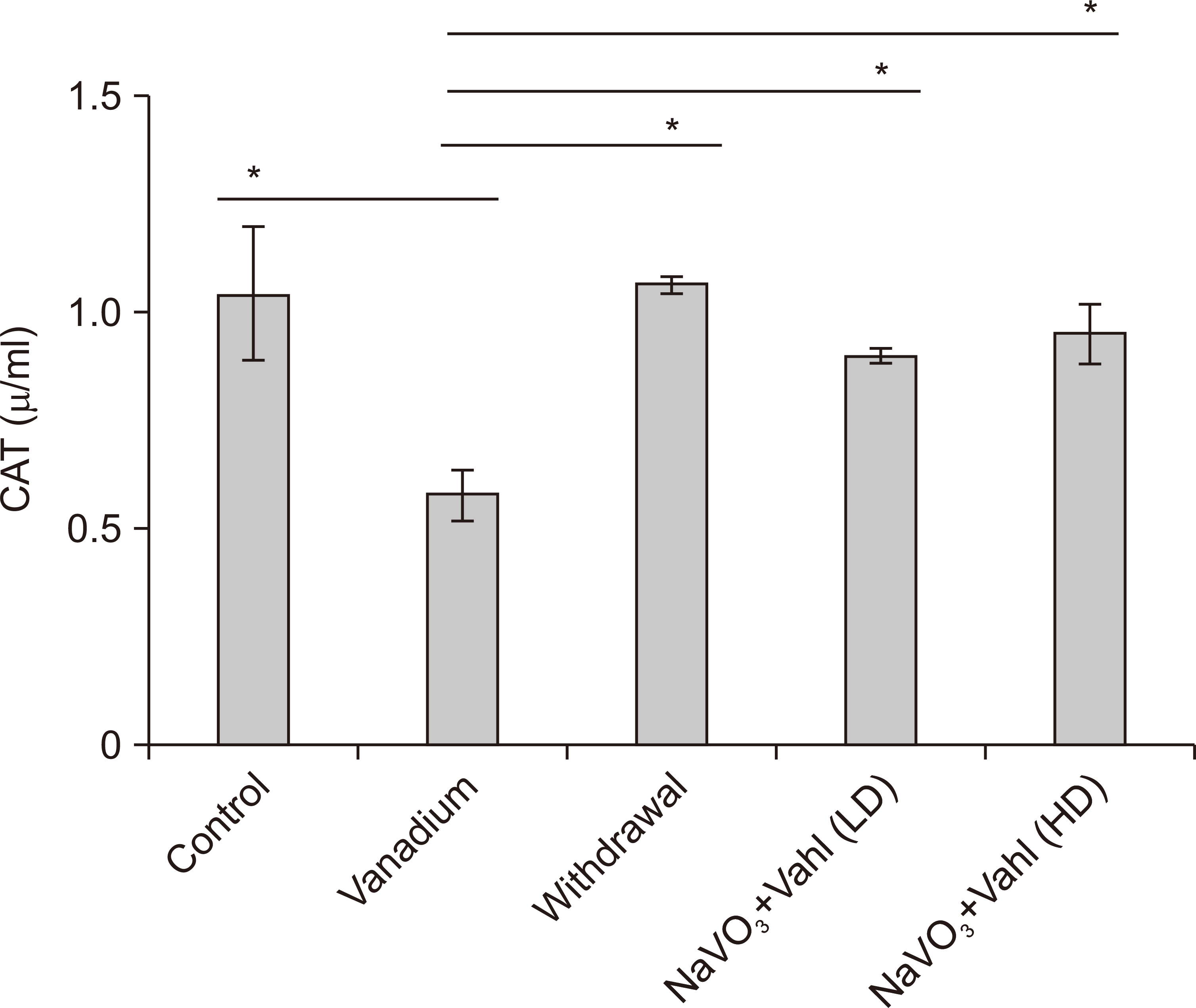 | Fig. 4Graph representing the concentration of CAT (units) per mg in brain tissue homogenate. There is statistical significant different in the CAT between the control and vanadium and Ficus exasperata treated groups. Sodium metavanadate and low dose of F. exasperata Vahl (NaVO3+Vahl LD), Sodium metavanadate and low dose of F. exasperata Vahl (NaVO3+Vahl HD). CAT, catalase; HD, high dose; LD, low dose. *P<0.05.
|
Glutathione
Vanadium group in Fig. 5 expressed significantly lower level of GPX when compared with the normal saline group (control), and when NaVO3+Vahl (low dose) group was compared with control group (*P<0.05).
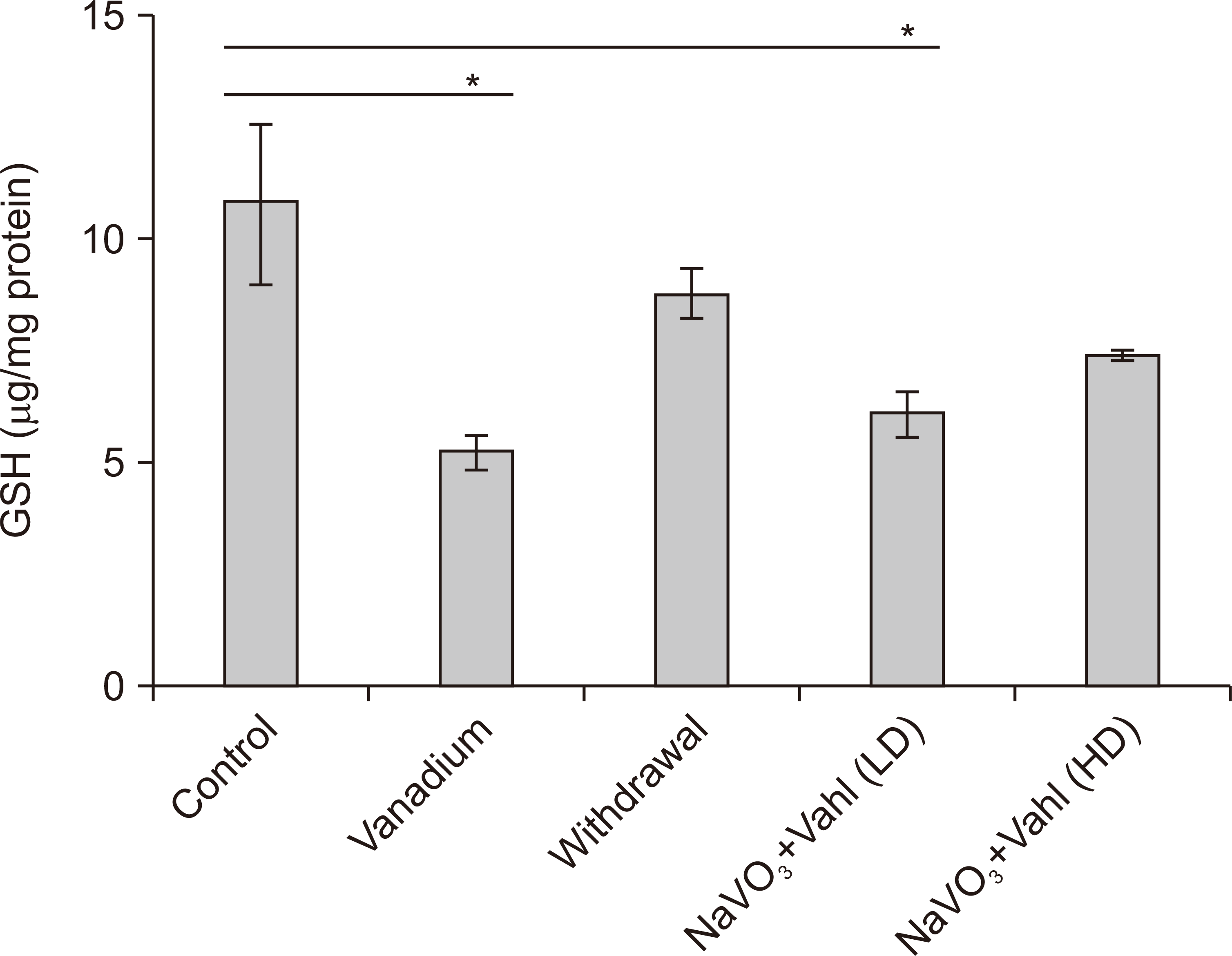 | Fig. 5Graph representing the concentration of GSH (units) per mg in brain tissue homogenate. There is statistical significant different in the catalase between the control and vanadium and Ficus exasperata treated groups. Sodium metavanadate and low dose of F. exasperata Vahl (NaVO3+Vahl LD), Sodium metavanadate and low dose of F. exasperata Vahl (NaVO3+Vahl HD). GSH, glutathione; HD, high dose, LD, low dose. *P<0.05.
|
Immunohistochemistry
Immunohistochemical studies evaluating the neuroinflammatory level and DA activities in a section of the SNc, and ameliorative roles of F. exasperata Vahl leaves extract after exposure to Vanadium was observed to be significant on the glia following the uses of primary antibodies directed against GFAP, DAT, and TH (Figs. 6–9).
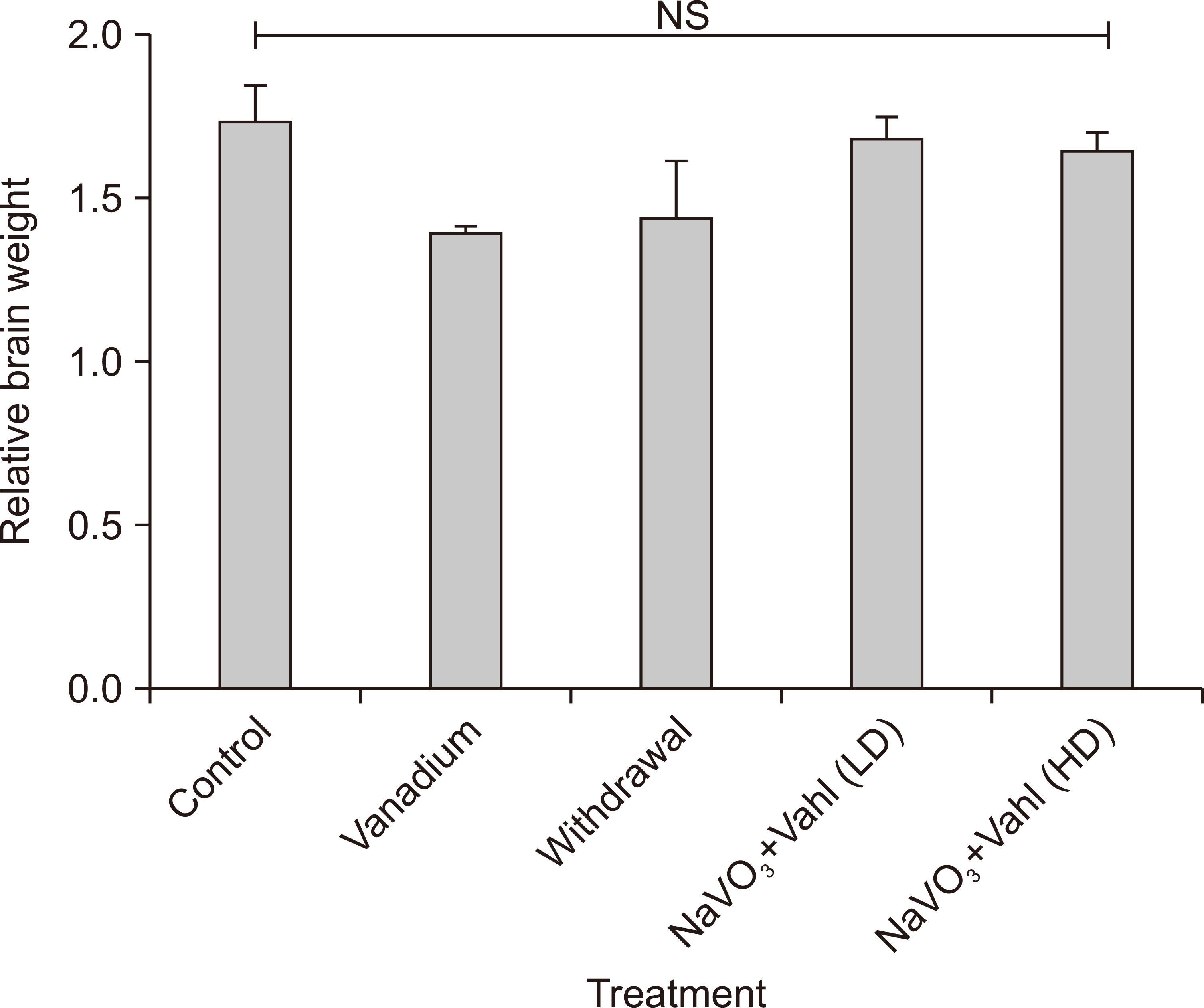 | Fig. 6Bar chart showing the relative brain weight of the experimental animals (P-value=0.458: F-value=1.918). There is no statistical significant different in the relative brain weight between the groups. Sodium metavanadate and low dose of Ficus exasperata Vahl (NaVO3+Vahl LD), Sodium metavanadate and low dose of F. exasperata Vahl (NaVO3+Vahl HD). HD, high dose; LD, low dose; NS, not significant.
|
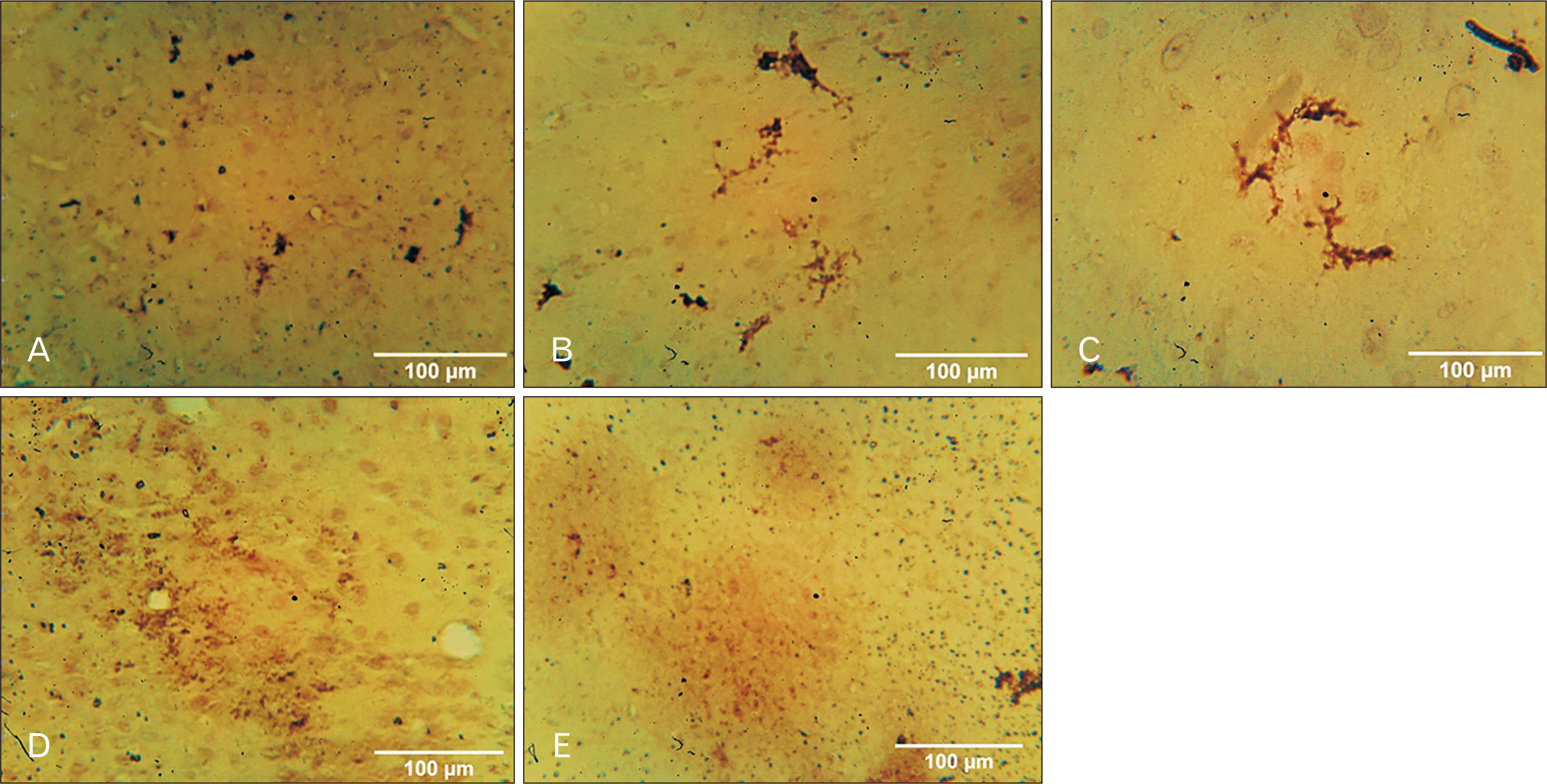
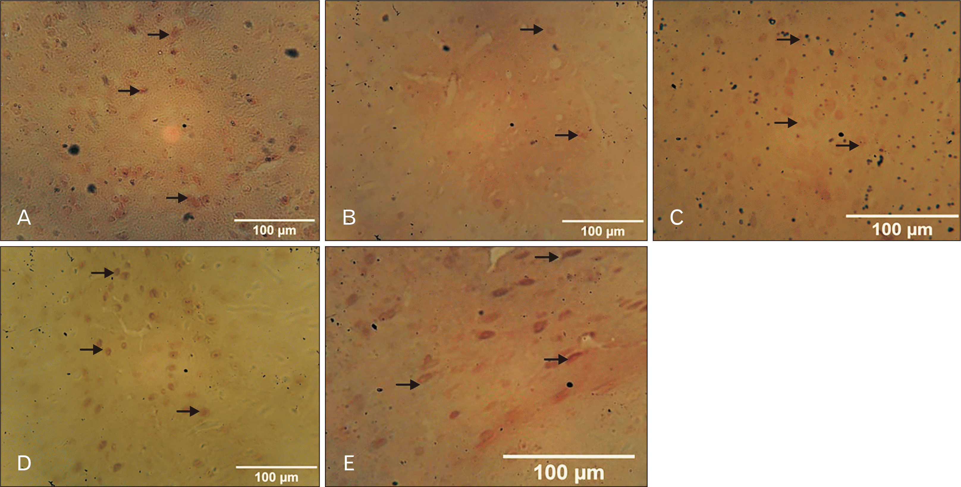 | Fig. 7Photomicrograph of the SNc immunostained for TH (×100). Control group (A) and NaVO3+Vahl group (D) low dose, showed normal DA neurons which are evenly distributed, while the vanadium exposed group (B) displayed degeneration and disruption of DA neurons, withdrawal group (C) shows the presence of scanty distribution of normal DA neurons, while NaVO3+Vahl group (E) high dose, revealed slight recovery of degenerated DA neurons. Black arrows indicate the TH cells (DA neurons). DA, dopaminergic; SNc, substantia nigra; TH, tyrosine hydroxylase.
|
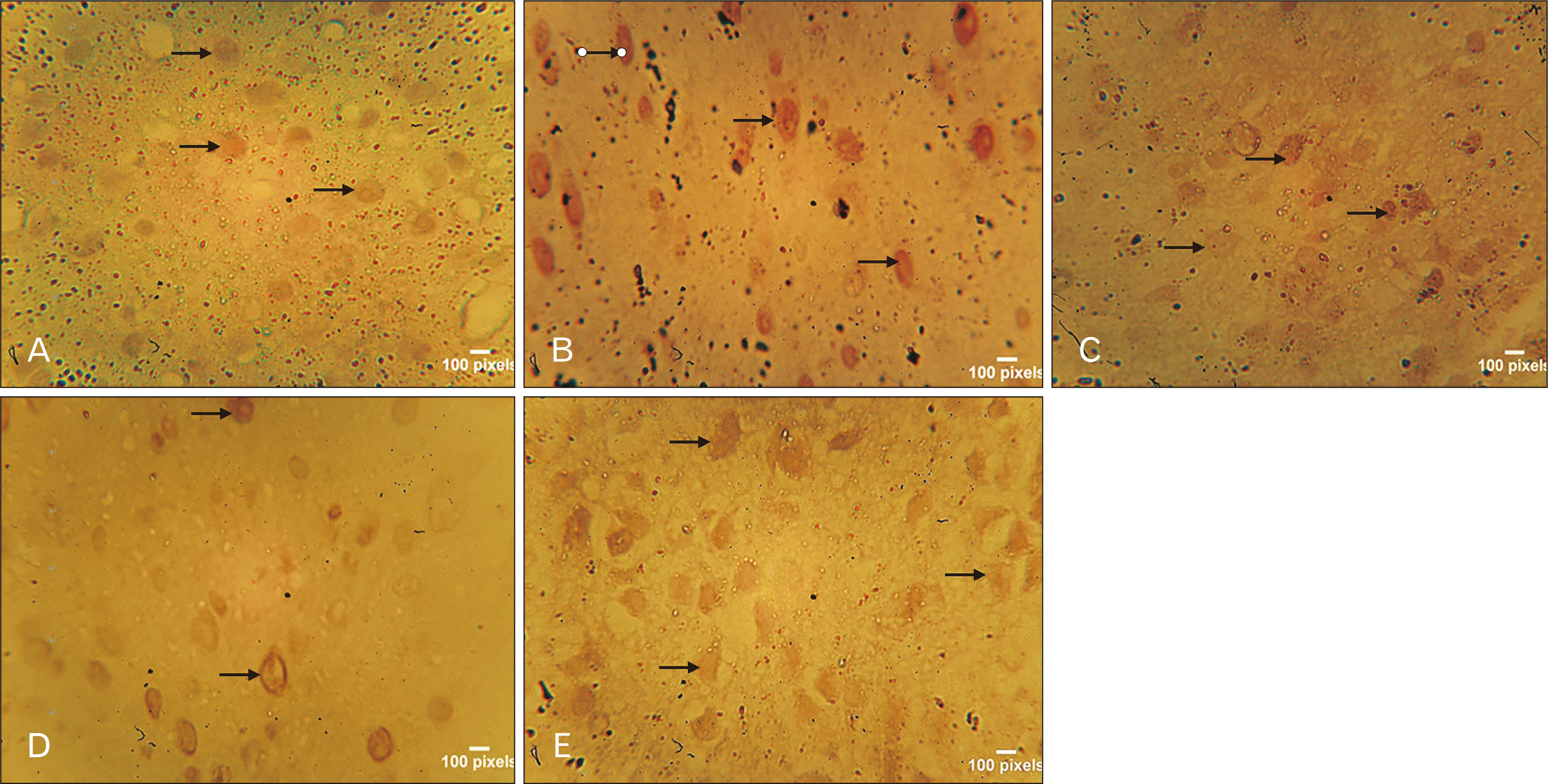 | Fig. 8Photomicrograph of the SNc immunostained for DAT (×100). Control (A) group revealed little expression of the DAT. Vanadium group (B) shows increased expression of DAT activity, while withdrawal group revealed little expression of DAT. However, NaVO3+Vahl group (D) low dose, revealed little expression of DAT, while NaVO3+Vahl group (E) high dose, revealed increased DAT activity. Black arrows indicat the dopaminergic transporter. DAT, dopamine transporter; SNc, substantia nigra.
|
 | Fig. 9Photomicrograph of the SNc immunostained for GFAP. From the vanadium Group (B) and withdrawal group (C), it can be observed that Vanadium-induced astroglia activation as shown by the presence of well-marked activated astrocytes, while control group (A) revealed no trace of astrocyte activation. NaVO3+Vahl group (D) low dose and NaVO3+Vahl group (E) high dose, revealed no traces of activated astrocytes. GFAP, glial fibrillary acidic protein; SNc, substantia nigra.
|
Tyrosine hydroxylase
Control group (A) and NaVO3+Vahl group (D) low dose, showed normal DA neurons which are evenly distributed, while the vanadium exposed group (B) displayed disruption of DA neurons, withdrawal group (C) shows the presence of scanty distribution of DA neurons, while NaVO3+Vahl group (E) high dose, revealed increase level of DA neurons as compared to the vanadium exposed group.
Dopamine transporter
Vanadium group (B) shows increased expression of DAT activity as compared to the control group, while withdrawal group (C) revealed little expression of DAT. However, NaVO3+Vahl group (D) low dose, revealed little expression of DAT, while NaVO3+Vahl group (E) high dose, revealed increased DAT activity. Black arrow indicates the DA transporter.
Glial fibrillary acidic protein
Vanadium (B) and withdrawal group (C) showed the presence of well-marked activated astrocytes, while control group (A) revealed no trace of astrocyte activation. NaVO3+Vahl group (D) low dose and NaVO3+Vahl group (E) high dose, revealed no traces of activated astrocytes.
Phytochemical analysis
The activities of phenol, tannin alkaloids, saponin and glycosides were more pronounced with the methanolic extract. However, saponin is seen to be more pronounced following the phytochemical of this extract (F. exasperata Vahl).
Go to :
Discussion
In the present investigation, we provide evidence for the motor benefit of the plant extract in a non-classical mouse model of Parkinson’s disease (PD). The benefit is related to the protective effect of the extract on DA neurons via an evidenced anti-oxidant activity. Our data suggest that the extract could have a beneficial effect in the treatment of PD.
As previously reported [22, 23], vanadium reduced locomotor activity and motor coordination using the RT [24]. The FEVL extract and withdrawal group demonstrate an increase in the retention time by spending a remarkable time on the rotating bar, which indicates an improvement in motor coordination. Also, another behavioral test used to assess motor coordination is a PBT which measures the LOT and the results obtained suggest a significant decrease in locomotor activities in the withdrawal group as well as when the extract is used after vanadium, and this suggests an ameliorative role of extract. Excessive exposure of the brain to amounts of trace elements like vanadium increase oxidative stress which later causes neurodegeneration and increased incidence of Parkinsonism and PD [5, 25].
Previous research done in our laboratory reviewed that FEVL extract alone at different dosages improves motor deficits in heavy metal consumption [4]. However, deleterious health effects induced by this transition element found in sodium metavanadate (NaVO3) compounds are seen in our environment especially due to heavy metal mining and in oil-producing communities where the burning of fossil fuel exist. This can be linked to dopaminergic neurons insult as a result of oxidative stress that leads to a decrease in dopamine levels in the brain region such as Striatum, SNc and globus pallidus which are implicated in vanadium toxicity [5].
The induction of lipid peroxidation in the brain following vanadium consumption is a testament to the role oxidative stress plays in the neurotoxicity of the metal [26, 27]. To ascertain this neurological effects being observed in humans [5, 28], MDA (an intermediate in the lipid peroxidation process) results showed an increased level in the vanadium-exposed group, and contribute to neuronal cell death and continued exposure to excess free radicals derived from some endogenous or exogenous neurotoxic species [29, 30]. However, when looking at the FEVL and those group that was withdrawn after some exposure, this in addition to the results above revealed a statistical reduction in MDA level. Reduction of lipid peroxidation may protect against oxidative neurodegeneration caused by vanadium exposure. Also, a significant increase in the activity of CAT level in vanadium-intoxicated mice was recorded, and this was seen to be reduced in the two groups that received FEVL extract. This is by following Shi and Dalal [31], which suggests that vanadium treatment may result in the increased formation of oxygen-free radicals, leading to oxidative stress. GSH as one of the markers of oxidative stress plays a major role in many cellular functions, which include detoxification of reactive oxygen species (ROS) in the brain, but abnormal levels of GSH is considered to be a sign of many diseases including neurotoxicity [32]. There was also a significant increase in the GSH level of the vanadium-exposed mice when compared with the control group, as seen in Fig. 5 (P<0.05).
However, the significant increase of GSH noticed in some treatment groups points to an ameliorative intervention of FeLV extract which indicates that the saponin fraction of FeLV extract has a strong antioxidant and anti-inflammatory properties to reduce the activities of these oxidative stress markers following the acute administration of vanadium. The changes in GSH and CAT, as well as increase in activities of MDA observed in the SNc suggest a correlation between loss of antioxidant response and an increase in oxidative stress caused by exposure to vanadium. MDA, GSH, and CAT constitute the main components of the antioxidant defense system, and modification in their expressions reflects potential oxidative stress [33]. The mechanism of action of the saponin fraction of FEVL extracts also affects the monoamine oxidase and DAT pathway to enhance and improve motor deficit in mice.
Dopamine, which is an important neurotransmitter in the brain, plays a modulatory role in the coordination of movement, reward, and cognition [34]. Alterations in DA neurotransmission are key hallmarks in the pathogenesis of several diseases such as Parkinson’s disease and addiction [35]. Results from this study can further substantiate that exposure to vanadium can lead to loss of dopaminergic neurons and a reduction of dopamine in the SNc [34]. While, withdrawal group showed a marked elimination of the distribution of noxious agent (vanadium) from the SNc, indicated by several dopaminergic neurons increase. However, the group with FEVL extract after exposure to vanadium revealed the perfect distribution of dopaminergic neurons, and this is by following the findings of Chinyere [36], which stipulate the possibility of the extract to have either inhibited the release or antagonize the action of these inflammatory mediators. On the other hand, the plasma membrane DAT which is considered to be a reliable marker of presynaptic dopaminergic terminal loss [37] that can transport potentially toxic agents into cells or elevate the intracellular concentration of dopamine to detrimental levels. Immunohistochemically, vanadium revealed increased expression of DAT when compared to the control group and withdrawal group, which revealed a little expression of DAT activity. However, the efficacy of the extract revealed little expression of DAT, which is dose-dependent. This finding agrees with numerous neurochemical studies of dopamine markers and is consistent with the hypothesis that those areas expressing the highest levels of DAT are most susceptible to neurotoxicity [37].
Inflammatory responses in the central nervous system are elicited by non-neuronal glial cells, chiefly astrocyte and microglia, whose activities underlie several neurodegenerative disease pathogeneses [38]. Astrocytes play a key role in the regulation of the neuronal activities, synaptic plasticity and also some key elements in the brain parenchyma defense against oxidative and toxic insults [39]. The number of visible astrocytes and the complexity of their processes may also be related to the extent of neural injury. In this current investigation, Vanadium-induced astrogliosis as shown by the presence of well-marked activated astrocytes, when compared to the control group, which revealed no trace of astrocyte activation. However, post-treated groups revealed no traces of activated astrocytes, which is as a result of the ameliorative effect of saponin fraction of FEVL extract. This corroborates with the findings of Garcia et al. [40] which stipulate that vanadium-induced astrogliosis has reported in the cerebellum and hippocampus after exposure to sodium metavanadate for five days, indicating a rapid response of astrocytes to this challenge.
In conclusion, taken together, This work has shown improved motor coordination with F. exasperata Vahl leave extract treatment after the vanadium-exposure. The results of the immunohistochemical and biochemical studies also revealed a well-marked expression of astrocytes and increased in the markers of oxidative stress, respectively, which become more pronounced during the process of neurodegeneration with vanadium. Therefore, from the present study, it can be deduced that vanadium accumulation in the brain can lead to astrocyte activation, which might induce neurotoxicity. However, inflammations actively contribute to neuronal damage and death, but the intervention of F. exasperata Vahl extract showed a remarkable improvement.
In conclusion, this study has shown that the saponin fraction of F. exasperata Vahl leaves has a powerful ameliorative effect on the motor deficits caused by vanadium in the SNc of male adult mice hence improve locomotor activity and motor coordination.
Go to :
 XML Download