

 PDF
PDF Citation
 Print
Print


Introduction
Doxorubicin (DOX) is broadly applied as an antineoplastic medication in the management of carcinomas such as hematological and solid malignancies. Nevertheless, DOX use was reduced due to the occurrence of toxicity in certain essential organs such as the heart, liver and kidneys [1]. In experimental studies, DOX induces nephrotoxicity in rodents marked by progressive glomerulosclerosis and tubulointerstitial disruption combined with hypoalbuminemia, hypercoagulability, dyslipidemia, proteinuria, edema, and ascites formation [2]. The DOX nephropathy model in rodents is therefore widely applied to induce experimental chronic renal diseases similar to that in human [3]. It is unclear how precisely DOX causes renal damage. Nevertheless, various mechanisms to explain nephropathy caused by DOX had been suggested. First, the nephrotoxicity of DOX may be caused through an immune system. Macrophages have a central role in the pathogenesis of DOX nephropathy via their T cell-stimulatory producing pro-inflammatory cytokines like tumor necrosis factor-alpha (TNF-α) [4]. One of the unusual vitamin D effects is its impact on the immune system. Originally, monocytes/macrophages from granulomatous disease patients are found to have a role in altering 25(OH) D3 to 1,25(OH)2D3 [5, 6]. Calcitriol has a determining role in the immune response, which was better known. The immune system may be modulated by calcitriol via intracrine, paracrine and endocrine pathways [5, 7]. Vitamin D3 (Vit D3) decreased the expression of hedgehog signaling target genes, which has a great role in regulating the immune system by controlling several T-cell properties as differentiation, proliferation, and activity [8]. Previous results showed that 1,25(OH)2D3 significantly decreased proteinuria and alleviated glomerulosclerosis of DOX-treated rats [9]. Dabak et al. [4] indicated that cholecalciferol causes satisfactory tubulointerstitial recovery in DOX-induced nephropathy in rats.
Another hypothesis is that the nephropathic effect of DOX may be caused by oxidative stress [10]. Therefore, the application of antioxidants could protect against DOX-induced toxicity [11]. Interestingly, overexpression of cyclooygenase-2 (COX-2) has a poor prognostic effect that makes cancer cells unresponsive to apoptotic stimuli [12]. A nonsteroidal anti-inflammatory drug (NSAID), meloxicam acts by inhibiting COX-2 activity [13]. In addition to their potential antioxidant properties, the drug has enhanced gastric and renal tolerability with increased therapeutic index when compared with traditional NSAIDs [14].
The objective of the current work is to assess the prospective ameliorating impacts of meloxicam versus Vit D3 against progressive DOX-induced nephropathy in rats trying to ascertain the possible mechanism underlying such amelioration
Go to : 
Materials and Methods
Chemicals and drugs
Vit D3 was ordered from Sigma Chemical Co. (St. Louis, MO, USA), DOX hydrochloride 10 mg vial from Pharmacia & Upjohn (Milan, Italy), Meloxicam (Mobic) 15 mg vial from Boehringer Ingelheim Pharma GmbH & Co KG (Biberach, Germany), polyclonal rabbit/anti-rat TNF-α antibody (Lab Vision Co., Fremont, CA, USA). Kits for malondialdehyde (MDA) reduced glutathione (GSH), protein and creatinine were bought from Spectrum Co. (Sigma-Aldrich, Cairo, Egypt).
Treatment protocol
Wistar Adult male rats weighing around 225–325 g were used in this study. Animals are held at 25°C±1°C temperature & 55 percent relative humidity with a daily 12-hour light/12-hour dark cycle in ordinary housing conditions in cages and for one week, they had been permitted to adapt for one week. All the research procedures had been performed according to the rules and regulations implemented by Mansoura University’s Institute Research Board.
Ninety rats were randomly distributed equally to five study groups as follows, each consisting of 18 animals which were further subdivided into 6 animals living for 7, 14, 21 days.
Group 1: considered as the negative control receiving saline intraperitoneal (2 ml/kg i.p.) once/day for 7, 14, 21 days consecutively, and represented as a negative control group
Group 2: rats were given meloxicam (2 mg/kg i.p) for 7, 14, 21 days [11].
Group 3: a single DOX dose (15 mg/kg i.p.) was injected to rats [15].
Group 4: a single DOX dose was injected to rats (i.p.) followed by Vit D3 orally (Devit-3, 50,000 IU/15 ml) daily for 7, 14, 21 days. Vit D3 had been given into the mouth by dropper (200 IU/day/rat) [4]
Group 5: a single DOX dose was injected to rats (i.p.) followed by meloxicam (2 mg/kg i.p.) for 7, 14, 21 days [11].
Urinary tests
Urine samples during 24-hours were collected from all rats at days 7th, 14th, and 21st of the study and analyzed to assess the urine protein [4].
Sample collection and assessment of renal functions
Animals were monitored daily basis and sacrificed after 7th, 14th, and 21st days for each assigned group. Blood was obtained and the kidneys were extracted and then prepared for evaluations. Serum creatinine on days 7, 14, and 21 of the assigned groups was assessed.
Evaluation of reduced glutathione and renal lipid peroxides
GSH kit had been used and reduced GSH was estimated in accordance with the method used by Moron et al. [16]. Resulting data had been presented as mmol/g tissue. The lipid peroxides in the kidney were measured by spectrophotometer established on the reaction between thiobarbituric acid and MDA according to Mihara and Uchiyama [17]. Results had been presented as nmol/gm tissue.
Histological assessment
Kidney specimens embedded in paraffin were sliced into 4–6 μm thickness sections and stained with hematoxylin and eosin (H&E) [18]. The slides had been assessed using light microscope (Olympus BH-2; Olympus, Tokyo, Japan) and photographed by two pathologists, blinded to the procedure of the study.
Immunohistochemical examination
Immunohistochemical staining for TNF-α was implemented using polyclonal rabbit/anti-rat antibody according to Côté [19].
Morphometric analysis
Glomerular geometry
Stained sections were observed under a light microscope and images were recorded at 400× magnification.
Glomerular volume: was recorded from the cross-sectional area using the formula according to Sanden et al. [20].
The glomerular tuft cross-sectional area (AG) was recorded from the tuft outlines using ImageJ program (×40 objectives, 0.12 µm/pixel).
Total glomerular surface area: The Bowman’s capsule internal edge was considered the limit of the whole glomerular surface using the program ImageJ (×40 objective, 0.12 µm/pixel)[21].
The peripheral urinary surface area: was achieved using the formula (total glomerulus surface area-AG). About 30 glomeruli from each kidney were measured [21].
Number of nuclei per glomerulus was counted in 3 adjacent non-overlapping fields levels and expressed as mean from 3 sections using QuPath program (version 0.1.2; Queen’s University Belfast, Belfast, UK).
Glomerular sclerosis and tubulointerstitial damage indices
Proposed grading system by El Nahas et al. [22] was used to assess glomerular sclerosis within the capillary tuft as an indicator of kidney injury. About 100 glomeruli for each animal at a magnification of ×400 were examined to establish the score. The scarring severity was presented on a grading system from 0 to 4.
Véniant et al. [21] proposed a tubulointerstitial score (0 to 4) based on histological findings of (tubular atrophy, dilation, interstitial inflammation, casts, and fibrosis).
Statistical analysis
The values were reported as means±SD using one way ANOVA and Dunnett multiple comparison tests. The graphs were plotted using Graph Pad Prism 7.00 (Graph Pad Software Inc., San Diego, CA, USA). The P<0.05 reflected statistical significance.
Go to :
Results
Effect on urinary protein
Fig. 1A shows that rats injected with DOX alone revealed a significant increase in urinary protein (4.3±0.21, 5.2±0.41, and 6.4±0.32 mg/24 hrs) compared to the control group (3.2±0.23, 3.4±0.11, and 3.3±0.2 mg/24 hrs.) after (1, 2, and 3 weeks respectively). Meloxicam alone treated group revealed no significant difference as compared with control saline group. Both Vit D3 and meloxicam treated groups following single-dose administration of DOX showed a significant reduction in urinary protein (3.7±0.3, 4.3±0.3, and 4.6±0.2 mg/24 hrs.) and (3.5±0.19, 3.8±10.17, and 4±0.45 mg/24 hrs.) after (1, 2, and 3 weeks respectively) compared to DOX treated rats but more than the saline group. Concomitant meloxicam administration with DOX showed a significant decrease in urinary protein after 2 and 3 weeks compared with Vit D3 treated groups.
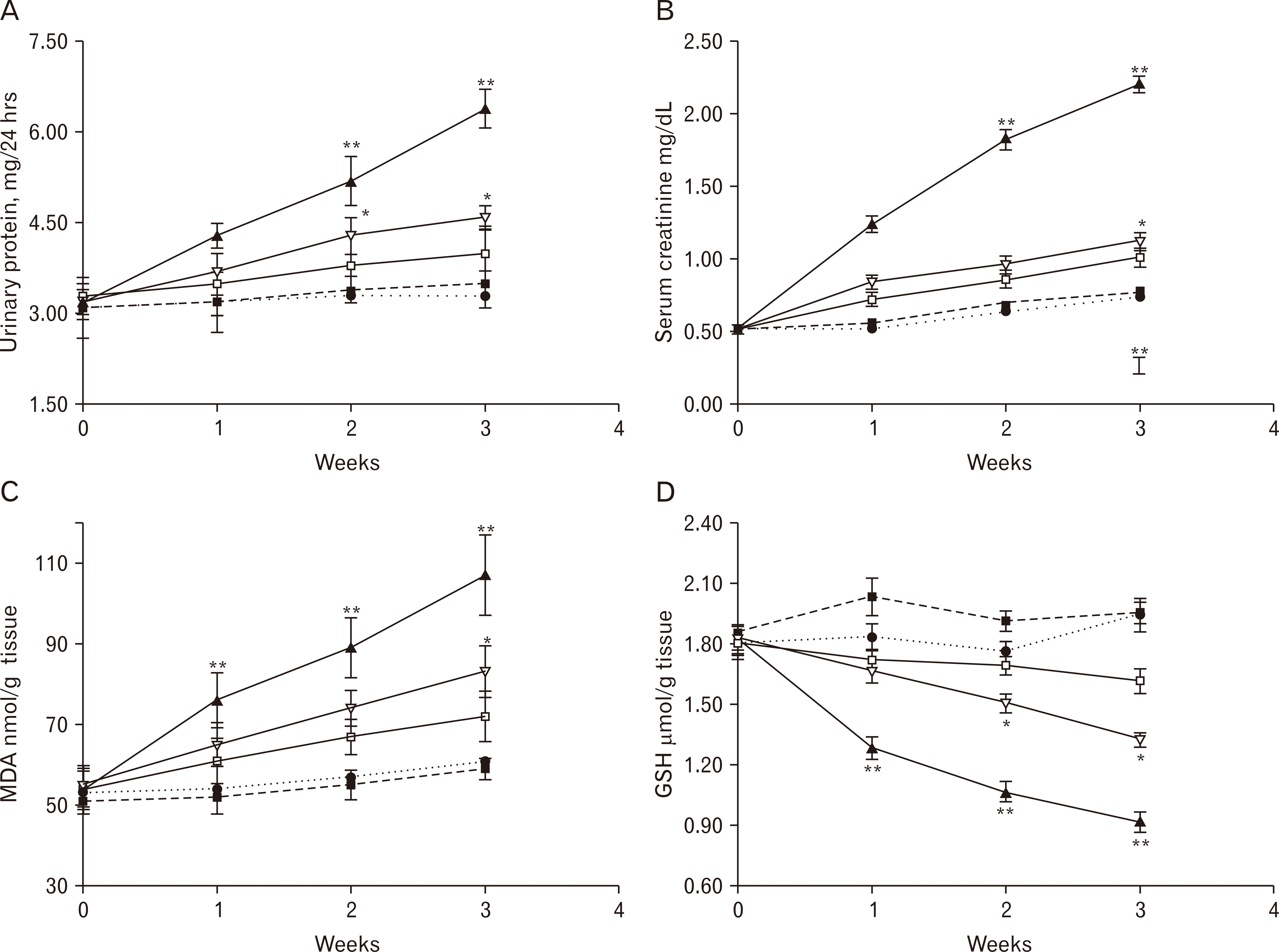 | Fig. 1Total urinary protein (A), serum creatinine (B), MDA level in renal tissue (C), and GSH in renal tissue (D) for groups with saline treated (●), meloxicam (■), DOX (▲), DOX+Vit D3 (▽) and DOX+meloxicam (□) treated groups graphed versus time. Values are presented as means±SD. DOX, doxorubicin; GSH, glutathione; MDA, malondialdehyde; Vit D3, vitamin D3. *P, significant difference between combined DOX+Vit D3 vs. combined DOX+meloxicam treated group; **P, significant in contrast to other groups.
|
Effect on serum creatinine
Fig. 1B shows that rats receiving DOX alone showed an increase in serum creatinine significantly (1.25±0.05, 1.83±0.07, and 2.21±0.06 mg/dl) compared to control group (0.53±0.06, 0.65±0.03, and 0.75±0.01 mg/dl) after (1, 2, and 3 weeks respectively). Meloxicam alone treated group revealed no significant difference as compared with control saline group. Serum creatinine was significantly decrease in Vit D3 and meloxicam treated groups following single-dose administration of DOX (0.85±0.05, 0.97±0.06, and 1.13±0.06 mg/dl) and (0.73±0.04, 0.87±0.07, and 1.02±0.06 mg/dl) after (1, 2, and 3 weeks respectively) compared to DOX treated rats but greater than the saline group. Concomitant meloxicam administration with DOX showed a decrease in serum creatinine compared with Vit D3 treated groups which were significant after 3 weeks.
Effect on lipid peroxidation
Renal kidney lipid peroxidation was assessed by MDA level in kidney tissue. As shown in Fig. 1C, meloxicam alone administration presented a reduction in the MDA content compared to the saline given group which is not significant. DOX significantly increased renal MDA (76±6.89, 89±7.47, and 107±9.98 nmol/g tissue) compared to control saline (54±4.26, 57±3.59, and 61±2.59 nmol/g tissue) which was progressive after (1, 2, and 3 weeks respectively). Administrating both Vit D3 and meloxicam to DOX treated rats significantly decreased MDA (65±5.47, 74±4.45 and 83±6.5 nmol/g tissue) and (61±5.65, 67±4.36, and 72±6.2 nmol/g tissue) after (1, 2, and 3 weeks respectively) compared to DOX injected rats but still greater than saline given group. Concomitant meloxicam administration with DOX showed a decrease in MDA renal content compared with Vit D3 treated groups which were significant after 3 weeks.
Time course variations of renal glutathione
As shown in Fig. 1D, meloxicam administration revealed an increase in the GSH content in contrast to the control group which is more evident after the first week (2.03±0.095) and consequently, its mean percentage value was higher than 100% of control (Table 1). Combined administration of Vit D3 and DOX shows a significant rise in the renal GSH content in contrast to control (Fig. 1D), with a subsequent increase in its mean percentage of control value (Table 1). Conversely, Co-administration of DOX and meloxicam revealed an improvement in the renal GSH content significantly (1.72±0.05, 1.69±0.05, and 1.61±0.03) after (1, 2, and 3 weeks) respectively in contrast to DOX-injected group (1.28±0.05, 1.06±0.05, and 0.91±0.02) and also combined Vit D3 and DOX (1.66±0.06, 1.50±0.04, and 1.32±0.03) as shown in (Fig. 1D) hence improvement in its mean percentage of control value reaching 82.10±3 after the third week compared to DOX (46.91±2) and combined Vit D3 and DOX (68.04±4) (Table 1).
Table 1
GSH levels in renal tissue as mean percentage of control value
| Group | 1st wk (%) | 2nd wk (%) | 3rd wk (%) |
|---|---|---|---|
| Saline (control) | 100a) | 100a) | 100a |
| Meloxicam | 110.93±6a) | 108.52±5a) | 100.52±8a) |
| DOX | 69.95±5b) | 60.23±4b) | 46.91±2b) |
| DOX+Vit D3 | 90.71±7c) | 85.23±6c) | 68.04±4c) |
| DOX+meloxicam | 93.99±7c) | 92.35±5d) | 82.99±3d) |
![]()
Morphometric results
The mean volume of individual glomeruli was evaluated on H&E stained renal sections. As shown in Table 2. After the first week, rats receiving DOX showed decreased glomerular volume in contrast to other groups reaching 1.90±0.96 ×105 µm3 with a significant difference. Regarding DOX+Vit D3receiving group, glomerular volume (3.25±2.35 ×105 µm3) was reduced compared with control and meloxicam groups but significantly higher than DOX group. In meloxicam+DOX receiving group, glomerular volume showed a significant increase (3.92±0.28 ×105 µm3) compared with DOX group and combined DOX and Vit D3 treated group though the difference is not significant. After the second week, mean volume of individual glomeruli was the least value in DOX treated group reaching 1.65±0.13 ×105 µm3 and attain the highest value in combined DOX and meloxicam treated group reaching 3.90±0.13 ×105 µm3 with a significant difference. After the third week, the values regarding the mean volume of individual glomeruli was similar to the second week regarding the lowest and highest values. Comparing the progress of the mean volume of individual glomeruli in different groups during weeks, it was observed a reduction in the glomerular volume with weeks especially in DOX treatment groups while in control and meloxicam treated groups the values were similar which was slightly higher in meloxicam treated group.
Table 2
Glomerular dimensions quantified by morphometry of the five study groups
| Group | 1st wk | 2nd wk | 3rd wk | |||||||||
|---|---|---|---|---|---|---|---|---|---|---|---|---|
|
|
|
|
||||||||||
| Mean volume of individual glomeruli (×105 μm3) | Glomerular surface area (×103 μm2) | Peripheral urinary surface area (×103 μm2) | Average number of nuclei per glomerulus | Mean volume of individual glomeruli (×105 μm3) | Glomerular surface area (×103 μm2) | Peripheral urinary surface area (×103 μm2) | Average number of nuclei per glomerulus | Mean volume of individual glomeruli (×105 μm3) | Glomerular surface area (×103 μm2) | Peripheral urinary surface area (×103 μm2) | Mean number of nuclei per glomerulus | |
| Saline (control) | 3.95±0.59a,c) | 6.10±0.31a,c) | 1.52±0.77a,c) | 49.00±9.90a,b) | 3.72±0.56a) | 6.01±3.90a,c) | 1.70±0.10a,c) | 43.50±3.43a,c) | 3.47±0.84a,c) | 6.37±1.02a) | 2.14±0.35a,c) | 47.33±1.53a) |
| meloxicam | 4.42±1.61c) | 6.27±4.05c) | 1.40±0.15c) | 55.50±0.71a) | 4.12±0.20a,c) | 6.32±2.45a,c) | 1.73±0.97a,c) | 42.00±3.00a,c) | 5.17±0.12a) | 7.64±1.14a) | 2.12±0.75a,c) | 48.50±4.95a) |
| DOX | 1.90±0.96b) | 6.76±4.03a,c) | 2.27±1.65b,c) | 39.75±7.32b,c) | 1.65±0.13b,c) | 6.86±2.72a,c) | 2.93±1.02a) | 37.00±9.61a,c) | 1.13±0.13b) | 4.67±2.42b) | 3.33±1.26a) | 32.00±32.00b,c) |
| DOX+Vit D3 | 3.25±2.35a) | 5.00±2.74a) | 1.53±0.96a,b) | 45.67±1.55b,c) | 3.27±0.23a) | 5.07±2.01b,c) | 1.18±0.70b,c) | 32.50±0.71a) | 3.60±0.22a,c) | 5.75±2.95a) | 2.27±0.98a,c) | 42.66±4.72a) |
| DOX+meloxicam | 3.92±0.28a) | 5.65±2.70a) | 1.22±0.78a) | 55.20±5.81a) | 3.90±0.13b,c) | 5.70±1.71b) | 1.08±0.72b,c) | 53.50±3.32b,c) | 3.82±0.16a,c) | 6.16±1.50a) | 1.07±0.40b,c) | 43.50±2.12a) |
![]()
Table 2 showed that after the first week, the glomerular surface area was highest in DOX treated group, which was not significant in contrast to other groups. After the 2nd week, glomerular surface area in DOX treated group showed an increase was significant compared to the combined DOX and meloxicam treated group. After the third week, the glomerular surface area was reduced significantly in DOX treated group in contrast to other groups.
In Table 2 DOX injected groups showed a significant increase in the peripheral urinary surface which was progressive (2.27±1.65, 2.93±1.02, and 3.33±1.26 ×105 µm3) after 1st, 2nd, and 3rd weeks respectively. On the other hand, the peripheral urinary space was significantly decreased in combined DOX+meloxicam treated groups (1.22±0.78, 1.08±0.72, and 1.07±0.40 ×105 µm3) after 1st, 2nd, and 3rd weeks respectively compared with other groups.
The average number of nuclei/glomerulus had decreased after the first week (39.75±7.32 n/glomerulus) in DOX treated group with a significant difference compared with combined DOX+meloxicam treated group (55.20±5.81 n/glomerulus). The average number of nuclei per glomerulus had decreased in all DOX treated groups with the progress of weeks but it was observed that combined DOX+meloxicam treated group attain the highest number compared with DOX and combined DOX+Vit D3 treated groups (Table 2).
Histological changes scoring were demonstrated in Table 3. No evidence of glomerulosclerosis or tubulointerstitial injury in group 1 and 2 in renal tissue. Glomerular sclerosis index was significantly greater in DOX injected group in contrast to DOX+Vit D3 and DOX+Meloxicam treated groups. Glomerular sclerosis index was progressively increased with weeks. When mean index score was compared between DOX+Vit D3 and DOX+Meloxicam treated groups, it was significantly higher in DOX+Vit D3 treated group especially after 1st and 2nd weeks. The picture of tubulointerstitial injury was coincident with glomerular sclerosis. The tubulointerstitial injury was evident in DOX treated group with a significant index increment compared with DOX+Meloxicam treated group and still higher than DOX+Vit D3 treated group. The tubulointerstitial index was progressively increased with weeks. Comparing DOX+Vit D3 and DOX+Meloxicam treated rats; the tubulointerstitial index was significantly greater in DOX+Vit D3 treated rats after 3rd weeks.
Table 3
Renal damage indices
| Group | Glomerular sclerosis | Tubulointerstitial damage | ||||
|---|---|---|---|---|---|---|
|
|
|
|||||
| 1st wk | 2nd wk | 3rd wk | 1st wk | 2nd w wk | 3rd wk | |
| Saline (control) | 0.0±0.0a,c) | 0.0±0.0a) | 0.0±0.0a) | 0.0±0.0a) | 0.0±0.0a) | 0.0±0.0a) |
| Meloxicam | 0.0±0.0a) | 0.0±0.0a) | 0.0±0.0a) | 0.0±0.0a) | 0.0±0.0a) | 0.0±0.0a) |
| DOX | 1.13±0.83b) | 1.67±0.43b) | 2.67±0.64b) | 1.50±0.38b) | 2.40±0.40b) | 3.60±0.45b) |
| DOX+Vit D3 | 0.75±0.26a) | 1.63±0.51b) | 2.30±0.15b,c) | 0.90±0.23b,c) | 1.43±0.20c) | 3.25±0.50b) |
| DOX+meloxicam | 0.25±0.10c) | 0.60±0.29a) | 1.50±0.18c) | 0.45±0.16a,c) | 0.90±0.18c) | 1.86±0.69c) |
![]()
Histopathological assessment
The control group histological pictures shown in Figs. 2A, B, Fig. 3A, B, and Fig. 4A, B after 1st, 2nd, and 3rd weeks respectively revealed the normal architecture of renal glomeruli. Similarly, renal sections of rats received meloxicam only showed no significant change in contrast to the control group as shown in Figs. 2C, D, Fig. 3C, D, and Fig. 4C, D after 1st, 2nd, and 3rd weeks respectively.
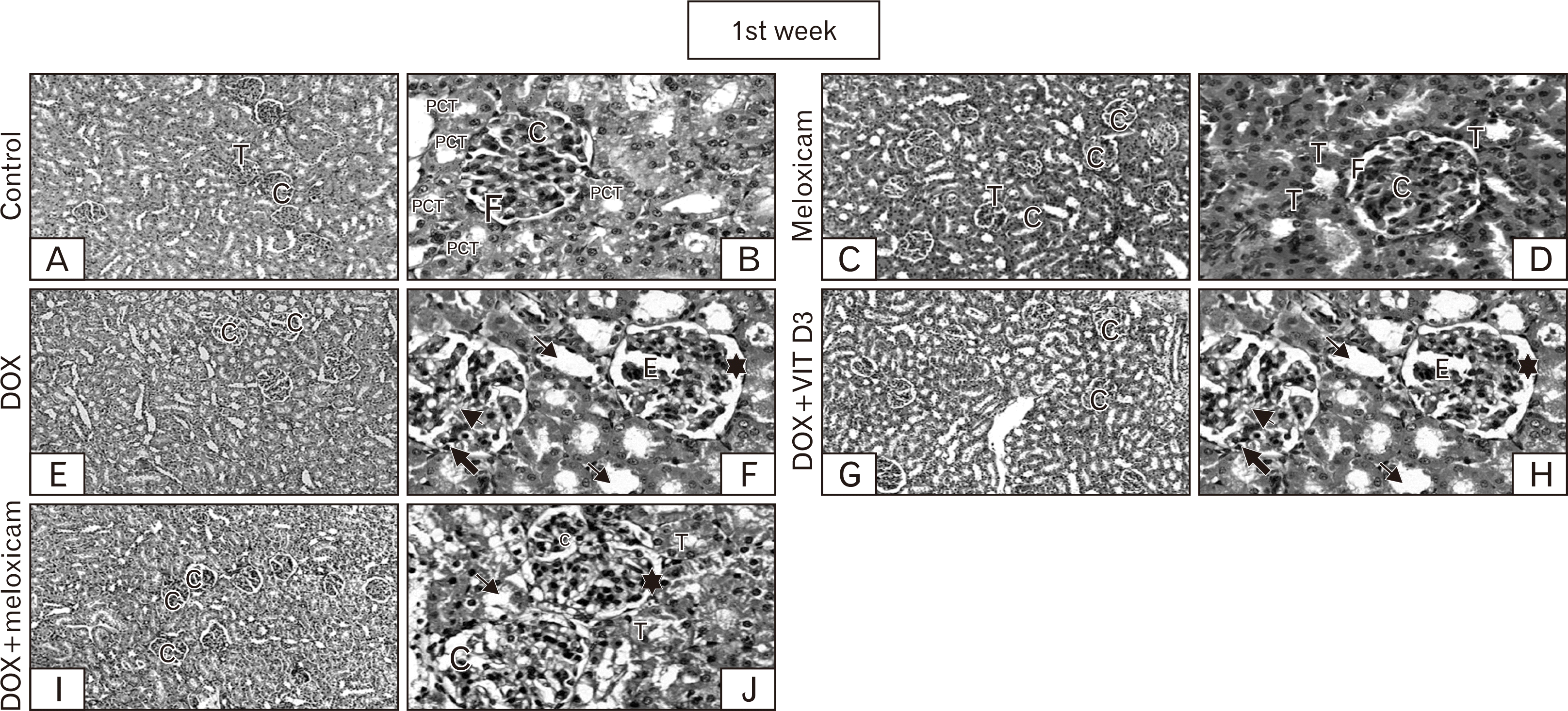 | Fig. 2Representative of cortical kidney sections stained by H&E from rats representing groups of control, meloxicam, DOX, DOX+Vit D3, and DOX+meloxicam after 1st week. Control sections revealed the normal histological architecture, renal T and renal C (A, ×100), F and PCT (B, ×400). Meloxicam revealed no changes (C, ×100). The renal glomeruli and tubules appear to great extent normal (D, ×400). DOX is showing decrease number of renal C in contrast to other groups (E, ×100), glomerular E, synechia of the glomerular tuft with Bowman’s capsule (➨), mesangial matrix increment (➤), and dilation of the F (🟌). Some cells of PCT loss its brush borders and some nuclei appear pyknotic (→) (F, ×400). Conversely, combination group revealed rare disruption of some renal C (G, I, ×100). Less dilation of F (🟌). Most of PCT appears normal (T) lower number of cells of PCT showed loss of brush border and pyknosis (→) compared to the DOX treated group (H, J, ×400). C, corpuscles; DOX, doxorubicin; E, edema; F, filtration space; PCT, proximal convoluted tubules; T, tubules; Vit D3, vitamin D3.
|
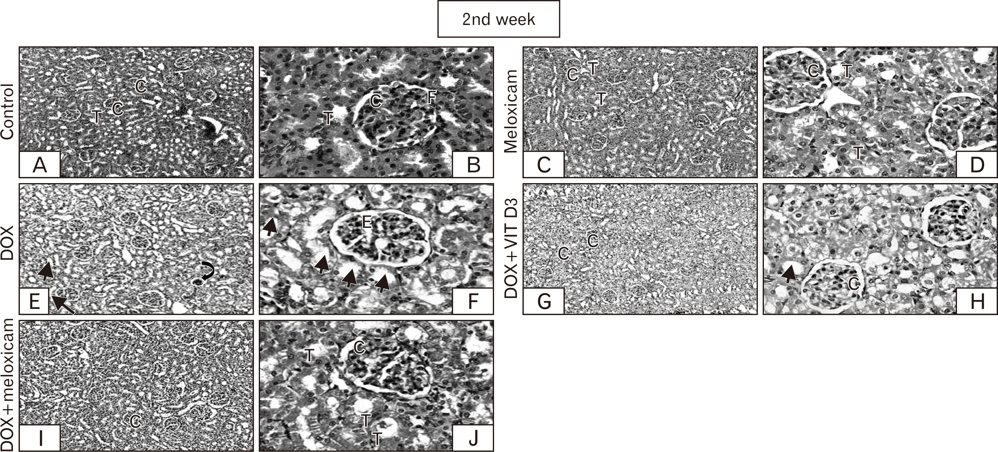 | Fig. 3After 2nd week: Sections of control group demonstrating the normal histological architecture, renal T, renal C (A, ×100), and F (B, ×400). No difference is shown in meloxicam (C, ×100). The renal glomeruli and tubules appear normal to great extent (D, ×400). DOX is showing shrinkage of renal C (⮏) compared to other groups (E, ×100), some showing glomerular E and most cells of PCT loss its brush borders and some nuclei appear psychotic (→) (F, ×400). However, combination group exhibiting less disruption of some renal C (G, I, ×100). Most of PCT appears normal (T) lower number of cells of PCT showed loss of brush border and pyknosis (→) compared to the DOX treated group (H, J, ×400), with better picture in DOX+meloxicam compared with DOX+Vit D3 group. C, corpuscles; DOX, doxorubicin; E, edema; F, filtration space; T, tubules; Vit D3, vitamin D3.
|
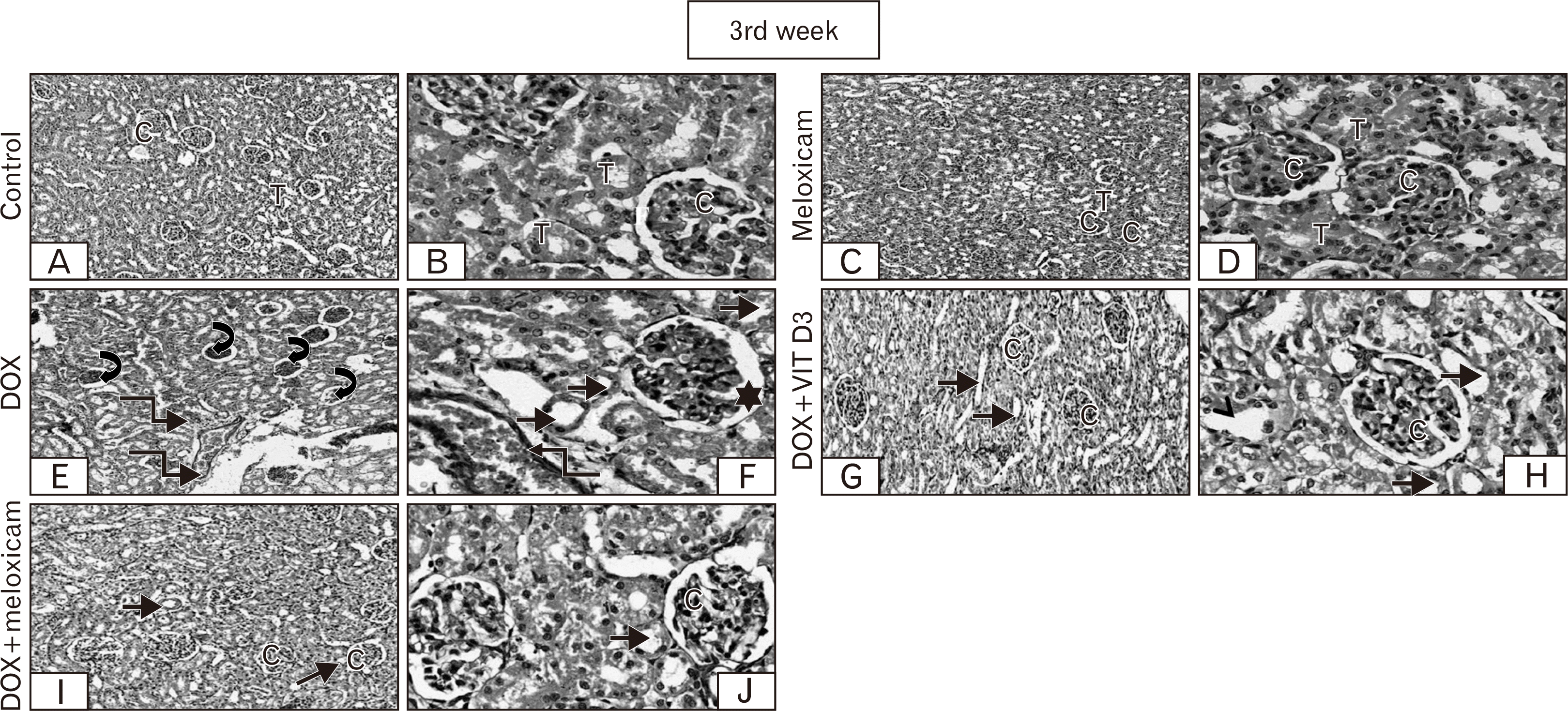 | Fig. 4After 3rd week Sections of control group exhibiting the normal histological architecture, renal C, renal T (A, ×100), and F (B, ×400). Meloxicam is showing no difference from the normal (C, ×100). The renal glomeruli and tubules appear to great extent normal (D, ×400). DOX is showing prominent shrinkage of renal C (⮏) compared to other groups and vascular congestion (↯) (E×100), and dilation of the F (🟌). Most cells of PCT loss its brush borders, dilated and some nuclei appear psychotic (→) (F×400). However, combination group showing less distortion of renal C (G, I, ×100). Most prominent finding is dilation of T and loss of brush borders (→) compared to the control group (H, J, ×400), with better picture in DOX+meloxicam compared with DOX+Vit D3 group. C, corpuscle; DOX, doxorubicin; F, filtration space; PCT, proximal convoluted tubules; T, tubules.
|
DOX treated group after 1st week (Fig. 2E, F) showing decrease number of renal corpuscles compared to other groups, glomerular edema, synechia of the glomerular tuft with Bowman’s capsule, dilation of the filtration spaces and mesangial matrix increment. Some cells of renal tubules loss their brush borders and some nuclei appear pyknotic. Combination groups (DOX+Vit D3 and DOX+meloxicam) showing rare distortion of some renal corpuscles (Fig. 2G–J). Less dilation of filtration spaces most of proximal convoluted tubules (PCT) appears normal. Lower number of cells of PCT showed a loss of brush border and pyknosis compared to the DOX treated group.
After 2nd week, DOX (Fig. 3E, F) is showing shrinkage of renal corpuscles compared to other groups, some showing glomerular edema and most cells of PCT loss its brush borders and some nuclei appear pyknotic. Combination groups (DOX+Vit D3 and DOX+meloxicam) showing better picture (Fig. 3G–J). Less dilation of filtration spaces most of PCT appears normal. Lower number of cells of PCT showed loss of brush border and pyknosis in contrast to the DOX injected group with an improvement of the picture in DOX+ Meloxicam compared with DOX+Vit D3 group.
After 3rd week, DOX (Fig. 4E, F) is showing prominent shrinkage of renal corpuscles compared to other groups, vascular congestion, and dilation of the urinary spaces. Most cells renal tubules lose their brush borders, dilated and some nuclei appear pyknotic. Combination groups showing less distortion of renal corpuscles (Fig. 4G–I) compared to DOX treated group. The most prominent finding is the dilation of renal tubules and loss of brush borders compared to the control group (Fig. 4H–J), with a better picture in DOX+Meloxicam compared with DOX+Vit D3 group.
Immunohistochemical results
Immunohistochemical results were analyzed and interpreted using an immunoreactive scoring system (IRS). IRS index was negative in groups 1 and 2 (Table 4, Fig. 5A–F) after (1st, 2nd, and 3rd weeks respectively). DOX administration showed a significant increase in the TNFα immunoreactivity (Table 4) which were displayed in both renal tubules and glomeruli with a higher score index in glomeruli compared with the tubules, which were progressively increased with weeks. IRS index in the glomeruli was moderate during the three weeks of treatment while ranged from mild to moderate in the tubules (Fig. 5G–I). Concomitant administration of DOX with Vit D3 and meloxicam decreased the expression of TNFα, in contrast to DOX group. IRS index in the glomeruli was ranged from mild to moderate in DOX+ Vit D3 treated group (Table 4, Fig. 5J–L) and negative to mild in DOX+Meloxicam treated groups (Table 4, Fig. 5M–O). IRS index in the tubules was ranged from negative to mild-moderate in DOX+Vit D3 and DOX+meloxicam treated groups. When the IRS index was compared between DOX+Vit D3 and DOX+meloxicam treated groups, it shows a significant increase in DOX+Vit D3 treated group.
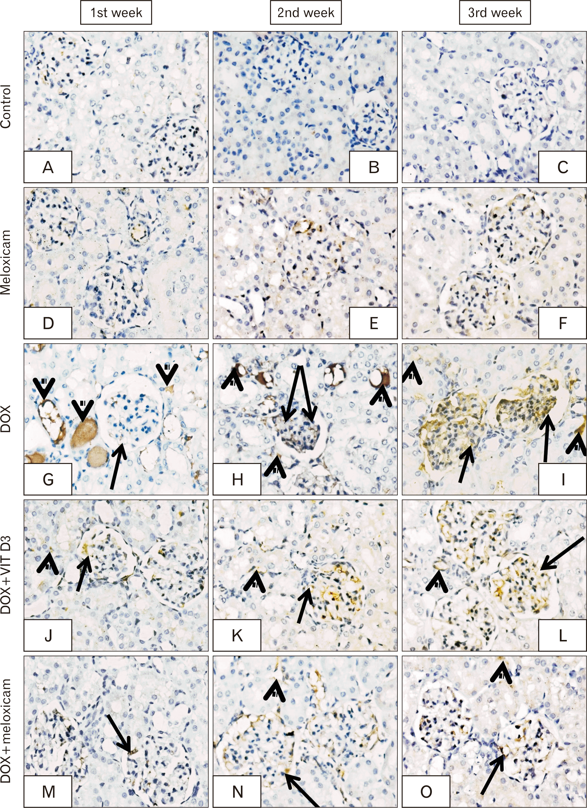 | Fig. 5Representatives of renal cortical tissue stained by TNF-α of: (A–C), control and meloxicam groups (D–F) after 1st, 2nd, and 3rd weeks, respectively displaying negative expression. DOX treated animals displayed intensive expression (G–I) after (1st, 2nd, and 3rd weeks respectively) in the renal glomeruli (→) and renal tubules (➤). Combined DOX-Vit D3 treated rats displayed moderate expression (J–L) after (1st, 2nd, and 3rd weeks respectively) within the glomeruli (→) while mild expression in the renal tubules (➤) .DOX/meloxicam treated groups demonstrated considerable improvement with mild expression (M–O) after (1st, 2nd, and 3rd weeks respectively) in the renal glomeruli (→) and renal tubules (➤). The expression is predominantly cytoplasmic, but with few nuclear expression. Immunohistochemistry counter stained with H&E, ×400. DOX, doxorubicin; TNF-α, tumor necrosis factor-alpha; Vit D3, vitamin D3.
|
Table 4
Immunoreactive score
| Group | Glomerular expression | Tubulointerstitial expression | ||||
|---|---|---|---|---|---|---|
|
|
|
|||||
| 1st wk | 2nd wk | 3rd wk | 1st wk | 2nd wk | 3rd wk | |
| Saline (control) | 1.75±0.50a) | 1.00±1.00a) | 0.50±0.21a) | 1.00±0.00a) | 1.00±0.00a) | 0.45±0.25a) |
| Meloxicam | 0.50±0.21a) | 1.00±1.00a) | 1.25±0.50a,c) | 0.80±0.30a) | 1.10±0.32a) | 1.11±0.29a) |
| DOX | 4.67±2.88b) | 5.60±2.19b) | 6.00±2.19b) | 3.13±1.55b) | 3.38±2.42b,c) | 4.37±1.85b) |
| DOX+Vit D3 | 3.57±1.72c) | 4.50±1.91b) | 4.67±2.30b,c) | 1.95±1.16a) | 2.21±1.22a,c) | 2.68±1.40a) |
| DOX+meloxicam | 1.67±0.57a) | 1.67±1.15a) | 2.50±1.22a) | 1.93±0.91a) | 2.00±0.33a,c) | 2.06±0.74a) |
![]()
Go to :
Discussion
DOX, a large-scale anti-cancer drug that prefers to be merged with new, targeted treatments to upgrading their efficacious response. Unfortunately, serious complications to DOX such as cardiotoxicity, nephrotoxicity, and liver toxicity are undermining its feasibility [25, 26].
The current study demonstrated progressive renal damage induced by DOX over three weeks that was clear from a relevant increase in urinary protein production and serum creatinine. This was consistent with Wang et al. [3] who observed that proteinuria occurred shortly after DOX injection and remained significantly higher for three weeks. Certain biochemical parameters were evaluated in our study; renal lipid peroxidation through measuring MDA and GSH level to ascertain oxidation. DOX has caused a significant progressive rise in renal MDA in comparison with saline control, which had been reported by previous studies [11]. In addition to, significant decrease in GSH content that has been observed in DOX-treated groups and because GSH has a remarkable role in antioxidation of ROS and free radicals and detoxification of xenobiotic compounds [27]. This makes the low level of GSH observed an indicator of the association of excessive oxidative stress. Based on our results, we can suggest that lipid peroxide and oxidative stress are triggered by DOX as a mechanism for kidney damage [28].
It is still unclear by which molecular mechanism DOX is responsible for renal damage. However, various mechanisms to explain nephrotoxicity caused by DOX were proposed. Firstly, nephrotoxicity induced by DOX can be mediated via an immune mechanism. Macrophages had a remarkable role in the pathogenesis of DOX nephropathy via their T cell-stimulatory producing pro-inflammatory cytokines like TNF-α [4]. Upon this fact, we tried to test the impact of combining vitamin D with DOX on renal damage as according to Di Rosa et al. [5], vitamin D had a nonconventional act on the immune system. Also, Szymczak and Pawliczak [6] noticed that monocytes/macrophages from granulomatous disease patients had a role in synthetizing 1,25(OH)2D3 from its precursor – 25(OH)D3. An alternative hypothesis had been proposed by Deepa and Varalakshmi [10] in which oxidative stress could result in the toxicity of the renal DOX. Oxidative stress is correlated with the development of COX2 and nephrotoxicity showing its overproduction [29, 30]. PGE2 was stated to have promoted cytokine production, like TNF-α, as a result of COX2; a prominent essential mediator of inflammatory disorders [31]. Hassan et al. [11] discovered a substantial increase in both PGE2 and TNF-α expression in the kidney tissue that also backed the DOX-induced oxidative stress hypothesis. DOX-induced nephrotoxicity was previously tested in compounds with which normal renal function and architecture were partially preserved via their antioxidant and anti-inflammatory activity as caffeic acid phenethyl ester [32], Zingiber officinale Roscoe [33], Solanum torvum [34] and Diacerein [15]. We had looked at the role of an anti-inflammatory drug as meloxicam with anti-COX2 and antioxidant effect in line with all the facts previously cited concerning the strong correlation between nephrotoxicity, oxidation, and inflammation induced with DOX. The combination of DOX with Vit D3 and meloxicam compared the two suggested pathways for DOX-induced nephrotoxicity in our research. Throughout our study, both Vit D3 and meloxicam treated groups were shown to reduce urinary and serum creatinine significantly compared with DOX injected rats but still higher than the saline control groups. Compared with Vit D3 treatment groups, consequent administration of meloxicam with DOX showed a significant fall in urinary protein and serum creatinine after 2nd and 3rd weeks. According to Komers et al. [35], COX2 protected from proteinuria and renal diabetic change, which supports our finding that the anti-inflammatory and antioxidant effects of meloxicam overlay Vit D3 action on oxidative stress induced by DOX injection. Furthermore, it was noticed that MDA kidney content was lowered in groups provided by meloxicam alone compared with control groups and reduced significantly in groups treated with DOX+meloxicam compared with DOX groups. Additionally, GSH content increased in meloxicam administration in contrast to the control group. The concomitant use of meloxicam with DOX also enhanced kidney GSH production in relation to DOX+Vit D3 and DOX alone.
The present study has demonstrated distorted glomerular morphometric proportions, showing a gradual decrease in the glomerular volume over weeks in DOX alone injected groups that accompanied by increase in glomerular surface area during the first and second weeks, which previously reported by Wang et al. [3] and related their observation to the early glomerular edema. In contrast, the glomerular surface area of the DOX treated group was significantly lower during the third week in comparison to other groups and this was consistent with results of previous studies that, after 4 weeks of the DOX injection [3] glomerular size and tuft were greatly reduced. The present study assessed the cause of the expansion of the glomerular region after a 2-week period either due to glomerular proliferation or due to edema, through counting the mean number of nuclei per glomerulus and measuring the peripheral urinary surface area. We found that peripheral urinary surface area of DOX injected groups have gradually increased significantly over several weeks. On the other hand, the peripheral urinary space was significantly reduced in DOX+meloxicam treated animals in contrast to other groups. Also, compiling Vit D3with DOX resulted in a decrease in peripheral urinary space compared to DOX alone which also observed by Xu et al. [36] when used Vit D3 as a pretreatment to attenuate LPS-induced renal damage and found that Vit D3 reduced the dilated renal capsular space. Our findings corresponded also to previous studies that reported dilated urinary space in renal corpuscle after DOX injection [37, 38]. Of relevance, in all the DOX injected groups, the average number of nuclei per glomerulus was decreased over weeks but combining DOX with meloxicam was found to have the highest number compared to DOX alone and DOX+Vit D3 treated groups. This is in line with Wang et al. [3] observation that, after the 2nd, 4th and 6th weeks of DOX injection, the number of nuclei/glomeruli has decreased. Upon our findings, we consider the glomerular edema to be the likely cause beyond the 2-week glomerular expansion after DOX injection. In our study, glomerular sclerosis and tubulointerstitial injury indices were documented to compare the degree of renal damage between different groups. The glomerular sclerosis index was significantly greater in DOX injected group in contrast to DOX+Vit D3 and DOX+meloxicam treated groups. The picture of tubulointerstitial injury was coincident with glomerular sclerosis. This is consistent with the evidence of DOX-induced nephrotoxicity and progressive glomerulosclerosis as well as tubulointerstitial damage, stated by Wang et al. [3]. Curiously enough, the degree of improvement of tubular injury in DOX+Vit D3 was more obvious than that of the glomerular but still lower than DOX+meloxicam. This finding supports results documented by Dabak et al. [4] that cholecalciferol reduced lesions induced by DOX injection in renal tubules and explained by the hypothesis proposed by old studies that vitamin D had a role in decreasing protein-based interstitial inflammation in renal damage [39].
Our histopathological findings reported glomerular edema, synechia of the glomerular tuft with Bowman’s capsule, dilation of the filtration spaces and increment of mesangial matrix after 1st and 2nd weeks of DOX injection reaching glomerular shrinkage after 3rd week. Also, tubular damage observed in the form loss of tubular brush borders and the appearance of some pyknotic nuclei. These findings seem to be consistent with previous studies that reported almost same histopathological changes induced by DOX injection [40, 41]. Some studies also reported the presence of small tuft-to-capsule adhesions in early stages of nephritis with dilatation of urinary space in renal corpuscle and renal tubular damage with the progress of DOX-induced nephropathy [37, 38].
In comparison to the DOX treated groups, the renal corpuscles were less distorted in combination groups and the most significant findings were dilation of renal tubules and loss of brush borders in contrast to the control group with a better picture in DOX+meloxicam compared with DOX+Vit D3 group. Previous studies had revealed that Vit D is nephroprotective [42]. He et al. [43] stated that paricalcitol reduced the damage to podocytes and the kidney damage through inhibition of WNT/β-catenin and reduction of pro-inflammatory cytokines. In addition, Panichi et al. [44] found that 1,25 (OH)2D3 therapy had lowered the apoptotic activity in kidney by decreasing glomerular hypercellularity and inflammatory cell infiltration in an experimental model of mesangioproliferative glomerulonephritis termed anti-Thy-1.1. nephritis. Dabak et al. [4] stated that combining vitamin D with DOX could partially reverse the histopathological changes in the kidney caused by DOX. An early research on dual use of meloxicam with DOX found that distorted corpuscles were decreased and vascular congestion was clearly reduced compared to DOX alone, and most histological features had been maintained in contrast to control group [11].
Our work revealed that DOX administration induced a significant progressive increase of TNF-α’s immunoreactivity over weeks in both glomeruli and tubules but the score index was higher in glomeruli compared with tubules. Similar results were established by Refaie et al. [15] and Al-Saedi et al. [41] which explained by the observation that TNF-α can cause the activation of nuclear factor (NF) by superoxide anion produced by DOX injection, resulting in over-expression for both NF and TNF-α [45]. TNF alpha is a cytotoxic factor that has a role in the pathogenesis of fibrosis, it is involved in many inflammatory reactions which are elicited by DOX. Also, had been proved to stimulate the glomerular hypertrophy, mesangial matrix expansion and thickening of the glomerular and tubular basement membranes, finally resulting in proteinuria, glomerulosclerosis and tubulointerstitial fibrosis [46]. Concomitant administration of Vit D3 and Meloxicam with DOX reduced TNF-α expression in contrast to DOX group. Reduction of TNF-α expression with combined use of Meloxicam and DOX was reported also by Hassan et al. [11] and considered Meloxicam a nephroprotective medication when statistical results showed drastic reduction in the renal damage induced by DOX injection to levels insignificant from the control group upon its concomitant use with DOX. Role of COX2 inhibitors as nephroprotective medications were previously reported by Sanchez et al. [47] when used selective COX2 inhibitor (NS-398), against changes caused by renal ablation in renal function. Also, previous studies observed that chronic COX2 inhibition reduced hyperfiltration, glomerulosclerosis, and proteinuria in remnant renal tissue from rats with subtotal nephrectomy [48] and renal changes in diabetes [35].
In conclusion, Vit D3 and meloxicam protected against nephropathy induced by DOX in rats. Meloxicam showed better results most likely due to anti-inflammatory and antioxidant activities superimposing the immune-modulatory effect of Vit D3. So, it is recommended to use meloxicam in patients receiving DOX as a renoprotective agent in addition to its analgesic effects required by cancer patients.
Go to :
 XML Download