Introduction
Materials and Methods
Setting and participants
Design
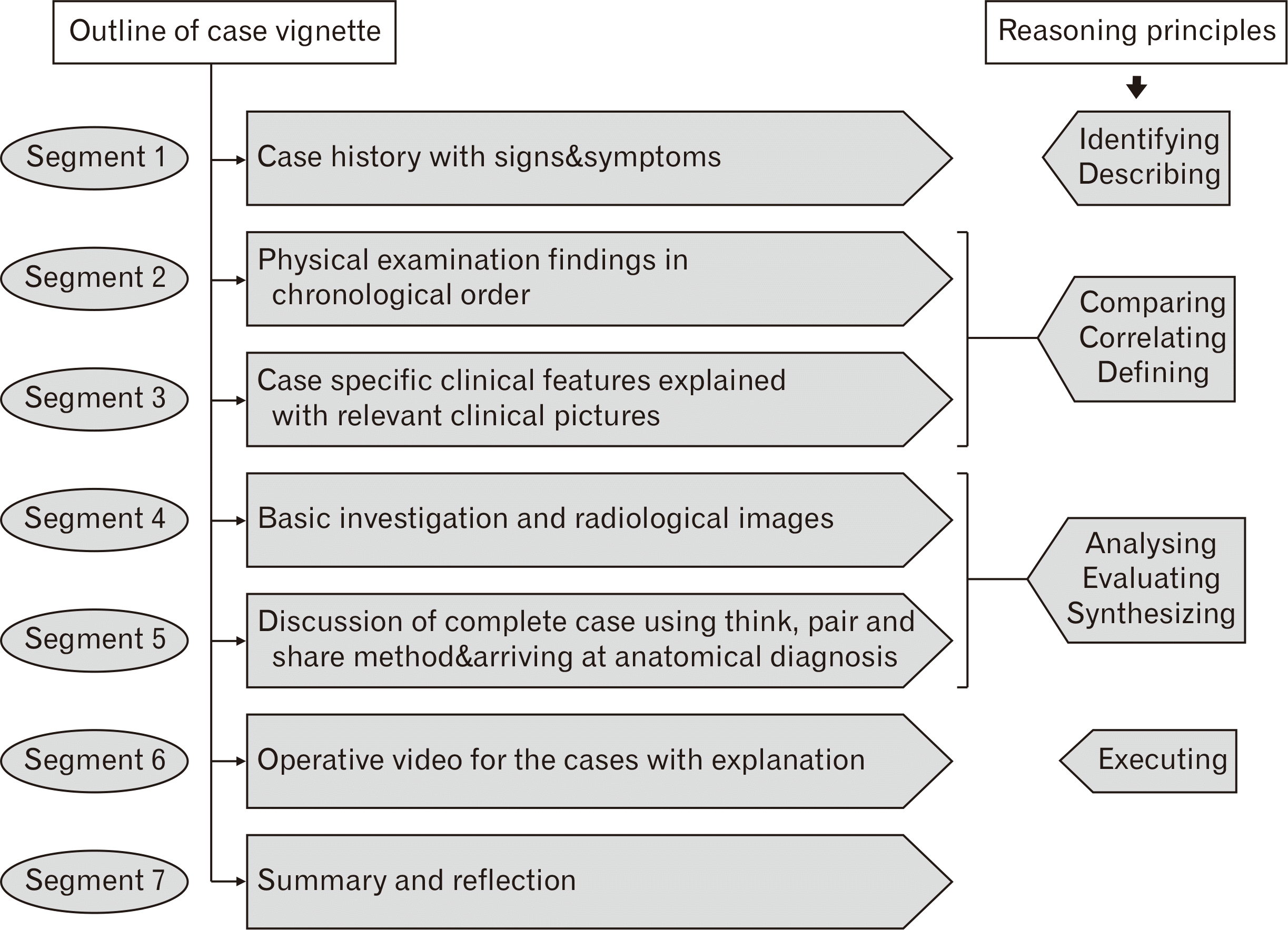
Reasoning principles of vignettes
Preparation of vignettes
Conducting the sessions
Overview of the modules used in the study
Varicose vein case module
-
- A 50 years old male came to outpatient department (OPD) with complaints of dull aching and heaviness over the left leg region. His symptoms are increasing throughout the day and relieved by taking rest with elevated legs
-
- His occupation is bus conductor; He is also complaining of swelling of ankle during prolonged standing and worm like long swellings in the leg region
-
- Picture showing engorged dilated veins over the medial aspect of leg and lower medial thigh was displayed
-
- Picture showing the skin over the lower part of leg was thick & darkly pigmented
-
- Surgeon inspected anterior abdominal wall and no abnormality seen in the anterior abdominal wall
-
- Surgeon checked the dorsalis pedis artery pulsation over dorsum of foot and found to be normal (with picture)
-
- Surgeon did per-abdominal and per-rectal examination and no abnormality was found
-
- The surgeon asked the patient to lie down and the veins are emptied. Then he compressed his thumb 3.5 cm below and lateral to pubic tubercle and asked the patient to stand up. He waited for 1 minute and the veins were gradually filling up from below
-
- The surgeon asked the patient to lie down and the veins are emptied. Then he tied a tourniquet around upper thigh and asked the patient to stand up, checked for venous filling. He repeated the test with placing the tourniquet in midthigh and over calf region.
-
- The surgeon asked the patient to lie down and the veins are emptied. Then he tied a tourniquet around upper thigh and asked the patient to stand up and do mild exercise, and the veins become distended and the distension increased with exercise
- Routine blood investigations were within normal limits (complete blood count, hemoglobin)/chest x-ray was normal
- Doppler ultrasonogram (USG) picture and videos of procedure and results were shown and discussed
- Medical and surgical managements for Varicose veins were discussed
- Surgical video of ligation and stripping procedure with labelling and probe questions and newer techniques of laser ablation/foam sclerotherapy were shown and discussed
Thyroid goiter case module
-
- Mr VM, a 45 year old male visits the surgical OPD, complaining that he could notice a visible swelling in the neck. He said that the swelling had gradually increased in size over the period of 6 months. He also complained that his voice has become hoarse over the period of 3 months
-
- On probing, he admits that he has difficulty in breathing at the times of intense work.
-
- The surgeon observed a swelling of size 2×3 cm in front of the neck.
-
- The surgeon asks the patient to put his tongue out and to swallow. The swelling ascended up and moved with deglutition.
-
- On further examination, the upper border of the swelling could be observed closer to the laryngeal prominence and the lower border of the swelling cannot be palpated (with picture)
-
- On asking the patient to elevate his arms, he had facial plethora and dilated veins over the neck (with picture)
-
- The trachea appears to be shifted towards the right side and there was a variation in the localization of carotid pulse on comparing both sides.
- The chest x-ray postero-anterior view of the patient was shown and asked to identify the abnormality
- USG picture was shown and asked to identify the structures and abnormality
- Labelled USG video on thyroid gland was shown and discussed
- To determine the extent and functional activity of thyroid gland scintigraphy was done – picture shown and the inference was discussed
- Fine needle aspiration cytology image was shown and asked to identify the procedure
- Computed tomography images – axial section, sagittal sections were shown and asked to identify anatomical structures related to the case
- Surgical incisions needed was explained with picture
- Surgical procedure explained step by step with pictures
- Surgical video with labelled structures was displayed with probe questions to identify the structures
Measuring the effectiveness of the clinical reasoning session
The evaluation was done at two levels
-
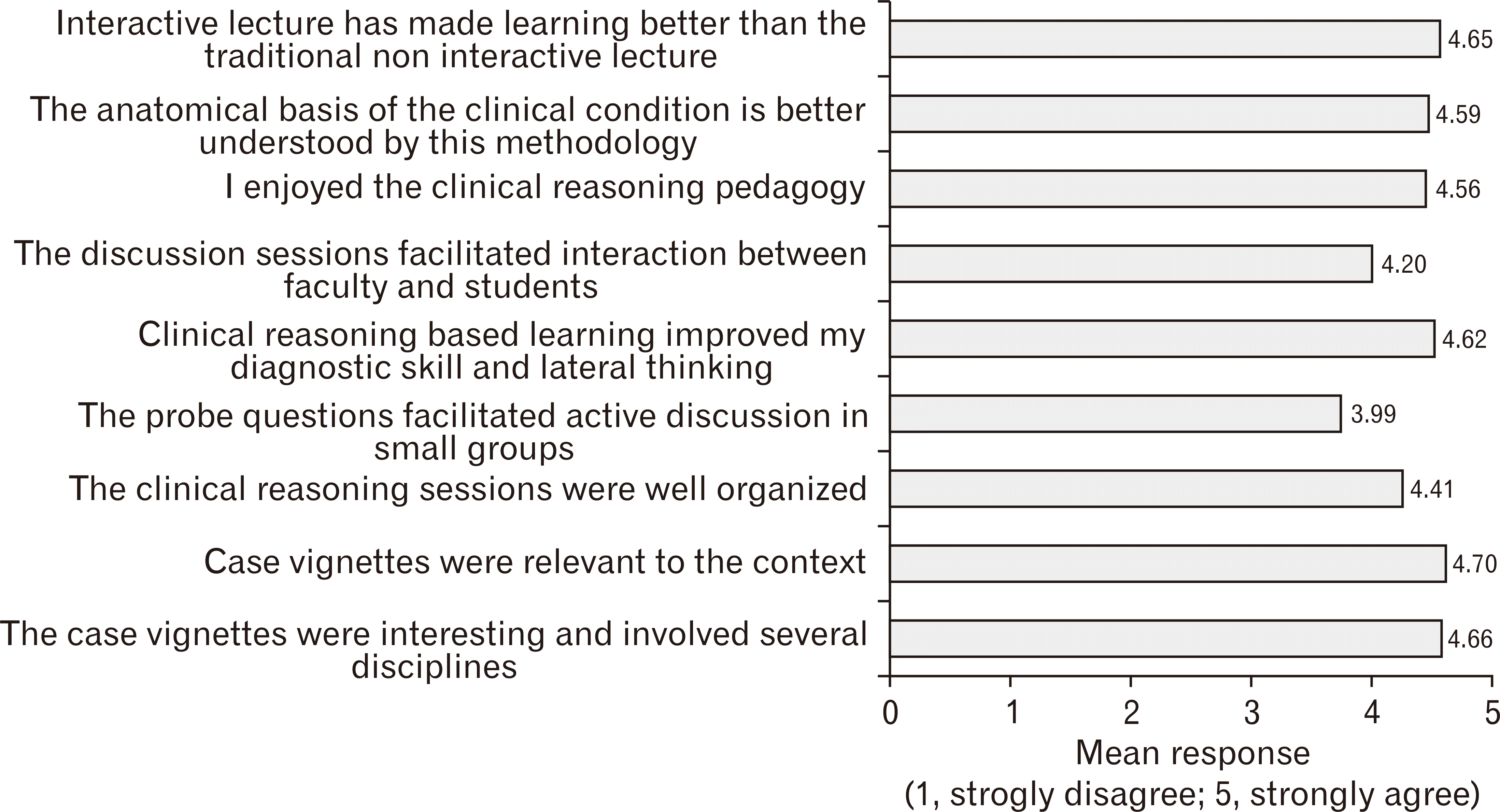
- Students reaction to the session (acceptance/perceived usefulness) was elicited
A) By asking them to rate the usefulness using quantitative items.Individual interest in clinical reasoning was measured and students could respond to these items on a five-point Likert scale ranging from ‘strongly disagree’ to ‘strongly agree’. The items were verified for internal construct reliability (Cronbach’s alpha - 0.84). B) By snapshotting perception of students about the proceedings of clinical reasoning session using nominal group technique. For this, students were divided into subgroups consisting of 15 students with one faculty facilitator each. Students were asked to reflect individually on the perceived benefits of clinical reasoning sessions and on the ways in which the session helped in solving clinical problems. In the second step, the individual responses are collated. Subsequently, the collated responses are rationalized and list of responses were prepared in each subgroup. The top five responses from each of the subgroup were collected and tabulated. After omission of replicative responses, the statements were arranged according to the frequency of representation.
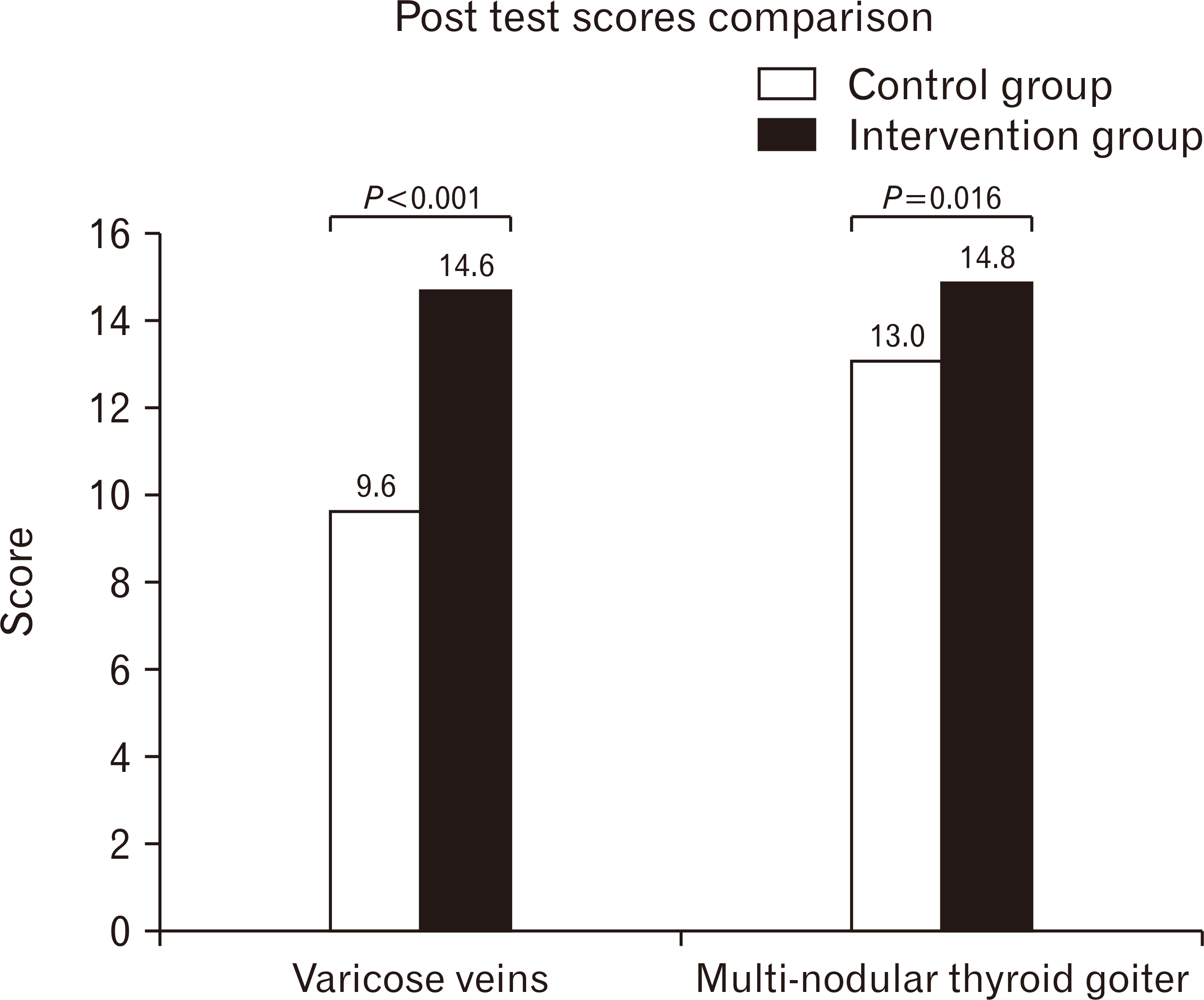
- The learning gains was measured by comparison of post-test scores (Kirkpatrick level 2: proof of benefit): The post-test question paper was prepared by one of the investigators who is blinded to the learning content. It had 12 questions [3-fill in the blank type (for factual knowledge); 3-clinical scenario based MCQs, 3-fill in the blank type (for clinical reasoning) and 3-true or false (scenario/factual)] with a maximum mark of 24.
Statistical analysis
Results
Student’s perceptions from free text responses on individual basis are collated as follows
Positives about the module
“Helped in applying theoretical knowledge in an applied way”
“Clinical videos which were displayed helped us to retain long term memory”
“Integrated clinical based learning helps us to correlate different pieces of knowledge”
“The interaction with peers helped us in effective team building”
“The clinical images and vignette showed the way in which diagnosis would be made in real life settings”
“Exposure to clinical based questions would help us in solving clinical scenarios in future”
The common reflections of students on the reasons for incorporating the clinical reasoning sessions in the anatomy curriculum
Do you feel that clinical reasoning sessions should be incorporated in anatomy curriculum? If so why?
It helped us in making better clinical correlation of the content learnt by conventional teaching
It kindled the curiosity in us
The interaction and reaching the diagnosis made us enthusiastic
It helped in gaining better and wholesome orientation of the subject
It was useful in developing my diagnostic skills
I feel it makes the subject to be learnt in an easier way
The sessions made me generate different ideas
Results
Merits of clinical reasoning sessions (top five responses after collation and arranged according to frequency)
Better correlation of content and analyzing it from various dimensions
Appropriate way for revising and consolidating the content
Makes us understand the relevance of the things learned
Very interactive and group discussion helps in team building
Long term retention of knowledge which would help in practical application in clinics


 PDF
PDF Citation
 Print
Print


 XML Download