

 PDF
PDF Citation
 Print
Print


Introduction
The C-shaped choroidal fissure is positioned centrally between the fornix and thalamus (Fig. 1) with the former forming its outer margin and the latter, its inner margin. At the level of the temporal horn, this fissure is situated between the stria terminalis superomedially and the fimbria of the hippocampus inferolaterally. Therefore, an understanding of this structure is essential role during various brain surgeries. Choroidal fissure cysts are related to this fissure and are often found incidentally on imaging. Patients are typically asymptomatic or, in some cases, symptoms either do not correlate with anatomical location or do not require surgical treatment.
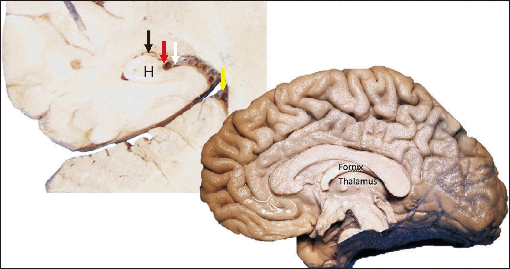 | Fig. 1Coronal brain slice (left) noting the ambient cistern (yellow arrow), the choroidal fissure (white arrow), fimbria (red arrow), hippocampus (H) and choroid plexus in the temporal horn (black arrow); The right sagittal image of the brain notes the relationship between the fornix and thalamus from a medial view.
|
Embryology
During the first stages of embryologic development, the primitive brain is comprised of a single lateral ventricle. During the 8th week of gestation, the vascular pia mater invaginates into the medial aspect of the cerebral hemisphere, giving rise to the formation of the choroidal fissure (Fig. 2). The choroidal fissure extends from the interventricular foramen (of Monro) to the posterior end of the primordial ventricular system [1]. Mesodermal tissue, mainly comprised of vascular structures, migrates into the newly formed ventricular cavity through the choroidal fissure to become the tela choroidea which will then mature and become more vascularized in order to form the choroid plexus. The choroid plexus attaches to the choroidal fissure in the lateral ventricles and no neural tissues develops between the pia mater and ependyma in this fissure making it the thinnest part of the lateral ventricular wall.
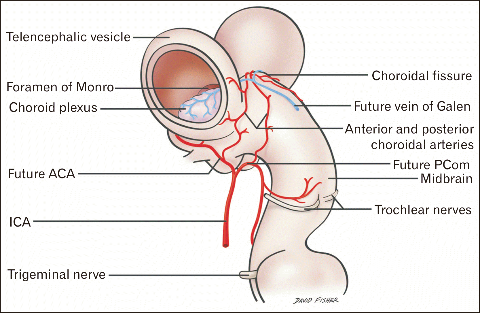 | Fig. 2Schematic drawing of the developing neural tube. As the telencephalic vesicles form from the prosencephalon, note the groove on the dorsal tube at the base of the vesicles, which is the primitive choroidal fissure. Observe that the choroidal vessels are entering and leaving the ventricle via this fissure. ACA, anterior cerebral artery; ICA, internal carotid artery; PCom, posterior communicating artery.
|
Anatomical and surgical significance
The choroidal fissure consists of three parts in the lateral ventricular wall abutting the roof of the third ventricle. The three parts are the body, an atrial part, and a temporal part. The limits of the choroidal fissure are the interventricular foramen, body and atrium of the ventricles, and the inferior choroidal point (Figs. 3 and 4) in the temporal horn. The absence of eloquent nervous tissue along this invagination makes it an important access route to the ventricles and basal cisterns. Important vascular structures run through the choroidal fissure. Arising from the internal carotid (anterior choroidal artery) (Fig. 4) or posterior cerebral arteries (posterior lateral choroidal artery) (Fig. 5), the choroidal arteries first pass through the basal cisterns and give off branches before entering the choroidal fissure. The anterior choroidal artery enters the choroidal fissure at the inferior choroidal point (Fig. 4) located at the posterosuperior edge of the uncus. At the choroidal fissure, the choroidal arteries supply the choroid plexuses of the temporal horn, body, roof of third ventricle, and atrium [2]. Due to the juxtaposition of the temporal horn and choroidal fissure, Yamada et al. [3] suggested that the choroidal fissure might act as an overflow location in cerebrospinal fluid (CSF) drainage in patients with normal pressure hydrocephalus.
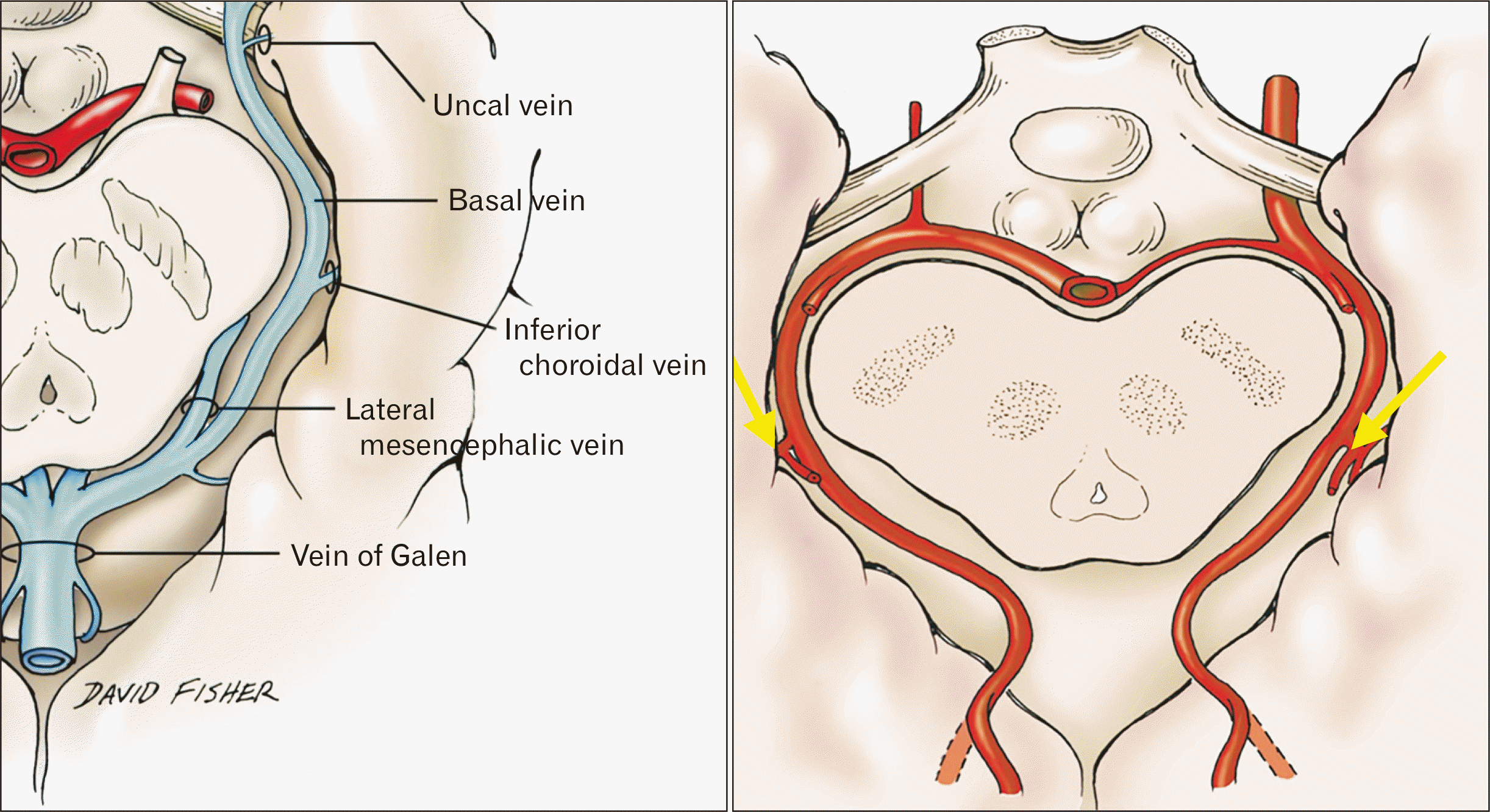 | Fig. 3Axial views of the inferior choroidal vein (left) and posterior lateral choroidal arteries (yellow arrows; right) at the choroidal fissure.
|
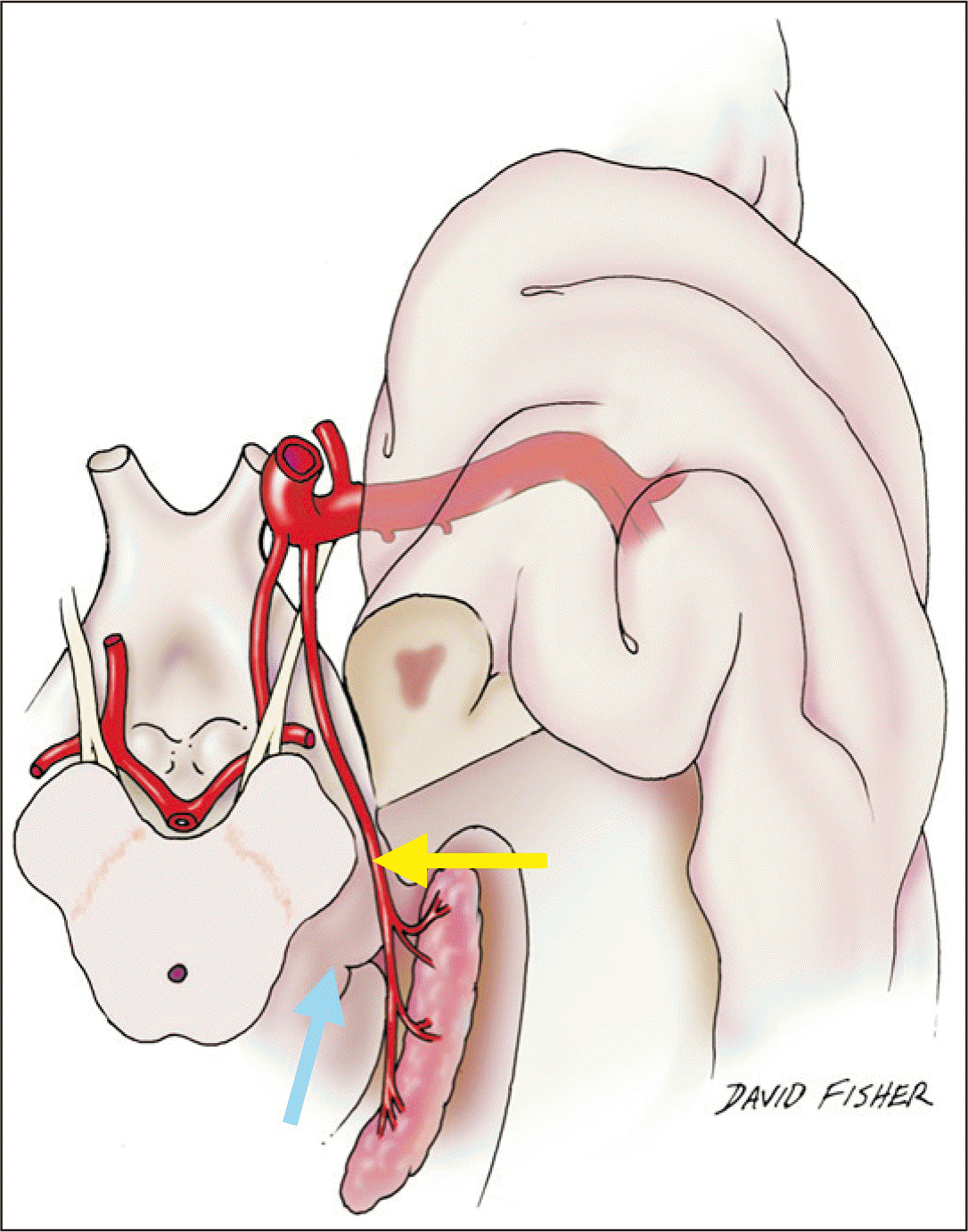 | Fig. 4Schematic drawing of a basal view of the brain (left) noting the choroidal fissure at the point of entrance (inferior choroidal point) of the anterior choroidal artery (yellow arrow) and for reference, the thalamus (blue arrow).
|
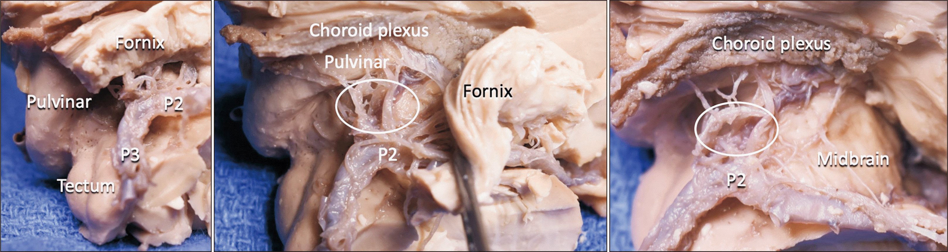 | Fig. 5Sequential dissection of the brain noting the relationship between the pulvinar and choroid plexus and posterior lateral choroidal arteries (white circle). Note that there are usually multiple (average 4) posterior lateral choroidal arteries as seen in this specimen. The P2 and P3 parts of the posterior cerebral artery are labeled.
|
Multiple surgical approaches have been developed in order to access the choroidal fissure and its adjacent structures. The interhemispheric approach allows access to the anterior circulation via the choroidal fissure. This is particularly helpful in some arteriovenous malformation cases, as demonstrated by da Costa et al. [4], and for removal of large tumors in the third ventricle [5]. Surgery involving the choroidal fissure should be conducted at the level of the taenia fornis and taenia fimbriae. Avoidance of the taenia choroidea avoids injury to the choroidal arteries as well as the thalamostriate, thalamocaudate, inferior-ventricular, and lateral-atrial veins, which enter and exit the choroidal fissure at this level [6, 7].
Accessing the body of the choroidal fissure can be accomplished through a corticectomy at the middle frontal gyrus or through the genu of the corpus callosum. This approach provides access to the lateral ventricles, third ventricle, and the velum interpositum in addition to the choroid plexus and ventricular structures. Approaches through the temporal horn, trans-sylvian-transchoroidal fissure, or transtemporal-transchoroidal fissure allow for access to structures in the crural and ambient cisterns as well as the P2–P3 junction of the posterior cerebral artery. A low temporal craniotomy is crucial in order to expose the inferior temporal gyrus and minimize retraction [8]. In comparison to the transtemporal-transchoroidal approach, the trans-sylvian-transchoroidal approach offers a shorter distance and better angle for exposing the mesial temporal area, if needed [9]. Depending on the hemisphere being treated, visual or speech deficits may occur with this approach [10]. Access through the choroidal fissure exposes the quadrigeminal cistern, pineal region, and posterior portion of the ambient cistern. A corticectomy of the superior parietal lobule through the intraparietal sulcus is preferred over a medial or superior temporal corticectomy in order to avoid the optic radiations [6].
Go to : 
Choroidal Fissure Cysts
A choroidal fissure cyst occurs in the plane of the choroidal fissure (Fig. 6). These have a neuroglial, neuroepithelial or arachnoid origin [11]. In addition, focal atrophy in the temporal lobe may cause choroidal fissure enlargement, mimicking a cyst due to its anatomical relationship [5, 12].
Diagnosis and clinical presentations
Choroidal fissure cysts are often incidental findings and, as a result, are frequently asymptomatic [13]. Growth of such lesions can lead to the appearance of symptoms [14]. In the temporal lobe, cysts can lead to seizures [15], although the severity of such seizures does not always correlate with imaging findings [16]. Some choroidal fissure cysts have been diagnosed prenatally [17].
These cysts can occur bilaterally and, in some cases, have been associated with septo-optic dysplasia, infantile spasms, late-onset diabetes insipidus, and bilateral club feet [18]. Genetic evaluation in one such case revealed interstitial deletion of bands 10q26.12 and 10q26.13 [18].
Commonly, magnetic resonance imaging (MRI) and computed tomography are used for studying cystic masses at the choroidal fissure. The coronal plane is most useful in distinguishing between choroidal fissure cysts (extra axial) and intraparenchymal cysts [19]. The primary goal is to differentiate between benign lesions, such as CSF-containing cysts, and lesions requiring active treatment, such as infectious cysts or cystic neoplasms.
Lesions at the level of the choroidal fissure can be classified into three different groups according to their intensities on MRI: 1) low intensity pattern (CSF), 2) intermediate intensity pattern (protein), 3) high intensity pattern (colloid cyst or hemorrhage) [20, 21]. Fluid-attenuated inversion recovery imaging has demonstrated superiority in cystic diagnostic accuracy as it is able to differentiate between maldevelopment and neoplasia or inflammation [22]. Other key characteristics suggesting CSF-containing cysts include homogenous consistency, undetectable wall or associated soft tissue mass, lack of surrounding gliosis or edema, and absence of contrast enhancement [23].
The location of the choroid plexus relative to the cyst can be used to differentiate temporal intraventricular cysts and choroidal fissure cysts. Intraventricular cysts will medially displace the choroid plexus, whereas choroidal fissure cysts will shift the plexus laterally [24]. However, the only way to identify CSF-containing arachnoid and neuroepithelial cysts at the choroidal fissure level is by histopathological investigation.
Treatment
Cysts of the choroidal fissure are generally asymptomatic and incidental findings. Recommended treatment consists of regular follow up examinations since a small patient subgroup may experience lesion growth over time [14, 25]. Surgical intervention is reserved for symptomatic cases that interfere with quality of life. The main goal of surgery is to establish communication between the cyst and the subarachnoid space [26]. Such can be achieved through endoscopically guided fenestration or an open craniotomy. Should this be insufficient, a shunt can be placed [27].
Go to :
Conclusion
The choroidal fissure is a key landmark used in neurosurgery. As a result, a thorough understanding of nearby anatomical structures is essential. Choroidal fissure cysts can be found incidentally, and well-known features allow one to differentiate these cysts from other lesions. Surgical treatment should be reserved for symptomatic patients, while asymptomatic patients require monitoring as although uncommon, these structures can grow over time.
Go to :
 XML Download