Journal List > Anat Cell Biol > v.53(2) > 1145470

Inchai, Vaseenon, Tanaka, and Mahakkanukrauh: The distribution pattern of the dorsal cutaneous nerves of the foot and its clinical implications

Abstract

Iatrogenic injury to subcutaneous nerves on the dorsum of the foot is an established risk factor during the surgical procedures resulting in postoperative pain, sensation loss and painful neuroma. Previous studies have reported on the distribution pattern of the superficial peroneal, deep peroneal and sural nerves (SNs) and their branches with various classifications about specific populations. The purpose of the present study was to evaluate the distribution pattern and classification of the nerves on the dorsum of the foot and analyze the location of these nerves into five zones with clinical implications. Fifty-four lower limbs of fresh frozen cadavers were used in the present study. The anatomical patterns of the superficial peroneal, deep peroneal, SN and their branches were classified into eight, two and five patterns respectively. Type VI pattern, a classic distribution pattern of the superficial peroneal nerve was the most frequent type with a prevalence of 13 limbs (25.00%). In Zone 5, where the arthroscopic portal placements for the first metatarsophalangeal joint arthrodesis, 29 limbs (55.77%) showed three nerve branches located in this zone. The results of the present study provide anatomical knowledge that may help the surgeon to choose the appropriate treatment for their patients and reducing the rate of complications in surgery.

Introduction

The deep peroneal nerve (DPN) and the superficial peroneal nerve (SPN) originate from the common peroneal nerve supplying the skin on the distal part of the anterior surface of the leg and dorsal part of the foot. The injury to cutaneous nerves adjacent to the ankle and dorsum of the foot are common complications resulting from the various treatment and procedures including regional or local anesthesia for foot and ankle surgery, hallux valgus-rigidus correction, cheilectomy and forefoot surgery [1, 2]. The SPN provides two terminal branches; the intermediate dorsal cutaneous nerve (IDCN) and medial dorsal cutaneous nerve (MDCN) [3-5]. Numerous studies focus on the point that the SPN emerges through the anterior crucial intermuscular septum pathway as well as the branching pattern of these nerves [5-9]. The DPN provides the motor supply to the muscles in the anterior compartment of the leg [10]. Finally, the DPN divides into two terminal branches; the medial sensory branch to innervate the skin of the dorsum of the foot and the lateral motor branch to supply the motor innervation to the extensor digitorum brevis muscle [11, 12]. The sural nerve (SN) provides the lateral dorsal cutaneous nerve (LDCN) to supply the sensory innervation to the lateral side of the dorsum of the foot. Previous studies have been focusing on the different patterns of formation of the SN in different populations and reported the various type of anatomical patterns [7,13-16].
The understanding of the exact distribution and branching pattern of the nerves related to the clinical implications may help the surgeon avoid iatrogenic injury and decrease the complication from surgery. The present study aims to evaluate the distribution pattern of the superficial peroneal, deep peroneal and SNs in the dorsum of the foot. Additionally, the researchers classify the zone on the dorsum of the foot into five metatarsal zones (1–5 toes) related to surgical procedure and clinical implications for better define the specific location of these nerves.

Materials and Methods

A total of 54 lower legs of fresh frozen cadavers; 10 females and 17 males were obtained from the Department of Anatomy, Faculty of Medicine, Chiang Mai University. The mean age at death was between 20 to 80 years. No history of lower extremity injury, pathology and ankle deformity was reported. Ethical approval for the present study was obtained from the Ethics Committee from the Faculty of Medicine, Chiang Mai University. The branching pattern of the SPN, DPN and the SN was performed in the present study by dissection. The SPN nerve branches at the dorsum of foot were noted and classified following the branching patterns from I to VIII types based on previous studies [6-9]. The present study described the SPN, DPN, and SN branches via divided the dorsum of the foot into five zones related to the location of dorsal cutaneous nerves to the metatarsal bone and interdigital space. Sketches and photographs were made for data collection. The data were analyzed with descriptive statistical analysis. All statistical analysis tests were performed using SPSS Statistics for Windows, Version 17.0 (SPSS Inc., Chicago, IL, USA). A value of P<0.05 was significant.

Results

The distribution and pattern of each cutaneous nerve identified in the present study and shown in Figs. 13.

Distribution of the SPN on the dorsum of the foot

The SPN distribution was classified into eight types based on previous research [6-9].
Type I the communicating branch from LDCN to IDCN was present in this type at the metatarsal bone level after that the IDCN divided into two branched on the dorsum of the foot. The result showed six limbs (11.54%) in this type.
Type II the IDCN supplied most of the areas in the dorsum of the foot while the MDCN was a single branch to supply the skin on the medial side of the first toe. The communicating branch from LDCN with IDCN was found in this type. The result showed one limb (1.92%) in this type.
Type III the IDCN was found to terminate at the ankle joint level while the LDCN supply the lateral 2.5 toes and the MDCN supply the medial 2.5 toes. The result showed eight limbs (15.38%) in this type.
Type IV the SPN passes downward and medially to the dorsum of the foot to supply the skin on the medial side of 2.5 toes. The MDCN and IDCN were not found in this type. The LDCN supplied the lateral 2.5 toes, the communicating branch was absent. The result showed 10 limbs (19.23%) in this type.
Type V the LDCN supplied the lateral 1.5 toes on the dorsum of the foot. The communicating branch from LDCN to IDCN was present. The result showed four limbs (7.69%) in this type.
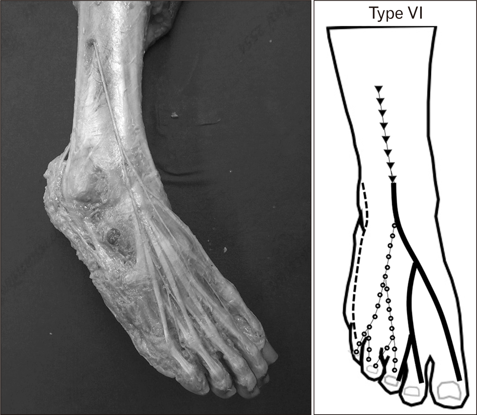
Type VI the SPN divided into MDCN and IDCN before passing the ankle joint line. The MDCN supplied the medial 1.5 toes except for the interdigital space between first and second toes. The IDCN supplied the third, fourth and medial side of the fifth toes. The LDCN supplies the lateral side of the fifth toe, the communicating branch was absent. The result showed 13 limbs (25.00%) in this type.
Type VII the IDCN supplied most of the areas in the dorsum of the foot from the lateral side of the second to the medial side of the fifth toe. The MDCN supplied the skin on the medial side of the first toe. The communicating branch from LDCN to IDCN was absent. The result showed one limb (1.92%) in this type.
Type VIII the LDCN supplied the skin on the lateral three toes. The communicating branch from the LDCN to IDCN was found in this type. The MDCN supplied the skin on the medial side of the first toe. The IDCN supplied the skin of the second toe except for the interdigital space between first and second toes. The result showed nine limbs (17.31%) in this type.

Distribution of the DPN on the dorsum of the foot

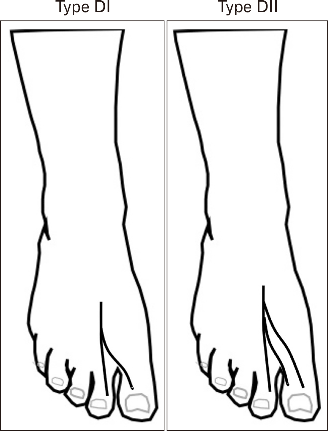
The DPN distribution was classified into two types (Fig. 2):
Type DI: The DPN supplied the skin on the lateral side of the first toe and the medial side of the second toe. The result showed 39 limbs (75.00%) in this type.
Type DII: The DPN supplied the skin on the lateral side of the first toe and the medial side of the second toe and provided the branch for supplied the medial side of the first toe while the branch from MDCN was absent. The result showed 13 limbs (25.00%) in this type.

Distribution of the SN on the dorsum of the foot

The SN distribution was classified into five types (Fig. 3):
Type SI: The LDCN supplied only the lateral side of the fifth toe. The communicating branch from LDCN to IDCN was absent. The results showed ten limbs (18.52%) in this type.
Type SII: The LDCN supplied the lateral two toes. The communicating branch from LDCN to IDCN was absent. the results showed nine limbs (16.67%) in this type.
Type SIII: The LDCN supplied the lateral side of the fifth toe and the communicating branch from LDCN to IDCN was present in this type. The result showed 15 limbs (27.78%) in this type.
Type SIV: The LDCN supplied the lateral three toes. The communicating branch from LDCN to IDCN was absent. the results showed 18 limbs (33.33%) in this type.
Type SV: The sural and LDCN was not found on the dorsum of the foot. The IDCN supplied the lateral and medial sides of the fifth toe. The results showed two limbs (3.70%) in this type.
The present study described the SPN, DPN, and SN branches by dividing their location into five zones via divided the dorsum of the foot into five zones related to the location of dorsal cutaneous nerves to the metatarsal bone and interdigital space (Fig. 4). The aim of the classification of the dorsum of the foot into specific zones was to evaluate the risk of dorsal cutaneous nerve damage as a complication from forefoot arthroscopic surgery. According to the fifth zone associated with the dorsomedial and dorsolateral portal placements, there are 29 limbs (55.77%) that three nerve branches located in zone 5. The present study demonstrated 45 limbs (86.54%) had one nerve branch in the medial part of zone 5 while the lateral part of zone 5 contained two nerve branches were found in 29 limbs (55.77%) indicating the risk of cutaneous nerve damage from arthroscopic portal placements should be considered.

Discussion

The researchers focused on the different formation and distribution patterns of these nerves as related to the clinical approach to improves the knowledge of exact anatomical patterns, to decrease the iatrogenic nerve injury from the surgical procedures in this region. The present study aimed to investigate the morphological pattern of cutaneous nerve formation and distribution on the dorsum of the foot and to map for provide the specific data of the Northern Thai population.
The nerve development of the lower limb region begins at 9 mm of the embryonic period around four weeks, the limb bud is filled with the mesenchymal tissue and the nerves to limb bud arise from the lumbosacral plexus. The peroneal nerve extends over the dorsal aspect of the limb bud and divided into a superficial and deep branch [17].
The location and distribution patterns of the SPN, DPN, and SN on the dorsum of foot were highly variable in this study. Despite this variability, in 1926, Kosinski [8] who was the first to report the description of dorsal cutaneous nerve pattern and distributions and performed the study of 229 feet and classified into 12 theoretical types (K1–K12). Type K1 was the most frequently observed type (54.4%). In 2001, Solomon and colleagues [9] evaluated 11 patterns of dorsal cutaneous nerve distribution of the dorsum of the foot in 68 patients. Type K2 was most commonly found in their study. Ikiz and colleagues [7] in 2006 reported the seven types of the distribution pattern of the SPN on the dorsum of the foot. Type 1 was the most common in these studies similar to the K4 and K6 of Kosinski’s classification. Jeon and colleagues [16] in 2017 performed the study of the SN pattern following the previous research of Madhavi and colleagues’ [14] classification. Type 3 was the most common by observed type (24.10%) [16]. In 2019, Heo and colleagues [6] clarified the distribution of dorsal cutaneous nerves on the dorsum of the foot, reported the classification of nerve pattern of the sural and DPN into five and four types respectively. Type 2 was reported as the most frequent type in their study (38.50%). Nayak and colleagues [18] in 2019 investigated and classified the different patterns of nerve distribution on the dorsum of the foot. The distribution pattern of the cutaneous nerve of the dorsum of foot was categorized into four groups. Group, I was the most common type (45%), the DPN innervated the skin at the interdigital space between the first and second toes, the SN innervated the skin at the lateral part of the fifth toe and the SPN was innervated the remaining part of the dorsum of the foot. In the present study to establish the morphological pattern of the dorsal cutaneous nerves formation on the dorsum of the foot and to classify the distribution pattern of SPN, DPN, and SN into eight types, two types and five types respectively. Type SVI of SPN distribution pattern was the most frequently found in the present study: 13 limbs (25.00%). The normal distribution pattern of the SPN was shown in the Fig. 5. This type also known as the classic distribution pattern of SPN which was similar to Canovas and colleagues report that K1 type was the most common type in their study. The K1 type of Kosinski’s classification and type 7 of Ikiz’s classification which was similar to type SVI in the present study. There are overlap in description of different type reported in previous studies related to the specific population. Comparing the present study of the DPN classification of Heo and colleagues [6], the present study showed two types of DPN distribution in the Northern Thai population. Type D1 was the most common by found in the present study in 39 limbs (75.00%). In addition, type 3 was not found in the present study.
Recently, arthroscopy is a minimally invasive technique and is the most commonly performed surgical procedure with minimal soft tissue damage, avoiding a neurovascular injury and decreased postoperative pain. There are two portal placements frequently used: dorsomedial and dorsolateral portals as to the extensor halluces longus tendon [19]. The dorsolateral and dorsomedial hallucal nerves, a nerve branches from the MDCN can be injured at the region of the first MTP joint. Vaseenon and Phisitkul [20] in 2010 reported the safe zones when performing the first metatarsophalangeal joint arthroscopy and suggested the medial and lateral to the extensor hallucis longus tendon for portal placements can avoid injury to the dorsolateral and dorsomedial hallucal nerves. Malagelada and colleagues [21] in 2019 reported that the complications in ankle arthroscopy can be decreased by the lectures on anatomical structures related to ankle arthroscopy during the cadaveric courses. Understanding the arthroscopic anatomy surrounding the arthroscopic portal sites is important for preventing neurovascular injuries. The limitation of the present study was the macroscopic cadaveric study of the cutaneous nerves related to surgical procedures. Further study with the microscopic study of the nerve is required to apply these data in clinical relevance.
In conclusion, the present study aimed to identify and investigate the nerves on the dorsum of the foot to provide the basic anatomical knowledge about the nerves on the ankle joint in the Northern Thai populations. The risk of iatrogenic nerve injury can be decreased by the knowledge of the exact anatomical pattern of the relevant structures. The result of the present study may raise awareness to surgeons regarding the anatomical variation and the location of the nerve and choose the appropriate treatment and surgical procedures for their patients.

Acknowledgements

This study was partially supported by research funding from the Faculty of Medicine, and research cluster of Chiang Mai University. The author would like to thank to those who donated the body for study and research.

Notes

Author Contributions

Conceptualization: CI, TV, YT, PM. Data acquisition: CI. Data analysis or interpretation: CI. Drafting of the manuscript: CI, YT. Critical revision of the manuscript: CI, PM. Approval of the final version of the manuscript: all authors.

Conflicts of Interest

No potential conflict of interest relevant to this article was reported.

REFERENCES

1. Kenzora JE. 1986; Sensory nerve neuromas--leading to failed foot surgery. Foot Ankle. 7:110–7. DOI: 10.1177/107110078600700209. PMID: 3770595.
crossref
2. Miller RA, Hartman G. 1996; Origin and course of the dorsomedial cutaneous nerve to the great toe. Foot Ankle Int. 17:620–2. DOI: 10.1177/107110079601701006. PMID: 8908488.
crossref
3. Blair JM, Botte MJ. 1994; Surgical anatomy of the superficial peroneal nerve in the ankle and foot. Clin Orthop Relat Res. (305):229–38. DOI: 10.1097/00003086-199408000-00028.
crossref
4. Canella C, Demondion X, Guillin R, Boutry N, Peltier J, Cotten A. 2009; Anatomic study of the superficial peroneal nerve using sonography. AJR Am J Roentgenol. 193:174–9. DOI: 10.2214/AJR.08.1898. PMID: 19542411.
crossref
5. Üçeyler N, Schäfer KA, Mackenrodt D, Sommer C, Müllges W. 2016; High-resolution ultrasonography of the superficial peroneal motor and sural sensory nerves may be a non-invasive approach to the diagnosis of vasculitic neuropathy. Front Neurol. 7:48. DOI: 10.3389/fneur.2016.00048. PMID: 27064457. PMCID: PMC4812111.
crossref
6. Heo Y, Jung TJ, Yang M, Kuk Y, Kim YD, Won HS. 2019; Aug. 20. Distribution patterns of the cutaneous nerves on the dorsum of the foot and their clinical significance. Clin Anat. [Epub]. http://dx.doi.org/10.1002/ca.23453. DOI: 10.1002/ca.23453. PMID: 31429988.
crossref
7. Aktan Ikiz ZA, Uçerler H, Bilge O. 2005; The anatomic features of the sural nerve with an emphasis on its clinical importance. Foot Ankle Int. 26:560–7. DOI: 10.1177/107110070502600712. PMID: 16045849.
8. Kosinski C. 1926; The course, mutual relations and distribution of the cutaneous nerves of the Metazonal region of leg and foot. J Anat. 60(Pt 3):274–97.
9. Solomon LB, Ferris L, Tedman R, Henneberg M. 2001; Surgical anatomy of the sural and superficial fibular nerves with an emphasis on the approach to the lateral malleolus. J Anat. 199(Pt 6):717–23. DOI: 10.1046/j.1469-7580.2001.19960717.x. PMID: 11787825. PMCID: PMC1468389.
crossref
10. Moore KL, Dalley AF, Agur AMR. 2006. Clinically oriented anatomy. 5th ed. Lippincott Williams & Wilkins;Philadelphia: p. 1–300.
11. Loveday DT, Nogaro MC, Calder JD, Carmichael J. 2013; Is there an anatomical marker for the deep peroneal nerve in midfoot surgical approaches? Clin Anat. 26:400–2. DOI: 10.1002/ca.22173. PMID: 23378070.
crossref
12. Causeret A, Ract I, Jouan J, Dreano T, Ropars M, Guillin R. 2018; A review of main anatomical and sonographic features of subcutaneous nerve injuries related to orthopedic surgery. Skeletal Radiol. 47:1051–68. DOI: 10.1007/s00256-018-2917-5. PMID: 29549379.
crossref
13. Mahakkanukrauh P, Chomsung R. 2002; Anatomical variations of the sural nerve. Clin Anat. 15:263–6. DOI: 10.1002/ca.10016. PMID: 12112352.
crossref
14. Madhavi C, Isaac B, Antoniswamy B, Holla SJ. 2005; Anatomical variations of the cutaneous innervation patterns of the sural nerve on the dorsum of the foot. Clin Anat. 18:206–9. DOI: 10.1002/ca.20094. PMID: 15768411.
crossref
15. Eid EM, Hegazy AM. 2011; Anatomical variations of the human sural nerve and its role in clinical and surgical procedures. Clin Anat. 24:237–45. DOI: 10.1002/ca.21068. PMID: 20949489.
crossref
16. Jeon SK, Paik DJ, Hwang YI. 2017; Variations in sural nerve formation pattern and distribution on the dorsum of the foot. Clin Anat. 30:525–32. DOI: 10.1002/ca.22873. PMID: 28281304.
crossref
17. Kelikian AS, Sarrafian SK. 2012. Sarrafian's anatomy of the foot and ankle: descriptive, topographic, functional. 3rd ed. Wolters Kluwer Health/Lippincott Williams & Wilkins;Philadelphia: p. 1–736.
18. Nayak VS, Bhat N, Nayak SS, Sumalatha S. 2019; Anatomical variations in the cutaneous innervation on the dorsum of the foot. Anat Cell Biol. 52:34–7. DOI: 10.5115/acb.2019.52.1.34. PMID: 30984449. PMCID: PMC6449594.
crossref
19. Siclari A, Piras M. 2015; Hallux metatarsophalangeal arthroscopy: indications and techniques. Foot Ankle Clin. 20:109–22. DOI: 10.1016/j.fcl.2014.10.012. PMID: 25726487.
20. Vaseenon T, Phisitkul P. 2010; Arthroscopic debridement for first metatarsophalangeal joint arthrodesis with a 2-versus 3-portal technique: a cadaveric study. Arthroscopy. 26:1363–7. DOI: 10.1016/j.arthro.2010.02.015. PMID: 20887934.
21. Malagelada F, Vega J, Guelfi M, Kerkhoffs G, Karlsson J, Dalmau-Pastor M. 2020; Anatomic lectures on structures at risk prior to cadaveric courses reduce injury to the superficial peroneal nerve, the commonest complication in ankle arthroscopy. Knee Surg Sports Traumatol Arthrosc. 28:79–85. DOI: 10.1007/s00167-019-05373-x. PMID: 30729253.
crossref

Fig. 1
Schematic illustration of the SPN branching pattern on the dorsum of the foot in the present study. IDCN, intermediate dorsal cutaneous nerve; MDCN, medial dorsal cutaneous nerve; SN, sural nerve; SPN, superficial peroneal nerve.
ACB-53-137-f1.tif
Fig. 2
Schematic illustration of the deep peroneal nerve branching pattern on the dorsum of the foot in the present study.
ACB-53-137-f2.tif
Fig. 3
Schematic illustration of the sural nerve branching pattern on the dorsum of the foot in the present study. SN, sural nerve; IDCN, intermediate dorsal cutaneous nerve.
ACB-53-137-f3.tif
Fig. 4
Schematic illustration of the 5 zones on the dorsum of the foot.
ACB-53-137-f4.tif
Fig. 5
Schematic illustration and normal dissected specimen; Type VI of superficial peroneal nerve (SPN) distribution pattern also known as the classic distribution pattern of SPN.
ACB-53-137-f5.tif
TOOLS
Similar articles