

 PDF
PDF Citation
 Print
Print


INTRODUCTION
Neuromuscular weakness often occurs in patients admitted to the intensive care unit (ICU), which increases the difficulty of weaning them off the mechanical ventilator.1 This clinical situation can be caused by various neuromuscular diseases such as Guillain-Barré syndrome, amyotrophic lateral sclerosis, myasthenia gravis, myositis, and myelopathy (Table 1); however, critical illness neuromyopathy (CINM) should be considered first.2-5 Olsen6 was the first to report peripheral neuropathy in comatose patients. Bolton et al.7 established the concept of critical illness polyneuropathy (CIP), which occurs in patients with sepsis or multiple organ failure (MOF) in the ICU. MacFarlane and Rosenthal8 reported myopathy in acute respiratory failure that was treated with steroids and neuromuscular blocking agents, and Lacomis et al.9 proposed the name of critical illness myopathy (CIM).
Table 1.
Causes of acute neuromuscular weakness in critically ill patients
![]()
Neuromuscular diseases in critically ill patients are largely classified into CIP and CIM. However, muscles and nerves are often affected simultaneously in practice, making it difficult to clearly differentiate between CIM and CIP.10-12 This has led to the two conditions being combined into CINM or critical illness polyneuromyopathy (CIPNM).13-17 and CINM is called by various names such as ICU-acquired weakness (ICU-AW), including ICU-acquied weakness (ICU-AW),13 ICU-acquired paresis (ICU-AP),18 critical illness myopathy and/or neuropathy (CRIMYNE),10 and critical illness neuromuscular abnormalities (CINMA).19 CINM results in a longer ICU stay, and even after recovery it often restricts motor function, thereby placing an additional economic burden on rehabilitation, which ultimately lead to significant problems in the prognosis of critically ill patients. The increasing clinical significance of CINM in critical care has prompted research into risk factors, pathogenesis, and diagnostic electrophysiological tests. This article summarizes recent findings related to CINM.
Go to : 
INCIDENCE AND RISK FACTORS
Prospective studies have found that CINM occurs in 25-87% of patients treated in the ICU for at least 1 week,4,5,11,13,15,20,21 with the reported incidence being higher in patients with sepsis, at 68-100%.13,15,17,22 CINM mostly occurs in adults older than 50 years and rarely in children, because sepsis occurs predominantly in the elderly. A meta-analysis found that the incidence of CINM was 57%, and the prevalence of subsets were 20% (CIM), 13% (CIP), and 9% (CINM) with the highest frequency of CIM.20
Since no specific treatment is available for CINM, prevention is currently the best strategy, which makes it very important to understand its triggering and risk factors.1 Systemic inflammatory response syndrome (SIRS) or MOF associated with sepsis is considered the most relevant risk factor.1,16,17 The various studies of risk factors for CINM have produced different results due to differences in the included patients or the applied diagnostic criteria. The risk factors reported so far are summarized below.
Severity of illness
Most studies have found that the longer maintenance of the mechanical ventilator or the longer stay in the ICU is related with the higher severity of the disease.1,10,13 The disease severity is scored on different scales, including Acute Physiology and Chronic Failure Evaluation (APACHE) III, Sequential Organ Failure Assessment (SOFA), and Simplified Acute Physiology Score (SAPS)-2. The incidence of CINM increases with the severity as scored on these scales.20
Hyperglycemia
Hyperglycemia has been found to be a risk factor in most studies.1,10,13,20,22 A meta-analysis found that active control of blood sugar with insulin significantly reduces the incidence of CINM and reduces the lengths of hospital stays and ventilator use.20 However, more research is required before recommending the taking of insulin for preventing CINM.1,17,20
Sepsis
CINM occurs in more than 50% of patients with sepsis, making this a very strong risk factor.20 A meta-analysis found that the risk of CINM was 3.7-fold higher in patients with sepsis at the first week of hospitalization and 2.5-fold higher in patients with sepsis at 30 days after using a ventilator.20
Steroids and neuromuscular blocking agents
Steroids increase the incidence of CINM 15-fold and therefore have also been regarded as a strong risk factor.4,20 Since the first reported case of CINM in an asthma patient taking a neuromuscular blocking agent, such blockers have been regarded as a potent risk factor for CINM.8-10 A meta-analysis found that neuromuscular blockers increased the incidence of CINM 16-fold, making them another strong risk factor.20,23
Malnutrition
The metabolism increases in most critically ill patients. Animal experiments revealed that protein production is reduced even if sufficient nutrition is supplied, and such an enhancement of catabolism of skeletal muscle can lead to CINM. Meta-analyses have found that parenteral nutrition increases the incidence of CINM 5-fold.20,23
Go to :
CLINICAL FEATURES AND DIAGNOSES
Critical illness polyneuropathy
A typical clinical feature of CIP is that it is difficult to wean off the ventilator due to neuromuscular weakness in patients older than 50 years who stay in the ICU for more than 5 days with respiratory failure and sepsis. This weakness is more severe in lower limbs than upper limbs, and involvement of the eye and facial muscles is rare.11,19 Distal weakness is more prominent.10,17 Muscle mass is lost in 30% of CIP cases.24 Deep tendon reflexes (DTRs) are often normal in the early stages and then gradually decrease as the disease progresses.24
The electrophysiological feature of CIP is axonal damage with reduced amplitudes of the compound muscle action potential (CMAP) and sensory nerve action potential (SNAP).10,15 The decreased CMAP amplitude first appears within 2 weeks after the onset of SIRS.25 Electrophysiological abnormalities were found in 63% of CIP patients with sepsis within the first 3 days, for which the mortality rate is generally high.26 Abnormal spontaneous potentials and polyphasic motor unit potentials (MUPs) are observed in electromyography (EMG).1,5,10 Electrophysiological findings vary depending on the stage of CIP: SNAPs are initially normal, become abnormal as the disease progresses, and then normalize again during the recovery phase.1 Therefore, performing repeated sensory examinations at different stages may be helpful for differentiating CIP from CIM.1 The diagnostic criteria of CIP include these clinical features and electrophysiological findings (Table 2).1
Table 2.
Diagnostic criteria for CINM
![]()
Weakness associated with the long-term use of neuromuscular blockers may be mistaken for CIP or CIM in the ICU, but this can be easily identified if the CMAP gradually reduces during repeated nerve stimulation.1 Normal muscle enzyme levels (creatine kinase [CK]), normal CMAP duration, and sensory abnormalities are important factors for differentiating CIP from CIM.5,10 While small-amplitude MUPs in EMG may help to differentiate between CIP and CIM, MUPs are difficult to measure accurately because most critically ill patients are uncooperative.27 This has prompted several tests being designed for uncooperative patients, and direct muscle stimulation (DMS) has recently been recommended as a diagnostic test.28
Critical illness myopathy
The main clinical features of CIM are flaccid paralysis of the limb and weakness of the neck flexor muscle. CIM is often accompanied by weakness of the facial and respiratory muscles, which makes weaning off the ventilator difficult.9,14 Proximal weakness is more prominent than distal weakness, and sensory function is normal.15 DTRs are either normal or decreased, and myalgia is rare.9
CIM is more common than CIP and has a faster recovery.10,12,29 Due to the variety of pathological findings and clinical patterns, studies often include different subtypes, which may lead to confusion. Generally, CIM includes all types of myopathy in critically ill patients, such as thick-filament myopathy, necrotizing myopathy, rhabdomyolysis, and cachectic myopathy.1,9,17 In addition, acute steroid myopathy and acute pancuronium myopathy may be included in CIM.9
The CMAP amplitude decreases in CIM like it does in CIP, but in CIM the duration of CMAP increases and the SNAP is normal.9,10 The conduction velocity of muscle fibers decreases with decreased excitability of the sarcolemma membrane, and the increased variability of muscle fiber conduction velocities increases the CMAP duration.1,5,18,30 It is important to confirm abnormal spontaneous potentials of short duration and small-amplitude MUPs in EMG. However, MUPs cannot be accurately evaluated in critically ill patients due to their non-cooperation.27 So DMS can be used instead to distinguish CIM from CIP.18,21,31 CK may be helpful in this differentiation, since the level is slightly elevated in CIM and normal in CIP.17
An electrophysiological examination and CK levels are the most important tools for diagnosing CIM (Table 2).1 While it is not always necessary to perform a biopsy to confirm CIM, a biopsy should be considered if inflammatory myopathy is suspected or if biopsy findings could significantly affect the treatment plan.1
Go to :
TREATMENT AND PROGNOSIS
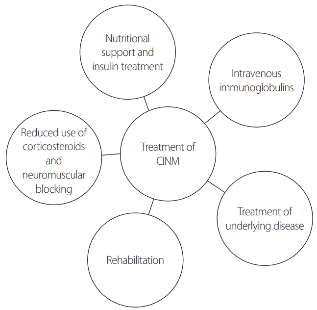
It has been reported that only preventive and supportive treatments are helpful for CINM. The treatment strategies include aggressively treating the underlying disease (sepsis or MOF), rehabilitation, nutritional support, reduced use of neuromuscular blocking agents and corticosteroids, and immunoglobulins (Fig. 1).32 Intravenous immunoglobulin G (IVIG) has been reported to prevent CIP when it is applied for Gram-negative sepsis within the first 3 days.33 IVIG may be useful a therapeutic agent in the future, but more studies are needed.1 Plasmapheresis was reported to be effective by removing various cytokines.1 Two studies found that intensive insulin therapy (targeted at producing a blood glucose of 80-110 mg/dL) may lower the incidence of CIM and CIP in patients staying in the ICU for at least 7 days,31,34 but this remains to be confirmed.
Most patients with CINM remain severely weak at discharge, but this weakness gradually recovers over time, especially in the upper limbs.20 Follow-up studies found that 68% of CINM patients were able to walk without help, whereas the sequela of severe weakness remained in 28% of CINM.13,35,36 Follow-ups after 3 months revealed clinical disability in 47% cases and electrophysiological abnormalities in 73% of CINM.20,35 The in-hospital mortality rate is much higher when accompanied by CINM (36-55%),20,23,37 with meta-analyses finding that CINM increases in-hospital mortality by 7.1-fold.20,23 It is difficult to wean off the mechanical ventilation in approximately 30% of CINM patients in the ICU.1,20 The occurrence CINM was found to increase the risk of long-term ventilator use 15- to 24-fold.20,23
Go to :
CONCLUSIONS
CINM is a major cause of neuromuscular weakness and is associated with difficulty of weaning off the ventilator in the ICU. Thus, CINM is a key factor in determining the prognosis of critically ill patients. The ability to accurately differentiate and rapidly diagnose CINM will lead to changes in treatment strategies and improvements in prognoses. Therefore, a clear understanding of CINM is important, and it is expected that many future studies on CINM will be conducted to improve the outcomes of critically ill patients.
Go to :
 XML Download