

 PDF
PDF Citation
 Print
Print


The Chiari network is characterized by a fenestrated, net-like structure connecting the edges of the inferior vena cava (IVC) and the coronary sinus valve with the crista terminalis.1 It has the potential to be misdiagnosed as other right atrial (RA) pathologies, such as a tumor, thrombi, vegetations, and Eustachian/Thebesian valves.2,3 Moreover, several researchers have reported that catheters, guidewires, and pacemaker leads have been entrapped in the Chiari network during intracardiac procedures.4–6 In recent years, three-dimensional (3D) echocardiography has been developed and its use has spread widely. We report two cases in which 3D transesophageal echocardiography (TEE) was used to confirm a Chiari network diagnosis, which helped to prevent the subsequent entrapment of intracardiac devices during the insertion of central and pulmonary artery catheters.
CASE
Case 1
A 60-year-old man presented to the hospital with aphasia which had started a week previous. A brain magnetic resonance image (MRI) showed a left basal ganglia infarction. The thrombus was suspected to be a fibroelastoma originating in the aortic valve (AV) and this was confirmed by transthoracic echocardiography (TTE). On TTE, a patent foramen ovale (PFO) was also identified. He was scheduled for a fibroelastoma resection with PFO closure.
In the operating room, he was intubated and anesthetized with sevoflurane and remifentanil. The left radial artery was cannulated. After insertion of a central venous catheter through the right internal jugular vein, the TEE probe was inserted. We found a linear structure in the RA cavity, which was suspected to be a Chiari network, an Eustachian valve, or a thrombus (Fig. 1A). A 3D TEE image was obtained and a net-like structure was identified, which connected the edge of the IVC to the interatrial septum and was diagnosed as a Chiari network (Fig. 1B). The fibroelastoma on the AV was resected and the PFO was closed successfully.
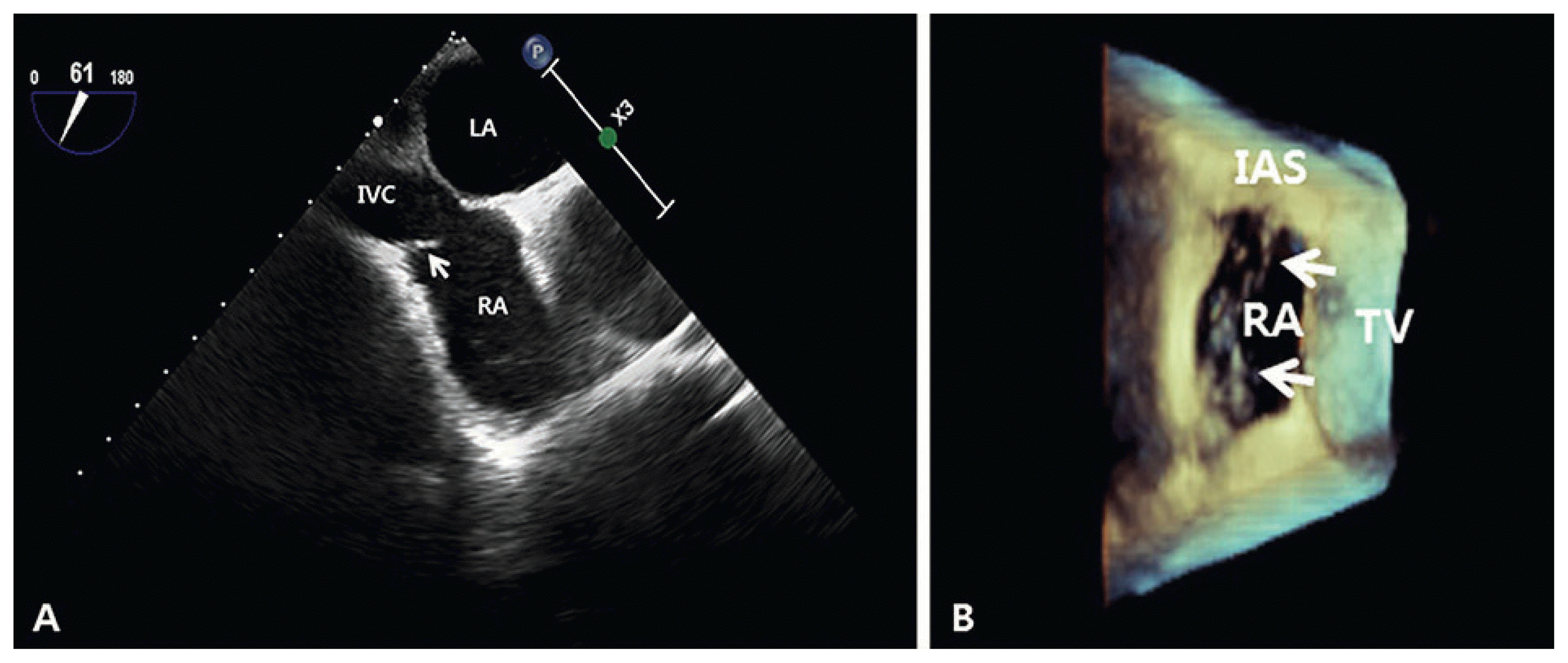 | Fig. 1(A) Two-dimensional transesophageal echocardiography (TEE) showing a linear structure in the right atrium (arrow). (B) Three-dimensional TEE clearly showing a net-like structure connected to the edge of the IVC and interatrial septum. IAS: interatrial septum; IVC: inferior vena cava; LA: left atrium; RA: right atrium; TV: tricuspid valve.
|
Case 2
A 60-year-old man presented to the hospital with repeated syncope. His electroencephalography and brain MRI showed normal results. For a more thorough cardiogenic examination, 24-hour Holter monitoring and TTE were performed and the TTE demonstrated severe aortic stenosis due to a bicuspid AV. Moreover, on TTE, whip-like structures extending from the opening of the IVC and moving freely within the RA cavity were observed, which were suspected to be a Chiari network. He was scheduled for AV replacement surgery along with resection of the Chiari network. Because he had a moderate to severe pulmonary hypertension, a pulmonary artery catheter (PAC) insertion was decided.
In the operating room, he was intubated and anesthetized with sevoflurane and remifentanil. The left radial artery was cannulated. Before central venous catheterization, a TEE probe was inserted, and the Chiari network was visualized using the midesophageal (ME) modified bicaval tricuspid valve (TV) view focused on the IVC (Fig. 2A). During ultrasound-guided internal jugular venous cannulation, a needle was inserted under the out-plane imaging of the internal jugular vein and a guidewire and catheter were carefully inserted using the ME modified bicaval TV view. Then, the PAC was inserted using the ME modified bicaval TV view, ensuring that the PAC was not entrapped in the Chiari network, in real time. After insertion of the PAC, we scanned the 3D TEE of the RA cavity to confirm that the PAC had passed through the TV without being entrapped in the Chiari network (Fig. 2B). The AV was replaced with a mechanical valve and the Chiari network was resected successfully.
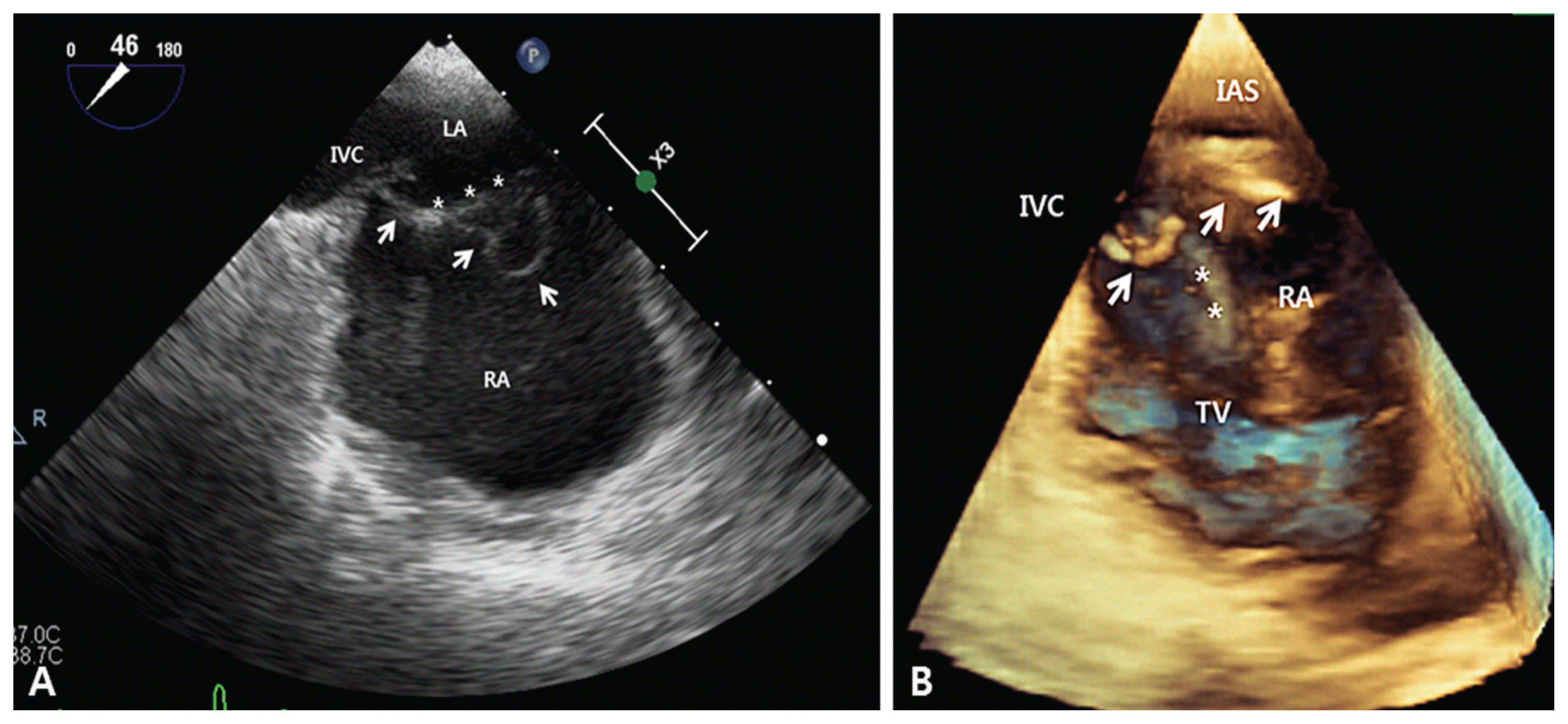 | Fig. 2(A) Two-dimensional transesophageal echocardiography (TEE) imaging does not clearly show the location of the Chiari network (arrow) in relation to the pulmonary artery catheter (PAC, *) (B) Three-dimensional TEE imaging shows that the PAC(*) successfully passed through the tricuspid valve without being entrapped in the Chiari network (arrow). IAS: interatrial septum; IVC: inferior vena cava; LA: left atrium; RA: right atrium; TV: tricuspid valve.
|
Go to : 
DISCUSSION
The Chiari network is an embryonic remnant of the sinus venosus valve which normally undergoes fenestration and disappears.7 It is usually found by accident and generally has no major clinical significance. However, sometimes it is difficult to differentiate the Chiari network from other RA pathologies that would require further management. Our first case patient had had a thromboembolic stroke but the origin of the thrombus had not been confirmed. On preoperative TTE, a fibroelastoma was identified on the AV and suspected to be the origin of the thrombus. During intraoperative TEE, we incidentally found a short linear structure which originated from the IVC and was freely moving in the RA cavity. Considering the patient’s history of thromboembolic stroke and PFO, we could not exclude a possible thrombus. On two-dimensional (2D) TEE, it was difficult to differentiate the Chiari network from the thrombus. However, with the 3D TEE view, a net-like structure attached at the edges of the IVC and interatrial septum could be clearly seen.
Intracardiac interventions have recently become more common and several case reports have cautioned that intracardiac devices could be entrapped in the Chiari network.4–6 In those studies, the entrapped intracardiac devices were removed using several techniques, including simple retraction under echocardiography, placement of a stiff catheter over the trapped guidewire to straighten the device, radiofrequency ablation of the entangled network strands, and even a thoracotomy.5,8 To prevent the entrapment of intracardiac devices in the Chiari network, real time echocardiography should be used to visualize the Chiari network and the intracardiac devices as they pass through the RA. Therefore, the IVC, the superior vena cava (SVC), the RA, TV, and right ventricle (RV) all should be visualized at the same time. As a result, we decided to observe the insertion of the central catheter and PAC using a ME modified bicaval TV view,9,10 which can be seen using the ME RV in-flow-outflow view and maintaining a transducer angle of 50 to 70 degrees while the probe is turned to the right until the TV is centered.5 In this case, we needed to focus on the IVC rather than the TV, so the transducer was slightly withdrawn and rotated to the left. The 2D TEE did not clearly show the location of the Chiari network in relation to the PAC (Fig. 2A). However, as Fig. 2B demonstrates, using the 3D TEE view, we could confirm that the PAC passed through the TV without being entrapped in the Chiari network.
Recently, real time 3D TEE guidance during catheter-based interventions have been introduced.11 However, there are still several limitations to the 3D TEE, such as issues with the frame rate, the spatial resolution, and the 3D color Doppler. Moreover, proficient use requires experience and practice to minimize these problems. During the insertion of catheters in our cases, we could not use the 3D TEE in real time because we lacked this experience and practice. Central catheterization and PAC insertion are not complex and time-consuming catheter-based interventions. If real time 3D TEE guidance during catheter insertion became standard practice, it would provide advantages such as clear visualization of the location of the catheter as it passed through the intracardiac structures and subsequent avoidance of potential complications.
Echocardiography is an excellent diagnostic imaging tool for the Chiari network. However, 2D echocardiography has a limited ability to differentiate structures and to clearly display the location of the Chiari network and intracardiac devices during invasive procedures. We suggest the use of 3D echocardiography when making a definitive diagnosis of Chiari network and to assist in avoiding entrapment and safely inserting a guidewire or catheter during intracardiac procedures.
Go to :
 XML Download