

 PDF
PDF Citation
 Print
Print


INTRODUCTION
MATERIALS AND METHODS
Inclusion criteria
General indication
Microsurgery (Fig. 1)
Table 1
![]()
Table 2
![]()
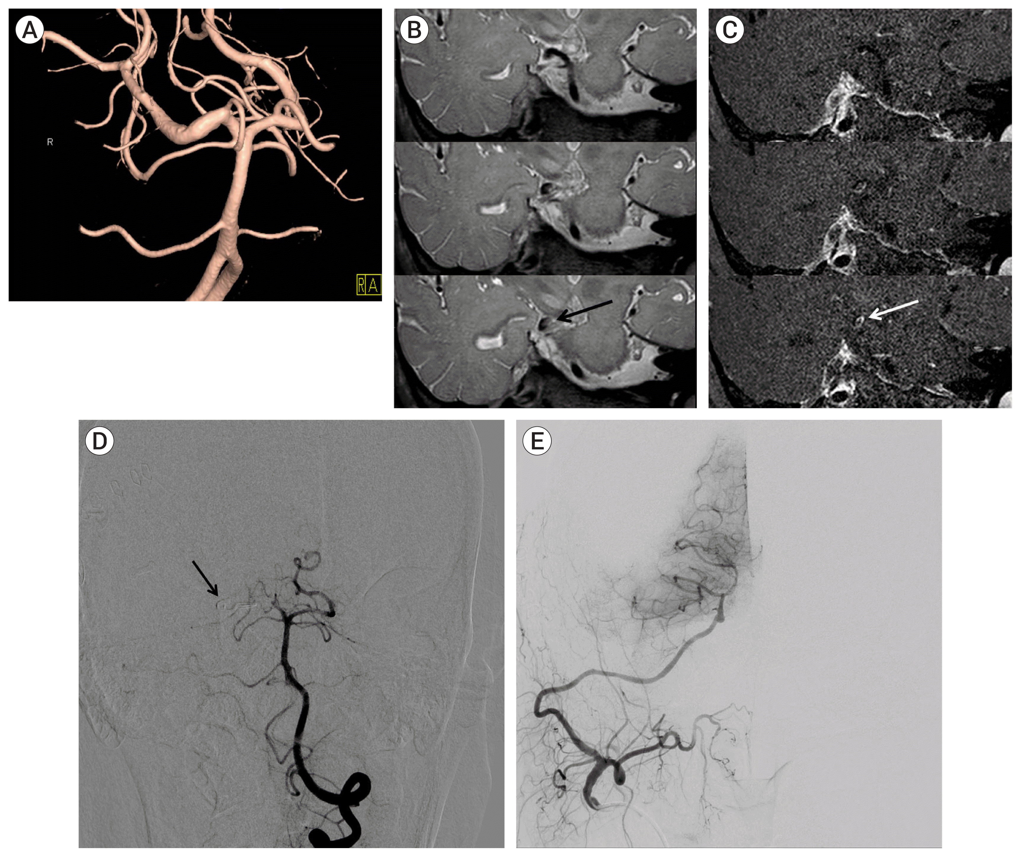 | Fig. 1(A) Three dimensional reconstruction of conventional cerebral angiography showing a fusiform aneurysm at the PCA. (B) A Coronal image with a T2 sequence at the level of the PCA dissecting aneurysm obtained through high-resolution magnetic resonance imaging. Black arrow indicates aneurysmal dilatation. (C) A Coronal image with a gadolinium-enhanced T1-weighted sequence at the level of the PCA dissecting aneurysm obtained through high-resolution magnetic resonance imaging. White arrow indicates intimal flap and double lumens. (D) Conventional cerebral angiography showing trapping of right proximal P2 segment of PCA. (E) Conventional cerebral angiography showing patent anastomosis of STA to the right distal P2 segment. PCA, posterior cerebral artery; STA, superficial temporal artery.
|
Endovascular procedure (Fig. 2)
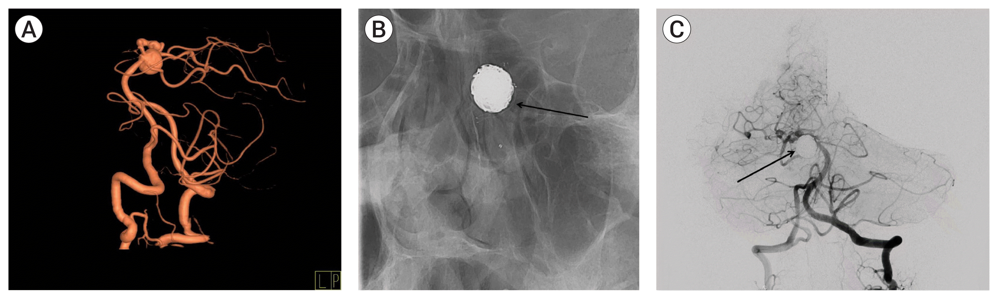 | Fig. 2(A) Three dimensional reconstruction of conventional cerebral angiography showing a superior cerebellar artery aneurysm. (B) Simple skull x-ray showing stent material (black arrow). (C) Conventional cerebral angiography following coil embolization showing complete obliteration of the aneurysm with coil mass (black arrow).
|
Clinical and radiologic outcomes and follow-up
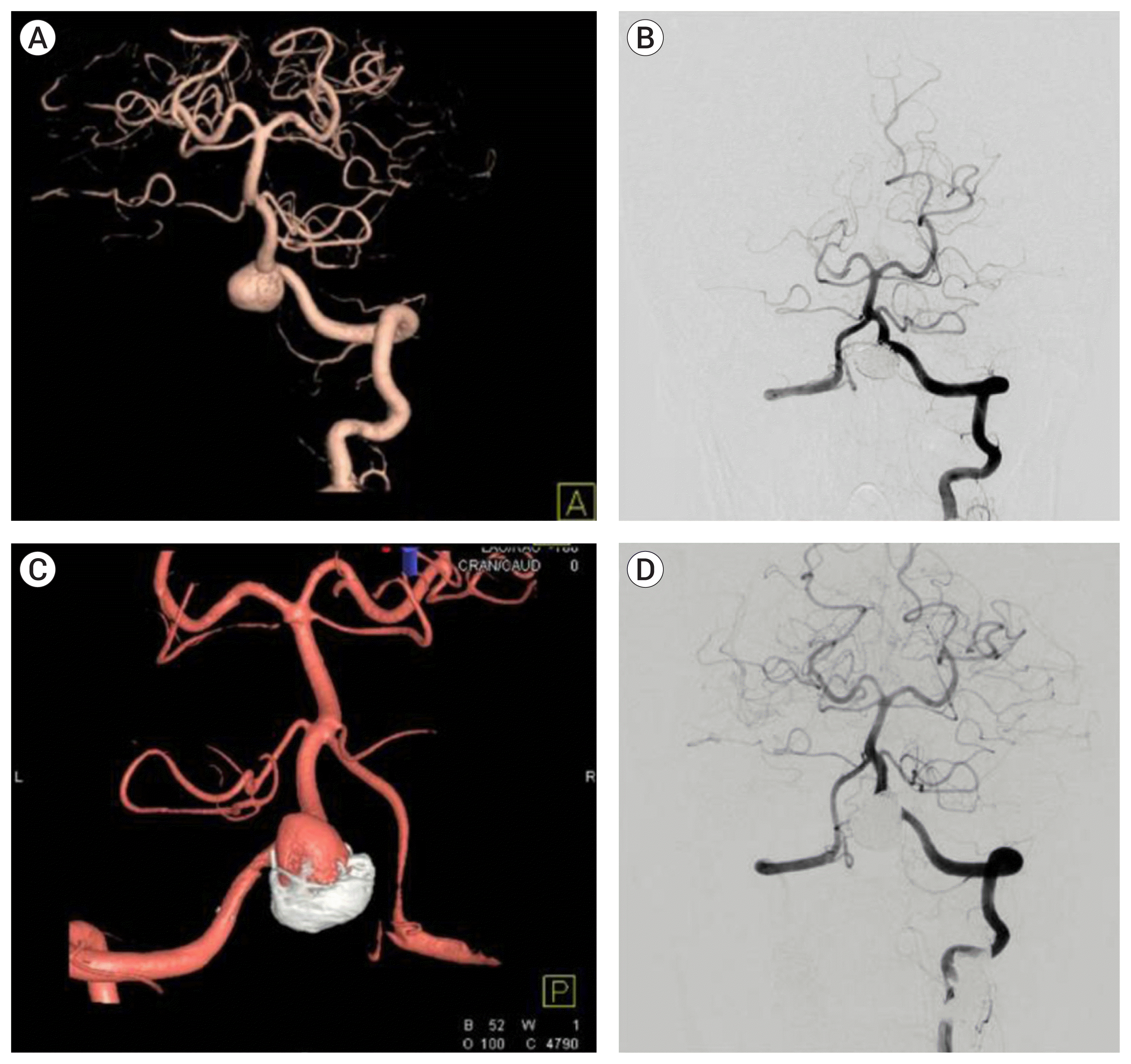 | Fig. 3(A) Three dimensional reconstruction of conventional cerebral angiography showing a vertebral artery aneurysm. (B) Conventional cerebral angiography after embolization showing complete obliteration of the aneurysm. (C) Follow-up angiography showing recurrence due to coil compaction. (D) Conventional cerebral angiography after additional coiling, showing complete obliteration of the aneurysm.
|
Data collection and statistical analysis
RESULTS
Characteristics of patients and aneurysms
Table 3
| Surgery type | Microsurgery | Endovascular | p value | Total patients |
|---|---|---|---|---|
| Total patients | 109 (28.1) | 281 (71.9) | 390 | |
| Age, yr | ||||
| Mean±SD | 56±11 | 56±11 | 0.52 | 56±11 |
| Sex | ||||
| Male:Female | 20:89 | 88:193 | 0.03 | 108:282 |
| Aneurysm size* | ||||
| Small (<10 mm) | 91 (83.4) | 222 (79.0) | 0.52 | 313 (80.3) |
| Large (10 mm to <25 mm) | 15 (13.8) | 39 (13.9) | 54 (13.8) | |
| Giant (≥25 mm) | 2 (1.8) | 4 (1.4) | 6 (1.5) | |
| HTN | 56 (51.4) | 106 (37.7) | 0.01 | 162 (42) |
| DM | 4 (3.7) | 19 (6.8) | 0.33 | 23 (6) |
| Coronary disease | 1 (0.9) | 7 (2.5) | 0.45 | 8 (2) |
| Comorbidity† | 3 (2.8) | 3 (1.1) | 0.36 | 6 (1.5) |
| Preoperative cranial nerve palsy | 0 | 0 | 0 | |
| Location | 0.09 | |||
| Basilar apex | 44 (40.4) | 145 (51.6) | 189 (48.5) | |
| PCA | 3 (2.8) | 16 (5.7) | 19 (4.9) | |
| SCA | 27 (24.8) | 32 (11.4) | 59 (15.1) | |
| AICA | 1 (0.9) | 4 (1.4) | 5 (1.3) | |
| Basilar trunk | 1 (0.9) | 8 (2.8) | 9 (2.3) | |
| VB junction | 2 (1.8) | 5 (1.8) | 7 (1.8) | |
| VA | 4 (3.7) | 56 (19.9) | 60 (15.4) | |
| PICA | 27 (24.8) | 15 (5.3) | 42 (10.8) | |
| Dissecting aneurysm‡ | 0.34 | |||
| SCA | 1 (0.9) | 0 | 1 (0.3) | |
| Basilar trunk | 1 (0.9) | 1 (0.4) | 2 (0.5) | |
| VA | 3 (2.8) | 20 (7.1) | 21(5.4) | |
| PICA | 3 (2.8) | 1 (0.4) | 4 (1.0) | |
| Complications | 13 (11.9) | 17 (6.1) | 0.05 | 30 (7.7) |
| Third nerve palsy | 7 (6.4) | 2 (0.7) | 0.01 | 9 (2.3) |
| Low cranial nerve palsy | 2 (0.9) | 0 | 0.28 | 2 (0.5) |
| Post operative bleeding | 0 | 4 (1.5) | 0.58 | 4 (1.0) |
| Cerebral infarction | 5 (4.6) | 11 (3.9) | 0.77 | 16 (4.1) |
| Immediate postoperative presence of residual sac | ||||
| Complete obliteration | 103 (94.5) | 181 (64.4) | 0.01 | 284 (72.8) |
| Incomplete obliteration (residual sac) | 6 (5.5) | 100 (35.6) | 106 (27.2) | |
| Recurrence | 6 (5.5) | 62 (22.1) | 0.01 | 71 (18.2) |
| After complete obliteration | 6 (5.5) | 5 (1.8) | 11 (2.8) | |
| Initial residual sac | 0 | 57 (20.3) | 57 (14.6) | |
| Recurrence period§ (Mean±SD[range], month) | 56.7±40.6 [6–102] | 32.8±36.2 [1–162] | 0.2 | 34.3±36.6 [1–162] |
| Retreatment|| | 1 (0.9) | 27 (9.6) | 0.01 | 28 (7.2) |
| Microsurgery | 0 | 0 | 0 | |
| Additional coiling¶ | 1 (0.9) | 20 (7.1) | 21 (5.54) | |
| Stent-assisted coiling** | 0 | 7 (2.5) | 7 (1.8) | |
| Retreatment period§ (Mean±SD[range], month) | 47.0 | 26.0±28.6 [1–138] | 0.5 | 26.8±28.3 [1–138] |
| GOS | 0.9 | |||
| 3 | 2 (1.8) | 5 (1.8) | 7 (1.8) | |
| 4 | 3 (2.8) | 6 (2.1) | 9 (2.3) | |
| 5 | 104 (95.4) | 270 (96.1) | 374 (95.9) | |
| GOS6mos | 0.2 | |||
| 2 | 0 | 4 (1.4) | 4 (1.0) | |
| 3 | 5 (4.6) | 5 (1.8) | 10 (2.6) | |
| 4 | 3 (2.8) | 17 (6.0) | 20 (5.1) | |
| 5 | 101 (92.7) | 255 (90.7) | 356 (91.3) | |
| Follow up period§ (Mean±SD[range], month) | 40.9±35.0 [6–141] | 50.0±41.3 [6–186] | 0.05 | 47.3±39.8 [6–186] |
* Missing values (in the absence of three dimensional reconstruction of conventional cerebral angiography, size measurements were not possible and were treated as missing).
† Liver cirrhosis, chronic kidney disease, end-stage renal disease (ESRD), pre-operative cerebral infarction, underlying cancer (gastric cancer, lung cancer), chronic obstructive pulmonary disease.
‡ Of all aneurysms, the number diagnosed as dissecting aneurysm. It is included in the total number of aneurysms and location of aneurysms.
![]()
Table 4
| Surgery type | Microsurgery | Endovascular | p value | Total patients |
|---|---|---|---|---|
| Total patients | 78 (33.8) | 153 (66.2) | 231 | |
| Age, yr | ||||
| Mean±SD | 54±12 | 51±13 | 0.76 | 52±13 |
| Sex | ||||
| Male:Female | 15:63 | 67:86 | <0.01 | 82:149 |
| Aneurysm size* | ||||
| Small (<10 mm) | 70 (89.7) | 108 (70.6) | <0.01 | 178 (76.1) |
| Large (10 mm to <25 mm) | 6 (7.7) | 36 (23.5) | 42 (17.9) | |
| Giant (≥25 mm) | 0 | 2 (1.3) | 2 (0.9) | |
| HTN | 19 (24.3) | 31 (20.3) | 0.5 | 50 (21) |
| DM | 3 (3.85) | 4 (2.6) | 0.7 | 7 (3) |
| Coronary disease | 0 | 4 (2.6) | 0.3 | 4 (2) |
| Comorbidity† | 0 | 6 (3.9) | 0.1 | 6 (2.5) |
| Preoperative cranial nerve palsy | 0 | 1 (0.7) | 0.47 | 1 (0.4) |
| Location | <0.01 | |||
| Basilar apex | 25 (32.1) | 57 (37.3) | 82 (35.5) | |
| PCA | 5 (6.4) | 15 (9.8) | 21 (9.1) | |
| SCA | 13 (46.7) | 19 (12.4) | 32 (13.9) | |
| AICA | 2 (2.6) | 5 (3.3) | 7 (3.0) | |
| Basukar trunk | 2 (2.6) | 5 (3.3) | 7 (3.0) | |
| VB junction | 2 (2.6) | 2 (1.3) | 4 (1.7) | |
| VA | 3 (3.8) | 31 (20.3) | 34 (14.7) | |
| PICA | 26 (33.3) | 19 (12.4) | 45 (19.5) | |
| Dissecting aneurysm‡ | 0.08 | |||
| PCA | 1 (1.3) | 1 (0.7) | 2 (0.9) | |
| VA | 2 (2.6) | 20 (13.1) | 22 (9.5) | |
| PICA | 2 (2.6) | 1 (0.7) | 3 (1.3) | |
| HH | 0.01 | |||
| 1 | 2 (2.6) | 6 (4.0) | 8 (3.4) | |
| 2 | 41 (52.6) | 63 (41.2) | 104 (45.0) | |
| 3 | 26 (33.3) | 43 (28.1) | 69 (29.9) | |
| 4 | 8(10.3) | 39 (25.5) | 47 (20.3) | |
| 5 | 1 (1.3) | 2 (1.3) | 3 (1.3) | |
| Fisher grade | 0.4 | |||
| 1 | 1 (1.3) | 0 | 1 (0.4) | |
| 2 | 7 (9.0) | 9 (5.9) | 16 (6.9) | |
| 3 | 24 (30.8) | 55 (35.9) | 79 (34.2) | |
| 4 | 46 (59.0) | 89 (58.2) | 135 (58.4) | |
| Complications | 12 (15.3) | 26 (17.0) | 0.14 | 38 (16.5) |
| Third nerve palsy | 6 (7.7) | 3 (2.0) | 0.01 | 9 (3.9) |
| Low cranial nerve palsy | 1 (1.3) | 1 (0.7) | 2 (0.9) | |
| Postoperative rebleeding | 4 (5.1) | 7 (4.6) | 0.26 | 11 (4.8) |
| Cerebral infarction | 2 (2.6) | 16 (10.45) | 0.23 | 18 (7.8) |
| Immediate postoperative presence of residual sac | ||||
| Complete obliteration | 77 (98.7) | 114 (74.5) | <0.01 | 191 |
| Incomplete obliteration (residual sac) | 1 (1.3) | 39 (25.5) | 40 | |
| Recurrence | 3 (3.85) | 27 (17.6) | <0.01 | 30 (13.0) |
| After complete obliteration | 3 (3.85) | 7 (4.6) | 10 (4.3) | |
| Initial residual sac | 0 | 20 (13.0) | 20 (8.7) | |
| Recurrence period§ (Mean± SD[range], month) | 43.0±40.5 [2–83] | 56.0±44.8 [2–170] | 0.5 | 54.7±44.0 [2–170] |
| Retreatment|| | 2 (2.6) | 7 (4.6) | 0.45 | 9 (3.9) |
| Microsurgery | 1(1.3) | 0 | 1 (0.4) | |
| Additional coiling¶ | 1(1.3) | 6 (3.9) | 7 | |
| Stent-assisted coiling** | 0 | 1 (0.7) | 1 (0.4) | |
| Retreatment period§ (Mean±SD [range], month) | 42.5±57.3 [2–83] | 32.7±16.6 [13–53] | <0.01 | 34.9±25.2 [2–53] |
| GOS | 0.3 | |||
| 2 | 5 (6.4) | 18 (11.8) | 23 (10) | |
| 3 | 10 (12.8) | 28 (18.3) | 38 (16.5) | |
| 4 | 11 (14.1) | 21 (13.7) | 32 (13.9) | |
| 5 | 52 (66.7) | 86 (56.2) | 138 (59.7) | |
| GOS6mos | 0.02 | |||
| 1 | 2 (2.6) | 12 (7.8) | 14 (6.1) | |
| 2 | 2 (2.6) | 12 (7.8) | 14 (6.1) | |
| 3 | 12 (15.4) | 10 (6.5) | 22 (9.5) | |
| 4 | 5 (6.4) | 21 (13.7) | 26 (11.3) | |
| 5 | 57 (73.1) | 98 (64.1) | 155 (67.1) | |
| Follow up period§ (Mean±SD [range], month | 43.0±36.7 [6–152] | 46.9±48.1 [6–185] | 0.03 | 45.6±44.5 [6–185] |
* Missing value (in the absence of three dimensional reconstruction of conventional cerebral angiography, size measurements were not possible and were treated as missing).
† Liver cirrhosis, chronic kidney disease, end-stage renal disease (ESRD), pre-operative cerebral infarction, underlying cancer(gastric cancer, lung cancer), chronic obstructive pulmonary disease.
‡ Of all aneurysms, the number diagnosed as dissecting aneurysm. It is included in the total number of aneurysms and location of aneurysms.
§ Because the number of follow-up patients is different, the follow-up period is sometimes smaller than the recurrence period and retreatment period.
PCA, posterior cerebral artery; SCA, superior cerebellar artery; AICA, anterior inferior cerebellar artery; VB, vertebrobasilar; VA, vertebral artery; PICA, posterior inferior cerebellar artery; HH: Hunt Hess grade; GOS, Glasgow outcome scale score; GOS6mos, Glasgow outcome scale score 6 months after surgery
![]()
 XML Download