

 PDF
PDF Citation
 Print
Print


INTRODUCTION
The anterior communicating artery (A-com) is known for the most common site for brain aneurysm formation and rupture as well [8,10]. The A-com aneurysm could be technically challenging from a surgical approach because of frequent anatomic variations, complex hemodynamics, unfavorable aneurysm morphology for clipping, and the presence of numerous critical perforators [4,7,11,16]. For the last 25 years, remarkable growth in endovascular technique and devices has led to a significant proportion of A-com aneurysms being successfully managed with endovascular approach [2-4,6]. However, one of the major drawbacks of endovascular treatment continues to be the risk of regrowth or rebleeding of these treated aneurysms regardless of location [4,12]. Although numerous studies have been conducted to compare the results between microsurgical clipping and endovascular coiling in the treatment of A-com aneurysms, but there is no clear answer for which treatment is superior [1,10,12,14,17]. Both treatment modalities have strengths and weaknesses which make them as complementary rather than competitive. Management of the A-com aneurysm often requires harmony between microsurgical clipping and endovascular therapy. Recently, dual training in endovascular and microsurgical surgery has become popular with vascular neurosurgeons around the world. To our knowledge, this is the first study that compare clipping and coiling of the A-com aneurysms, where both were conducted by a single hybrid vascular neurosurgeon. Therefore, the purpose of this study is to provide the best management protocol for these conditions based on our results.
Go to : 
MATERIALS AND METHODS
This study was done in 70 cases of an unruptured A-com aneurysm treated by a dual-trained vascular neurosurgeon in our institution from March 2012 to December 2019. Due to the retrospective nature, the present study is exempt in accord with the Institutional Review Board standards of our institution. All aneurysms were diagnosed based on the findings of digital subtraction angiography. All patients were classified into two groups depending upon the therapeutic modality performed:
(a) Microsurgical clipping (33)
(b) Endovascular coiling (37)
Multiple aneurysms, ruptured aneurysms, aneurysms associated with a brain arteriovenous malformation, and other than A-com aneurysm were excluded. A coiling was considered first in selecting treatment methods for each case. The reasons not to treat a patient with coiling were the followings: aneurysms that had an unfavorable dome-to-neck ratio for coiling or that were expected to be amenable for clipping; and patients with vascular anatomy unfavorable for endovascular navigation (for example severe tortuosity of proximal vessels and carotid stenosis). However, endovascular procedure was often chosen for the patients with old age, or poor general condition for general anesthesia, regardless of the above criteria. In addition, aneurysms with an unfavorable dome-to-neck ratio for coiling were occasionally treated with coiling using stent, balloon, or multiple catheters. Actually, the pros and cons of both procedures were thoroughly explained to the patients and their families, and one procedure was chosen. In this retrospective study, the authors examined the treatment-related complications, angiographic and clinical outcomes, and the results between the two groups were compared.
Surgical approaches
Two types of surgical approaches were used in clipping of the A-com aneurysms, namely pterional approach and interhemispheric approach. Pterional approach was used mainly, because damage of the olfactory nerve can be minimized and bilateral parent arteries of the proximal side can be secured in early stage of the procedure. However, this approach cannot be used without partial resection of rectus gyrus, when the aneurysm is located high within interhemispheric fissure (distance from the planum sphenoidale to the neck of the aneurysm ≥10 mm) and directed postero-superiorly. Optimal surgical direction, right or left side, concerning the pterional approach to the A-com aneurysms was decided according to many factors, such as, dominant feeding artery, shape, size, and direction of aneurysm, vascular anomaly, and variation around the A-com aneurysm, dominant hemisphere, and another aneurysm. The monitoring of motor evoked potential and somatosensory evoked potential was routinely performed in all cases. The patency of the parent artery, major branches, and visible perforators was confirmed with Doppler ultrasonography and indocyanine green angiography.
Endovascular procedure
Dual antiplatelet, defined as the use of clopidogrel (P2Y12 receptor inhibitor) and acetylsalicylic acid, was prescribed patients, who will be given endovascular procedure, before one week for prevention of excessive thrombogenesis and thromboembolism. Coiling the aneurysms was executed under local anesthesia, and intravenous drip infusion of heparin was conducted employing catheter pressure infusion systems with continuous infusion of 1,000 U of heparin per 1,000 mL of saline. The aim of the coiling procedure was to obtain a packing of the aneurysm as attenuated as possible. Various technical measures and precautions were taken to overcome specific problems. Remodeling techniques using a balloon, a stent, or multiple catheters were unavailable for the aneurysms treated during the early part of our series. However, we have recently favored these remodeling techniques to achieve higher postoperative occlusion rates. In the case of intraprocedural thromboembolic complication, various strategies for thrombolysis were applied, such as mechanical thrombolysis and intra-arterial or intravenous aggrastat or heparin infusion during or after the procedure. Unless the thromboembolic complication occurred or a stent was used, further antiplatelet or anticoagulant medications were not administered. The indications for long-term antiplatelet therapy of acetylsalicylic acid and/or clopidogrel were as follows; formation of a thrombus at the end of the coil or thromboembolic events, stent-assisted coiling, and coil protrusion into the parent artery. Immediately after both procedures, a complete neurological examination was performed on all patients by a vascular neurosurgeon. Furthermore, all patients underwent a non-enhanced brain computed tomography (CT) scan to evaluate possible hemorrhagic complications. And 24-48 hours after the procedures, diffusion-weighted magnetic resonance imaging of the brain was performed to assess possible acute cerebral infarction.
Clinical and Angiographic follow-ups
Clinical results for both groups were assessed upon at 6 months of follow-up using the modified Rankin Scale (mRS). Poor clinical outcome was defined as a mRS score of 3 to 6. The follow-up conventional angiographies for both groups were performed for 12 months after initial treatment. Non-invasive methods (i.e., Magnetic resonance angiography and CT angiography) were also used for imaging follow-ups. However, the follow-ups using these non-invasive imaging modalities were excluded from this study, because it was difficult to accurately assess whether aneurysm has recurred or not, due to metal artifacts. So only the follow-up digital subtraction angiographic results were analyzed. Immediate angiographic results were included only in coil group. On follow-up angiography of coil group, occlusion rate was classified, as follows: “stable” (i.e., no contrast filling of the aneurysm sac and neck), “minor recanalization” (i.e., minimal coil compaction at the aneurysm neck), “major recanalization” (i.e., contrast filling within the sac of the aneurysm because of significant coil compaction), and “regrowth” (i.e., appearance of a new aneurysm dilatation or daughter sac). Re-treatment was required in a case of major recanalization or regrowth. As in clip group, major recurrence, which required retreatment, defined as contrast filling within the aneurysm fundus or appearance of a new aneurysm dilatation on follow-up angiography. Complications were defined as all adverse events associated with the procedure, and these were evaluated retrospectively through medical records. Subclinical complications that did not cause symptoms, were excluded from the analysis of this study.
Go to :
RESULTS
Patient demographic data and characteristics of the aneurysms in each group are shown in Table 1.
Table 1.
Patient demographic data and characteristics of the aneurysms
![]()
Clip group
The clip group was composed of 16 (48.5%) men and 17 (51.5%) women with mean age of 57.6 years (range, 29-71 years). In this series, except for the two cases using the interhemispheric approach, the pterional approach was applied in all cases, and aneurysm neck clipping was possible in all cases. Two cases of interhemispheric approach were applied to large A-com aneurysm and very high-positioned A-com aneurysm cases. The mean of the measurable distance from the planum sphenoidale to the neck of the aneurysm in the clip group was 6.6 mm. There was just 6 patients (18.2%) of high positioned A-com aneurysm (distance from the planum sphenoidale to the neck of the aneurysm ≥10 mm) in this group. In this group, 22 (59.5%) aneurysms were directed anteriorly or inferiorly, and 15 (40.3%) posteriorly or superiorly. Thirty (90.9%) aneurysms were less than 7 mm in diameter. Twenty-nine (87.9%) aneurysms had a wide-neck (dome-to-neck ratio ≤1.5 or neck ≥4 mm). Twenty-three (69.7%) aneurysms had a left dominancy of A1, and 5 (15.1%) aneurysms showed an agenesis of the contralateral A1.
Coil group
The coil group consisted of 23 (62.2%) men and 14 (37.8%) women with a mean age of 64.8 years (range, 43-78 years). The mean measurable distance from the planum sphenoidale to the neck of aneurysm in this group was 9.6 mm. There was just 17 patients (46.0%) of high positioned A-com aneurysm (distance from the planum sphenoidale to the neck of the aneurysm ≥10 mm) in this group. In this group, 36 (78.8%) aneurysms were directed anteriorly or inferiorly, 7 (21.2%) posteriorly or superiorly. Thirty (81.1%) aneurysms were less than 7 mm in diameter, and 28 (75.7%) aneurysms had a wide-neck. Twenty-five (67.6%) aneurysms had a left dominancy of A1, and 4 (10.8%) aneurysms had an agenesis of the contralateral A1.
The treatment results for the coil and clip groups are presented in Tables 2 and 3, respectively.
Table 2.
Treatment results in the coil group
![]()
Table 3.
Treatment results in the clip group
![]()
Complications and clinical results
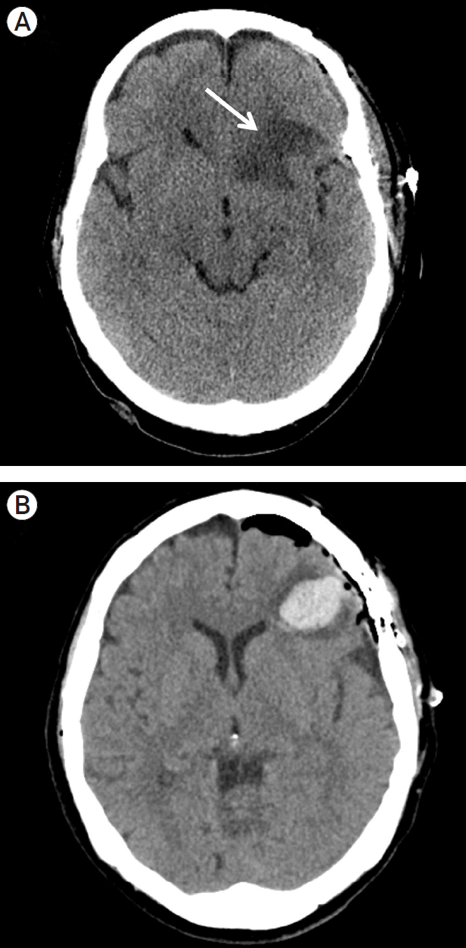
Procedure-related symptomatic complications occurred in 2 patients (2/37, 5.4%) in the coil group and 3 patients (3/33, 9.1%) in the clip group. Coiling-related symptomatic complications were an intra-procedural rupture in both cases. Diagnosis of intra-procedural rupture was made based on the angiographic visualization of contrast material extravasation in both cases. Fortunately, hemostasis was possible with prompt additional coil insertion and injection of protamine sulfate in both cases. One of them had complained of subarachnoid hemorrhage-related severe headache immediately after procedure, but was finally discharged without any neurological deficits (Fig. 1). While the other remained serious disabled (mRS 4) due to rebleeding that occurred three hours after the procedure. Procedure-related symptomatic complications developed in 3 patients (3/33, 9.1%) in the clip group (Fig. 2). Two of them were accompanied by a perforator infarction and the other by a remote cortical hemorrhage immediate after the surgery. Perforator infarction was presumed to have been caused by the emboli which was originated from several trials of clipping, or direct compression of perforator origin by blade of clip. On the basis of radiologic findings, it appeared to be hypothalamic branches among the perforators around A-com. And remote cortical hematoma was presumed to be caused by careless cortical injury during the surgery. These caused a mild cognitive impairment for all three of them (mRS 1). Several patients complained of anosmia immediately after craniotomy, but the symptoms were not addressed in this study because they were somewhat subjective and sometimes slowly recovered. To sum up, the periprocedural mortality in all patients was 0% (0/70) and morbidity was 7.1% (5/70). Poor clinical outcome (mRS of 3 to 6) at months of follow-up was seen in only one patient (1/37, 2.7%) for the coil group. None had a poor clinical outcome in the clip group. The one poor outcome was the result of intra-procedural rupture during the coiling.
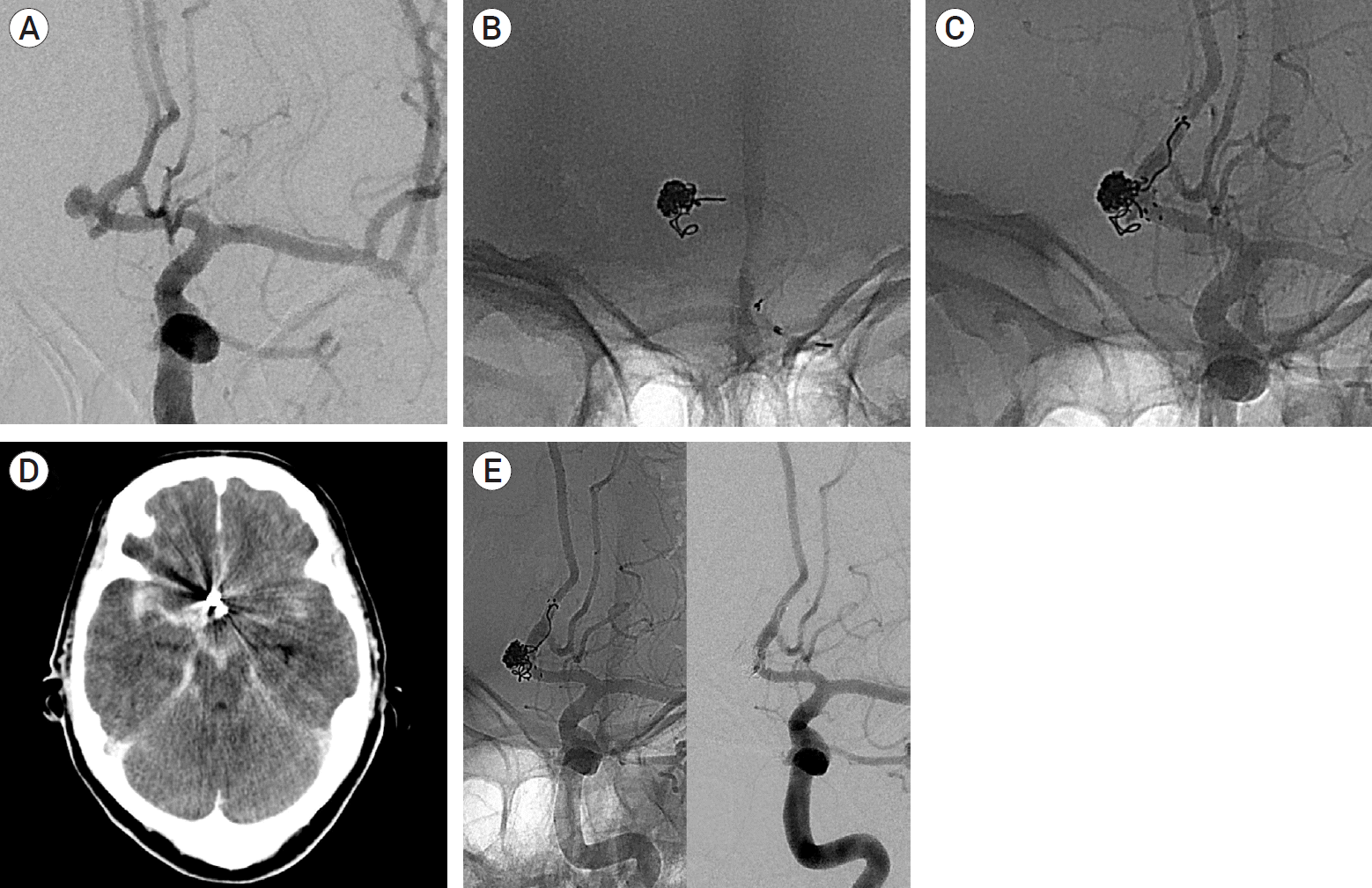 | Fig. 1.(A) Left ICA angiography showing an unruptured ACOM aneurysm (4.6×3.0 mm in size). (B) During placement of the coil, coil extrusion from the aneurysm sac was identified with minimal contrast leakage. (C) Additional coils and stent could prevent further contrast leakage. (D) Computed tomography scan obtained immediately after the procedure showed diffuse subarachnoid hemorrhage in the basal cistern and minimal contrast leak hyperdensity. Fortunately, the patient was discharged without any neurologic deficits. (E) Follow-up angiogram obtained 24 months after the initial intervention shows a stable aneurysm occlusion and patent stented artery. ICA, internal carotid artery; ACOM, anterior communicating artery.
|
Immediate and Follow-up angiographic results
In the coil group, the endovascular approach was technically possible in all 37 patients. The treatment of 11 aneurysms was possible with a single microcatheter, but the other 26 required an adjunctive technique, that is, double catheter, a balloon-assisted, or a stent-assisted technique. Immediate post-procedural angiograms showed complete aneurysm occlusion in 29 (29/37, 78.4%) and near-complete occlusion in 8 (8/37, 21.6%). Follow-up conventional angiography data (mean duration, 15.0 months) were available for 18 (18/37, 48.7%) in the coil group and 10 (10/33, 30.3%) in the clip group. Major recanalization or recurrence, which required retreatment, was identified in one patient each in both groups. The initially coil-embolized patient, who was later identified to have a major recanalization, was treated with craniotomy, because the recanalized aneurysm had an unfavorable dome-to-neck ratio for recoiling and the patient was very young (Fig. 3). Contrarily, the initially clipped patient, who was later confirmed to have a major recurrence, was treated endovascularly, because of concerns about postoperative tissue adhesion (Fig. 4). Therefore, the major recanalization rate is 5.6% for the coil group (1/18) and 10.0% for the clip group (1/10). To sum up, the major recanalization rate in both groups was 7.1% (2/28). Representative cases are shown in Fig. 5, 6.
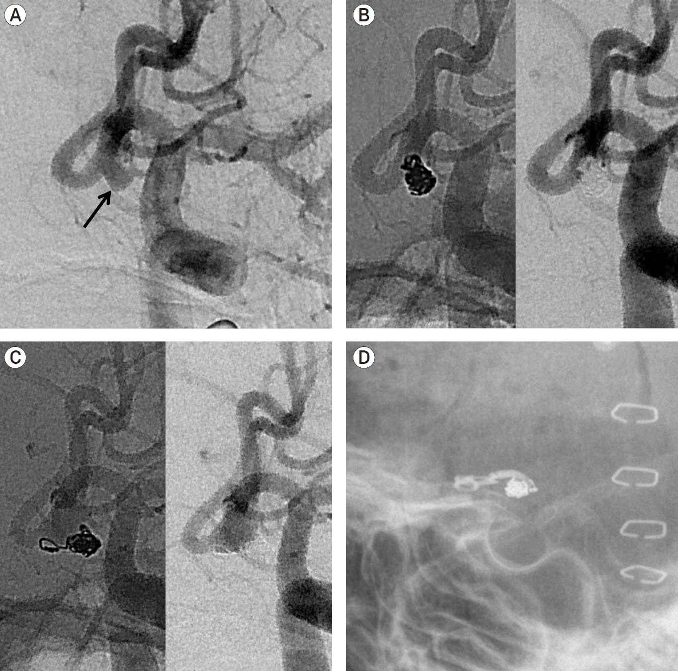 | Fig. 3.(A) Angiogram obtained in a 27-year-old man who had a chronic migraine reveals an ACOM aneurysm (2.5×4.5 mm in size, arrow). (B) Final angiogram just after uneventful coiling shows complete occlusion of the aneurysm. (C) A 12-month routine follow-up angiogram shows coil loosening and recurrence of the aneurysm. (D) The patient underwent a left-sided craniotomy for clip ligation of the residual aneurysm. Postoperative radiograph shows a clip on the previously coiled aneurysm. ACOM, Anterior communicating artery.
|
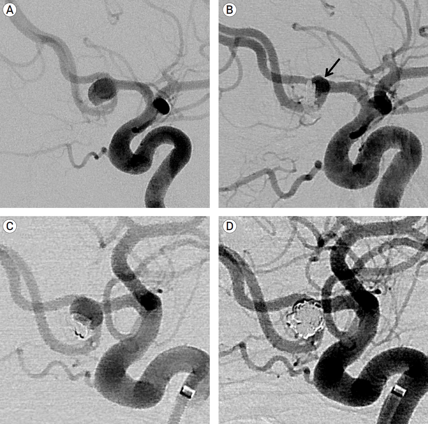 | Fig. 4.(A) A 62-year-old female with an ACOM aneurysm. Left ICA angiogram demonstrates a left ACOM aneurysm (6.7×6.2 mm in size). (B) Subtracted images acquired 2 weeks after microsurgical clipping reveal a residual aneurysm (arrow) due to incomplete clipping. (C) Follow-up angiogram obtained 24 months after the craniotomy shows a regrowth of the remnant aneurysm. (D) Left ICA angiogram just after simple coiling shows a successful complete occlusion of the recurred aneurysm. ACOM, anterior communicating artery; ICA, internal carotid artery.
|
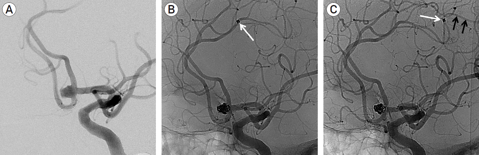 | Fig. 5.(A) A 71-year-old male with an ACOM aneurysm (4.6×3.0 mm in size). (B) Final angiogram just after stent-assisted coiling shows migration of the last coil (1.5 mm×2.0 cm, arrow) into the distal anterior cerebral artery, which is patent. The patient was discharged in good neurological condition. (C) Routine control carotid angiogram performed at 1 year from the coiling shows the migrated coil (white arrow) observed in the more distal artery, which is also patent (black arrows). ACOM, anterior communicating artery.
|
| Fig. 6.(A) A 63-year-old male with an ACOM aneurysm (6.6×5.8 mm in size). (B) Final angiogram just after simple coiling shows complete occlusion of the aneurysm. (C) Computed tomography 5 days after the procedure discloses silent infarction (arrow) secondary to perforator occlusion. ACOM, anterior communicating artery.
|
Go to :
DISCUSSION
The A-com aneurysm is the most commonly treated cerebral aneurysm, accounting for about 39% [8,14]. The A-com complex has a peculiar anatomic structure, and accompanying anatomic variations, multiple critical perforators, and complex regional flow dynamics, often put the neurosurgeon in trouble by making surgical approach difficult [4,7,11,16]. Over the past 2 decades, considerable development in neurointervention has led to a significant proportion of A-com aneurysms being successfully managed with endovascular approach [2-4,6]. However, one of the major weaknesses of this treatment continues to be the risk of regrowth or rebleeding of these aneurysms after coiling [4,12].
Traditionally, when it comes to the treatment of A-com aneurysms, craniotomy was preferred to coil embolization. The inherent small diameter of A-com aneurysms as well as the propensity to be small in size or wide-neck are the features that can make endovascular treatment difficult. Since the mid-1990s, advances in technology such as 3D rotational angiography and newer microguidewire/catheters, in combination with adjunctive devices such as balloon-assisted and stent-assisted coiling, have revolutionized treatment of aneurysms once deemed unfeasible to coiling. Based on my personal experiences, it seems that there are few A-com aneurysms that cannot be treated with coil embolization in recent years except for some aneurysms such as a large-sized aneurysm with an ultra-wide neck or a blister like aneurysm.
When the fundus of an aneurysm is superiorly or posteriorly directed or the A-com aneurysm is located far from the planum sphenoidale, wide resection of the rectus gyrus is needed to expose the A-com aneurysm through the pterional approach. Sometimes, an excessive retraction of the frontal lobe may cause terrible complications postoperatively in these situations. The results of this study also show that more coiling was chosen than craniotomy in these situations. In the clip group of this series, superiorly or posteriorly directing aneurysms accounted for only 21.2%, and high positioned A-com aneurysms accounted for only 18.2%. It is thought to be a recent global trend to prefer coiling to craniotomy to treat the above complex aneurysms.
Until now, unruptured A-com aneurysm has been generally managed with either endovascular coiling or microsurgical clipping. These preventive treatments require an acceptable morbidity and mortality to justify the intervention against that of the expected natural history. When intervention is deemed appropriate, understanding both the safety and effectiveness of treatment is essential to inform clinical decision making and discussion with the patient before treatment. Numerous studies have been conducted to compare microsurgical clipping and endovascular coiling in the management of A-com aneurysms [1,10,12,14,17]. So far, since one treatment modality cannot be said to be superior in all results, the choice of one treatment over the other requires careful consideration of various factors for the aneurysm and patient. Solution of that problem could be mutual complementary application of two treatment modalities. The proper harmonization of these two treatment modalities in the treatment of the A-com aneurysm can lead to the best results. It’s because A-com aneurysms present unique challenges for both approaches. The microsurgical clip application in this area is sometimes expected to be a significant undertaking because of unfavorable aneurysm morphology for clipping, the deep midline location, and frequent anatomic variations that are inherent to this region [5,7,9,13-15]. Endovascular treatment techniques for A-com aneurysms can be complicated as well by inherently unfavorable vascular morphology (multiple critical perforators, small sizeddome, and unfavorable dome-to-neck ratio, etc [1,2,4]. In recent decades, dual training in endovascular and microsurgical surgery has become popular for vascular neurosurgeons in several developed countries [18]. As a result, there might be more favorable circumstances of each modality in treatment of the A-com aneurysm. These hybrid neurosurgeons who have a great deal of practical experience in both modalities are expected to make more optimal selection and utilization of two treatment modalities.
This study had been conducted to compare microsurgical clipping with endovascular coiling in treatment of unruptured A-com aneurysms in terms of clinical and angiographic results. In particular, this study is meaningful in that both treatment modalities were performed by a single hybrid neurosurgeon. To our knowledge, this is the first study that evaluates the results of A-com aneurysms treated in two modalities, where both were done by a single hybrid vascular neurosurgeon. As a result of this study, there was no significant difference in results between the two treatment modalities performed by a single neurosurgeon. The small number of cases is a critical limitation of this study, and a selection bias is bound to exist in our study as well.
Recently, O’Neill et al. reported a systemic review of the existing literature data on the clinical and radiological outcomes following coiling, clipping, and stent-assisted coiling of unruptured A-com aneurysms [12]. Their study included 14 previous reports with 862 treated unruptured A-com aneurysms. The morbidity from simple coiling of an unruptured A-com aneurysm was significantly lower (<1%), compared with clipping (4.4%) or stent-assisted coiling (7.9%). However, clipping showed the most solid durability, with significantly lower rates of angiographic recurrence (clipping 0%, coiling 7.2%, stent-assisted coiling 12.3%) and retreatment (clipping 0%, coiling 4.9%, stent-assisted coiling 6.8%). In the other previous systematic review assessing outcomes of endovascular treatment of both ruptured and unruptured A-com aneurysms, Fang et al. reported an overall procedure morbidity (8%) and mortality (2%) for unruptured aneurysms (87 patients) [4]. In our cohort including only coil group, the periprocedural mortality was 0% (0/37) and morbidity 5.4% (2/37). The overall results seem to be a little better for us in comparison with their study, but this discrepancy may be explained by a lower number of patients included in our series (37 patients) and the selection bias that might have precluded patients unsuitable for coiling by the hybrid neurosurgeon. In our study analysis, the major recanalization rate is 5.6% for the coil group (1/18) and 10.0% for the clip group (1/10). There was no significant difference in follow-up angiographic results for both groups. To sum up, the major recanalization rate in both groups was 7.1% (2/28). The reported angiographic outcomes seem a little better than our results, with a reported retreatment rate of 3% [4]. However, in their report, Fang et al. did not include the patients underwent clipping, and our digital subtraction angiography follow-up rate was very low, which makes direct comparison difficult [4]. Anyhow, our radiological results are also considered acceptable, as retreatment does not necessarily mean poor prognosis.
As explained earlier, previous studies that demonstrated the superiority of coiling over clipping for the treatment of A-com aneurysms had just compared dedicated neurointerventionists versus open vascular neurosurgeons. This is the first study to evaluate the outcome of coiling versus clipping of A-com aneurysms conducted by a single hybrid vascular neurosurgeon. We hope that this protocol will help in bringing about better results in these conditions. However, this study is limited by the short-term follow-up, small number of cases, and patient selection bias. In particular, a small number of patients who underwent follow-up angiography is a fatal weakness in this study. It is because this study was designed retrospectively and non-invasive methods (i.e., Magnetic resonance angiography and CT angiography) were excluded from the analysis.
Go to :
CONCLUSIONS
Management of A-com aneurysms requires more collaboration between microsurgical clipping and endovascular therapy. In addition, it is important to evaluate patient and aneurysm characteristics by considering the advantages and disadvantages of both techniques to provide an optimal treatment modality. Therefore, a hybrid vascular neurosurgeon is expected to be a proper solution for the management of these conditions.
Go to :
 XML Download