Abstract
Objective
Implantation of drug-eluting stents (DES) for extra- and intracranial atherosclerotic stenoses is an emerging topic. It has the potential benefit of preventing recurrent stroke with a reduced rate of in-stent restenosis (ISR).
Methods
Patients who underwent extra- or intracranial stenting using DES in a single institution were retrospectively reviewed with long-term angiographic and clinical follow-up data.
Results
Twenty-one patients, 9 (42.9%) with extracranial lesions and 12 (57.1%) with intracranial lesions, were included. The most common symptom was cerebral infarction (71.4%), followed by vertebrobasilar insufficiency (19.1%) and transient ischemic attack (9.5%). All patients achieved technical success, with the mean degree of stenosis of 85.9±6.3% before the procedure and 19.5±5.9% after the procedure. All patients showed clinical improvement and no symptomatic recurrence was reported during the mean clinical follow-up period of 45.5±8.9 months. The significant ISR was observed in one patient (4.8%) during the mean radiological follow-up period of 42.8±10.0 months.
Extra- and intracranial atherosclerotic stenoses are responsible for recurrent ischemic strokes, and the endovascular approach with percutaneous transluminal angioplasty and stenting (PTAS) has been suggested for the treatment. However, PTAS has not been yet recommended as the first-line treatment for patients with symptomatic extra- and intracranial stenoses, because evidence showing better outcomes from PTAS compared to the aggressive medical treatment without endovascular procedures is lacking from large-scale randomized clinical trials [4,5,21,30]. Nevertheless, PTAS has still been considered in a certain group of patients with extra- and intracranial stenoses who present recurrent strokes despite medical therapy, and several studies have shown favorable outcomes [7,20,25].
On the other hand, the development of novel devices and techniques in recent decades contributed to a more effective and safer treatment in endovascular neurosurgery. One of them is the implantation of drug-eluting stents (DES). It has the potential benefits of reducing instent restenosis (ISR). Several reports revealed that DES used for extra- and intracranial stenoses showed lower rate of both ISR and symptomatic recurrence, compared with conventional bare-metal stents [1,3,8,12,16,22-24,26-28]. However, to the best of our knowledge, the long-term efficacy and safety of DES implantation has been rarely discussed. In this study, we present our experience with DES implantation in patients with symptomatic extra- and intracranial atherosclerotic stenoses.
Patients who underwent DES implantation for extra- and intracranial atherosclerotic stenoses in our institution between July 2014 and November 2016 were retrospectively reviewed. We performed DES implantation for extra- and intracranial atherosclerotic stenoses if all of the following conditions were met: 1) a symptomatic stenosis with a degree of more than 70%, as measured by digital subtraction angiography (DSA); 2) hypoperfusion on the same side of the lesion, confirmed by at least one perfusion imaging study, either computed tomography perfusion (CTP) or single photon emission computed tomography (SPECT); and 3) recurrent ischemic events despite medical therapy. This study was approved by the institutional review board of our institution, and the requirement for informed consent was waived.
All procedures were performed by board-certified neurosurgeons with a strict, standardized protocol set in place by our institution. The procedure was performed through femoral artery access under general anesthesia. An 8 Fr introducer sheath was inserted into the right femoral artery. The patient received an intravenous bolus of 3,000 IU of heparin shortly after insertion of the femoral sheath. A 6 Fr shuttle sheath (Cook, Bloomington, IN, USA) was placed in the distal common carotid artery or near the vertebral artery orifice. An intermediate catheter, 5.2 Fr Digital Access Catheter (DAC; Stryker, Kalamazoo, MI, USA) or 5 Fr Soft torqueable catheter Optimized For Intracranial Access (SOFIA; MicroVention, Tustin, CA, USA), was positioned through the shuttle sheath as close to the stenosis site as possible. Through an intermediate catheter, the Orsiro (Biotronik AG, Blüch, Switzerland) DES delivery device was advanced over a 0.014-inch microwire and positioned to the targeted lesion under road map fluoroscopy. The stent diameter was selected to the size that was slightly smaller than the diameter of the adjacent normal artery, and the stent length was selected to cover 2 or 3 mm of the end of both sides of the stenosis. DES implantation was performed by a single balloon inflation with a slow rate of 1 atm per 10 seconds, which never exceeded the nominal pressure of 8 atm. The balloon was also deflated slowly while the patient’s systolic blood pressure was strictly controlled below 130 mmHg, which is also maintained in one or two post-procedural days until discharge. After the procedure, all patients continued dual antiplatelet medications for at least 12 months.
The clinical outcomes were evaluated using the modified Rankin Scale (mRS) score at admission before the procedure and at the last outpatient visit after discharge. Any periprocedural complications, such as thromboembolism, arterial dissection, or hemorrhage, were evaluated. During the follow-up period, recurrent events, such as cerebral infarction, transient ischemic attack (TIA) or vertebrobasilar insufficiency (VBI) in the territory of the treated artery, were also reviewed.
The degree of stenosis was measured according to the following equation: the diameter at the lesion of the greatest stenosis divided by the diameter of the adjacent normal part of the same artery. Technical success was defined as achievement of less than 30% of residual stenosis of the lesion without periprocedural complications. Follow-up studies with computed tomography angiography (CTA) or DSA were performed at 6 months, 18 months and yearly thereafter. The significant ISR was defined as more than 50% luminal stenosis within the stent on DSA. In cases with follow-up using CTA, significant ISR was designated to be absent if the stented segment and adjacent parent vessel were clearly observed and patent on CTA scans.
A total of 21 patients were enrolled in this study. The baseline characteristics of patients are summarized in Table 1. Sixteen patients (76.2%) were male and the mean age was 64.8±10.5 years. The most common symptom was cerebral infarction (71.4%), followed by VBI (19.1%) and TIA (9.5%). Of 21 patients, 9 (42.9%) had extracranial lesions and 12 (57.1%) had intracranial lesions.
Most patients had the risk factors of strokes, including hyperlipidemia (90.5%), hypertension (81.0%), a history of smoking (57.1%), diabetes mellitus (47.6%), and coronary artery disease (9.5%). All patients received aspirin (100 mg/day) and clopidogrel (75 mg/day) with appropriate medications for each risk factor (diabetes mellitus, hypertension, and hyperlipidemia). P2Y12 reaction unit (PRU) values, measured by VerifyNow (Accumetrics, San Diego, CA, USA) on the day before the procedure, were available for 15 patients, and patients with clopidogrel resistance, in which the result was greater than 220 PRU, received a modified antiplatelet regimen [15].
Technical success was achieved in all 21 cases. No periprocedural complication was reported. A representative case is described in Fig. 1. The mean degree of stenosis was 85.9±6.3% before the procedure and 19.5±5.9% after the procedure. The mean clinical follow-up duration was 45.5±8.9 months. The mean mRS score was 2.2±0.8 before stenting and 1.3±0.9 after stenting at the last follow-up. All patients demonstrated clinical improvement, and no symptomatic stroke events were observed during the follow-up. Radiological follow-up data was available in all patients. The mean radiological follow-up duration was 42.8±10.0 months. On the last follow-up, significant ISR was observed in one patient (4.8%) who underwent stenting in the vertebral artery orifice; however, no lesion-associated symptoms were observed.
Previous large-scale randomized clinical trials, including the Stenting and Aggressive Medical Management for Preventing Recurrent Stroke in Intracranial Stenosis (SAMMPRIS) and the Vitesse Intracranial Stent Study for Ischemic Stroke Therapy (VISSIT) trials, demonstrated the inferiority of PTAS compared with aggressive medical treatment in patients with symptomatic intracranial stenosis, but the risk of recurrent stroke or death at one year was still high despite aggressive medical treatment: 12.6% in the SAMMPRIS trial and 15.1% in the VISSIT trial [4,30]. Thereafter, additional endovascular treatments to prevent recurrent stroke have been suggested. Several studies revealed that performing PTAS under specific conditions might be helpful for patients with recurrent ischemic symptoms who fail medical therapy, although periprocedural complications and ISR still remain major issues in PTAS, affecting the longterm prognosis [7,20,25].
The poor outcome of PTAS in these previous trials may be attribute to the high rate of periprocedural complications. These studies had several limitations regarding complications that need to be addressed. First, these studies were limited in patient selection. Ischemic strokes associated with extra- and intracranial atherosclerotic stenoses can be demonstrated by three mechanisms: 1) thromboembolism from atherosclerotic plaques, 2) hypoperfusion, and 3) direct occlusion of perforators [6]. If hypoperfusion is the cause of strokes, improvement of perfusion by PTAS should be helpful in prevention of stroke recurrence; otherwise, strokes that occur by the other two mechanisms might be best treated with medication. In the SAMMPRIS and VISST trials, PTAS was performed without confirmation of ipsilateral hypoperfusion; hence, they might include some cases that unnecessarily underwent PTAS even in situations where medication should have been considered first, resulting in a high rate of periprocedural complications. In our study, PTAS was performed in highly selected patients after detailed analysis of the cerebral vasculature, in which hypoperfusion was confirmed by perfusion images on the same side of symptomatic stenosis, with a degree of more than 70%.
Second, according to a study with the subset analysis of periprocedural strokes in the SAMMPRIS trial, perforator occlusion, rather than stent occlusion or failed perfusion augmentation, was the most common cause of periprocedural strokes [9]. Those perforator infarctions frequently occurred in the basilar artery or middle cerebral artery. A well-known mechanism of perforator occlusion after PTAS is the displacement or disruption of atheromatous debris, or snow-plowing [17]. Therefore, for those high-risk locations, preprocedural and intraprocedural radiological analyses of cerebral vasculature, including precise information of perforators from DSA or vessel wall imaging, is important for the prevention of complications. Our study included only two cases of basilar artery stenting, which might attribute to the favorable outcomes. Nevertheless, these two cases, which we clearly identified perforating arteries around the targeted lesion during the procedure, underwent the stenting procedure without complications.
In previous studies with DESs, the periprocedural complication rates varied from 0.0% to 25.0% [1,3,8,12,16,22-24,26-28]. The differences in rates of procedural complications among studies might be related to the biases in patient selection and the inconsistent procedural processes. In our study, the rates of periprocedural complications and recurrent stroke events were near zero levels. Our theory is that a strictly controlled protocol for PTAS could prevent most complications.
There are several tenets for stenting to reduce complications: First, under the optimized antiplatelet preparation, PTAS can be safely performed without thromboembolic events. In our institution, patients generally receive either standard or modified antiplatelet preparation regimen, based on their clopidogrel responsiveness from PRU test before the procedure [13,15]. Second, the shuttle sheath and intermediate catheter provide a stable support during PTAS. Although the DES delivery system used in our study, Orsiro, is designed to be flexible, it is stiffer than the self-expandable stent system; thus, to overcome the tortuous vascular path and to maintain a stable support during the stent implantation, it is important to place the intermediate catheter as close to the targeted lesion as possible. This approach achieved 100% technical success and 0% vascular injury in our study.
In addition, there are other several considerations during stent insertion. To reduce the risk of disruption of atherosclerotic plaques or vessel injuries, the diameter of the stent should be selected to the size that is slightly smaller than that of the parent artery, and the stent should be deployed in a single try with a very slow balloon inflation, as described above. To reduce the risk of hyperperfusion injury or cerebral hemorrhage by sudden high-pressure blood flow, the balloon should also be deflated in a slow fashion while the patient’s systolic blood pressure was strictly controlled below 130 mmHg.
ISR after PTAS significantly increases the risk of recurrent ischemic events that affect long-term prognosis, which is another major concern for PTAS [14]. Incidence of ISR has been reported with a wide range, 0.0-32.3%, which might be due to the various lesion location or type of stents [1,3,5,8,10,12,16,21-24,26-28,30]. Although the role of DES in neurovascular intervention has not been established, several studies using DES, as listed in Table 2, reported promising outcomes in reducing the overall or symptomatic ISR [1,3,8,12,16,22-24,26-28]. In a recent meta-analysis report on DES for intracranial atherosclerotic disease, the rate of ISR was 4.1%, and the symptomatic ISR rate was only 0.5%, which is surprisingly lower than those from conventional studies: approximately 29.7% in the Wingspan stent study and up to 26.5% at one year in VISSIT trial [19,29,30]. It is similar to our result with the ISR rate of 4.8% without recurrent symptoms.
Although studies using DES for extra- and intracranial stenoses have shown good clinical and radiological outcomes, most studies have only shown short- to mid-term results (up to 18 months); otherwise, one study presented an angiographic outcome of 52 months but included only 8 cases [29]. Therefore, the long-term effects of reducing the risk of recurrent stroke via DES in patients with extra- and intracranial atherosclerotic stenoses remain unclear. However, in our study, there were no periprocedural complications or recurrent stroke symptoms with a low ISR rate of 4.8% during a mean follow-up period of 45.5 months. This long-term success of DES implantation for symptomatic extra- and intracranial stenoses may be due to the achievement of adequate perfusion augmentation where perfusion is lacking, as well as the maintenance of medical management, such as antiplatelets, to prevent late stent thrombosis. These results support the long-term efficacy of DES implantation for symptomatic extra- and intracranial atherosclerotic stenosis.
This study also demonstrates the long-term safety and durability of DES implantation for symptomatic extra- and intracranial atherosclerotic stenoses. The neurotoxicity of drugs used in DES may be of some concern. These drugs are known as anticancer drugs or immunosuppressants. Theoretically, these drugs might be slowly released into the cerebrovascular system, thereby affecting the central nervous system.
In our study, Orsiro, the sirolimus-eluting stent with a 60 μm-sized thin stent strut was used, and the dose of sirolimus on this stent was 1.4 μg/mm2. Sirolimus is an immunosuppressant which is often used after organ transplantation, and it is a well-known trigger of reversible cerebral vasospasm syndrome or posterior reversible encephalopathy syndrome at high doses [2,11]. To our knowledge, there have been no studies on the neurotoxicity of sirolimus-eluting stents to the central nervous system in human; however, according to a recent canine experiment, no neurotoxicity was documented after implantation of sirolimus-eluting stents during 6-month observations [18]. There have been no reports of neurotoxic complications in previous studies with DES and our series [1,3,8,12,16,22-24,26-28]. Based on these results, DES are not only effective in reducing the rates of ISR, but also safe and feasible for implantation in extra- and intracranial atherosclerotic stenosis.
Implantation of DES for symptomatic extra- and intracranial atherosclerotic stenoses has the potential benefit of reducing the rate of ISR without increasing the risk of periprocedural complications. Further randomized prospective studies under a strictly controlled procedural process and the appropriate selection of patients are needed to confirm the long-term efficacy and safety of DES implantation for extra- and intracranial atherosclerotic stenosis.
REFERENCES
1. Abou-Chebl A, Bashir Q, Yadav JS. Drug-eluting stents for the treatment of intracranial atherosclerosis: initial experience and midterm angiographic follow-up. Stroke. 2005; Dec. 36(12):e165–8.
2. Ban SP, Hwang G, Kim CH, Kwon OK. Reversible cerebral vasoconstriction syndrome combined with posterior reversible encephalopathy syndrome after heart transplantation. J Clin Neurosci. 2017; Aug. 42:118–21.
3. Boulos AS, Agner C, Deshaies EM. Preliminary evidence supporting the safety of drug-eluting stents in neurovascular disease. Neurol Res. 2005; 27(Suppl 1):S95–102.
4. Chimowitz MI, Lynn MJ, Derdeyn CP, Turan TN, Fiorella D, Lane BF, et al. Stenting versus aggressive medical therapy for intracranial arterial stenosis. N Engl J Med. 2011; Sep. 365(11):993–1003.
5. Compter A, van der Worp HB, Schonewille WJ, Vos JA, Boiten J, Nederkoorn PJ, et al. Stenting versus medical treatment in patients with symptomatic vertebral artery stenosis: a randomised open-label phase 2 trial. Lancet Neurol. 2015; Jun. 14(6):606–14.
6. Derdeyn CP. Mechanisms of ischemic stroke secondary to large artery atherosclerotic disease. Neuroimaging Clin N Am. 2007; Aug. 17(3):303–11. vii-viii.
7. Dumont TM, Sonig A, Mokin M, Eller JL, Sorkin GC, Snyder KV, et al. Submaximal angioplasty for symptomatic intracranial atherosclerosis: a prospective Phase I study. J Neurosurg. 2016; Oct. 125(4):964–71.
8. Fields JD, Petersen BD, Lutsep HL, Nesbit GM, Liu KC, Dogan A, et al. Drug eluting stents for symptomatic intracranial and vertebral artery stenosis. Interv Neuroradiol. 2011; Jun. 17(2):241–7.
9. Fiorella D, Derdeyn CP, Lynn MJ, Barnwell SL, Hoh BL, Levy EI, et al. Detailed analysis of periprocedural strokes in patients undergoing intracranial stenting in Stenting and Aggressive Medical Management for Preventing Recurrent Stroke in Intracranial Stenosis (SAMMPRIS). Stroke. 2012; Oct. 43(10):2682–8.
10. Fiorella DJ, Turk AS, Levy EI, Pride GL Jr, Woo HH, Albuquerque FC, et al. U.S. Wingspan Registry: 12-month follow-up results. Stroke. 2011; Jul. 42(7):1976–81.
11. Fugate JE, Rabinstein AA. Posterior reversible encephalopathy syndrome: clinical and radiological manifestations, pathophysiology, and outstanding questions. Lancet Neurol. 2015; Sep. 14(9):914–25.
12. Gupta R, Al-Ali F, Thomas AJ, Horowitz MB, Barrow T, Vora NA, et al. Safety, feasibility, and short-term follow-up of drug-eluting stent placement in the intracranial and extracranial circulation. Stroke. 2006; Oct. 37(10):2562–6.
13. Hwang G, Huh W, Lee JS, Villavicencio JB, Villamor RB Jr, Ahn SY, et al. Standard vs modified antiplatelet preparation for preventing thromboembolic events in patients with high on-treatment platelet reactivity undergoing coil embolization for an unruptured intracranial aneurysm: a randomized clinical trial. JAMA Neurol. 2015; Jul. 72(7):764–72.
14. Jin M, Fu X, Wei Y, Du B, Xu XT, Jiang WJ. Higher risk of recurrent ischemic events in patients with intracranial in-stent restenosis. Stroke. 2013; Nov. 44(11):2990–4.
15. Kim CH, Hwang G, Kwon OK, Ban SP, Chinh ND, Tjahjadi M, et al. P2Y12 reaction units threshold for implementing modified antiplatelet preparation in coil embolization of unruptured aneurysms: a prospective validation study. Radiology. 2017; Feb. 282(2):542–51.
16. Kurre W, Aguilar-Pérez M, Fischer S, Arnold G, Schmid E, Bäzner H, et al. Solving the issue of restenosis after stenting of intracranial stenoses: experience with two thin-strut drug-eluting stents (DES)-Taxus Element™ and Resolute Integrity™. Cardiovasc Intervent Radiol. 2015; Jun. 38(3):583–91.
17. Levy EI, Hanel RA, Boulos AS, Bendok BR, Kim SH, Gibbons KJ, et al. Comparison of periprocedure complications resulting from direct stent placement compared with those due to conventional and staged stent placement in the basilar artery. J Neurosurg. 2003; Oct. 99(4):653–60.
18. Levy EI, Hanel RA, Tio FO, Garlick DS, Bailey L, Cunningham MR, et al. Safety and pharmacokinetics of sirolimus-eluting stents in the canine cerebral vasculature: 180 day assessment. Neurosurgery. 2006; Oct. 59(4):925–33. discussion 933-4.
19. Levy EI, Turk AS, Albuquerque FC, Niemann DB, Aagaard-Kienitz B, Pride L, et al. Wingspan in-stent restenosis and thrombosis: incidence, clinical presentation, and management. Neurosurgery. 2007; Sep. 61(3):644–50. discussion 650-1.
20. Ma N, Zhang Y, Shuai J, Jiang C, Zhu Q, Chen K, et al. Stenting for symptomatic intracranial arterial stenosis in China: 1-year outcome of a multicentre registry study. Stroke Vasc Neurol. 2018; May. 3(3):176–84.
21. Markus HS, Larsson SC, Kuker W, Schulz UG, Fird I, Rothwell PM, et al. Stenting for symptomatic vertebral artery stenosis: the vertebral artery ischaemia stenting trial. Neurology. 2017; Sep. 89(12):1229–36.
22. Natarajan SK, Ogilvy CS, Hopkins LN, Siddiqui AH, Levy EI. Initial experience with an everolimus-eluting, second-generation drug-eluting stent for treatment of intracranial atherosclerosis. J Neurointerv Surg. 2010; Jun. 2(2):104–9.
23. Park S, Lee DG, Chung WJ, Lee DH, Suh DC. Long-term outcomes of drug-eluting stents in symptomatic intracranial stenosis. Neurointervention. 2013; Feb. 8(1):9–14.
24. Qureshi AI, Kirmani JF, Hussein HM, Harris-Lane P, Divani AA, Suri MF, et al. Early and intermediate-term outcomes with drug-eluting stents in high-risk patients with symptomatic intracranial stenosis. Neurosurgery. 2006; Nov. 59(5):1044–51. discussion 1051.
25. Shao JX, Ling YA, Du HP, Zhai GJ, Xu Y, Cao YJ. Comparison of hemodynamic changes and prognosis between stenting and standardized medical treatment in patients with symptomatic moderate to severe vertebral artery origin stenosis. Medicine (Baltimore). 2019; Mar. 98(13):e14899.
26. Song L, Li J, Gu Y, Yu H, Chen B, Guo L, et al. Drug-eluting vs. bare metal stents for symptomatic vertebral artery stenosis. J Endovas Ther. 2012; Apr. 19(2):231–8.
27. Steinfort B, Ng PP, Faulder K, Harrington T, Grinnell V, Sorby W, et al. Midterm outcomes of paclitaxel-eluting stents for the treatment of intracranial posterior circulation stenoses. J Neurosurg. 2007; Feb. 106(2):222–5.
28. Vajda Z, Aguilar M, Göhringer T, Horváth-Rizea D, Bäzner H, Henkes H. Treatment of intracranial atherosclerotic disease with a balloon-expandable paclitaxel eluting stent: procedural safety, efficacy and mid-term patency. Clin Neuroradiol. 2012; Sep. 22(3):227–33.
29. Ye G, Yin X, Yang X, Wang J, Qi P, Lu J, et al. Efficacy and safety of drug-eluting stent for the intracranial atherosclerotic disease: A systematic review and meta-analysis. J Clin Neurosci. 2019; Jan. 59:112–8.
30. Zaidat OO, Fitzsimmons BF, Woodward BK, Wang Z, Killer-Oberpfalzer M, Wakhloo A, et al. Effect of a balloon-expandable intracranial stent vs medical therapy on risk of stroke in patients with symptomatic intracranial stenosis: the VISSIT randomized clinical trial. JAMA. 2015; Mar. 313(12):1240–8.
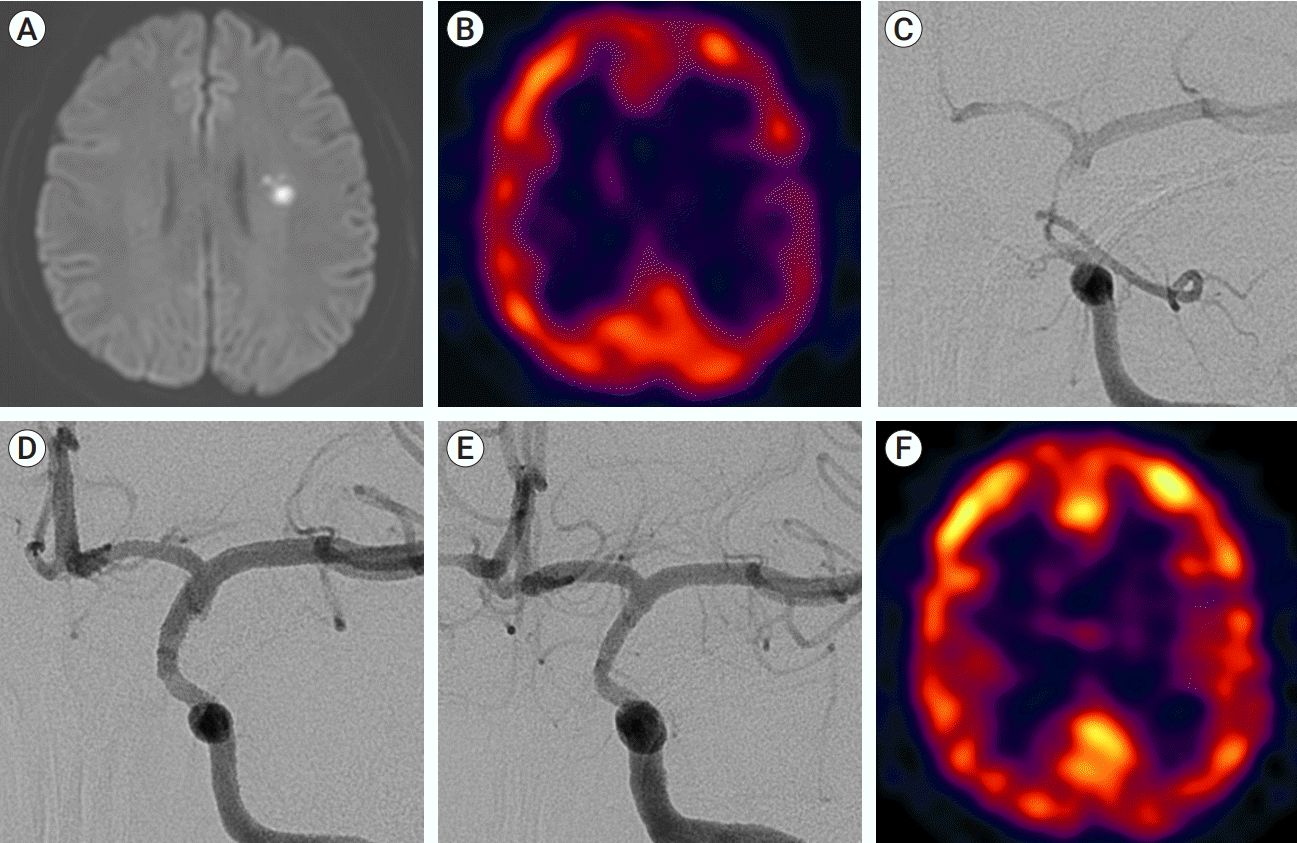
Table 1.
mRS, modified Rankin Scale; ISR, in-stent restenosis; TIA, transient ischemic attack; VBI, vertebrobasilar insufficiency; ICA, internal carotid artery; VAO, vertebral artery orifice; BA, basilar artery; V4, intradural segment of vertebral artery; CTA, computed tomography angiography; DSA, digital subtraction angiography
Table 2.
Study | Study population | Technical success rate | Periprocedural complication rate | Mean clinical follow-up duration (months) | Mean radiological follow-up duration (months) | Overall rate of ISR (≥50%) | Rate of symptomatic ISR |
---|---|---|---|---|---|---|---|
Abou-Chebl et al. (2005) [1] | Intracranial | 100.0 (8/8) | 25.0 (2/8) | 11.1 | 9.6 | 0.0 (0/8) | 0.0 (0/8) |
Boulos et al. (2005) [3] | Extra- and intracranial | 100.0 (19/19) | 0.0 (0/19) | NA | NA | NA | 7.1 (1/14) |
Gupta et al. (2006) [12] | Extra- and intracranial | 95.4 (62/65) | 3.2 (2/62) | 4.0 | 4.0 | 6.0 (3/50) | 2.0 (1/50) |
Qureshi et al. (2006) [24] | Intracranial | 85.7 (18/21) | 5.6 (1/18) | 14.0 | 6.0 | 14.3 (1/7) | 0.0 (0/7) |
Steinfort et al. (2007) [27] | Intracranial | 100.0 (13/13) | 23.1 (3/13) | 10.9 | 5.4 | 0.0 (0/9) | 0.0 (0/9) |
Natarajan et al. (2010) [22] | Intracranial | 100.0 (6/6) | 16.7 (1/6) | NA | NA | 0.0 (0/5) | 0.0 (0/5) |
Fields et al. (2011) [8] | Extra- and intracranial | 100.0 (27/27) | 11.1 (3/27) | 11.3 | NA | 27.3 (6/22) | 9.1 (2/22) |
Song et al. (2012) [26] | Extra- and intracranial | 98.3 (119/121) | 2.7 (3/121) | NA | NA | 6.3 (7/112) | 3.6 (4/112) |
Vajda et al. (2012) [28] | Intracranial | 93.4 (99/106) | 5.1 (5/99) | 16.1 | 16.1 | 3.8 (3/78) | 0.0 (0/78) |
Park et al. (2013) [23] | Intracranial | 100.0 (11/11) | 9.0 (1/11) | 67.0 | 55.0 | 0.0 (0/8) | 0.0 (0/8) |
Kurre et al. (2015) [16] | Intracranial | 85.5 (100/117) | 12.0 (10/83) | 11.7 | 11.7 | 3.6 (3/83) | 0.0 (0/83) |
Current study | Extra- and intracranial | 100.0 (21/21) | 0.0 (0/21) | 45.5 | 42.8 | 4.8 (1/21) | 0.0 (0/21) |