Abstract
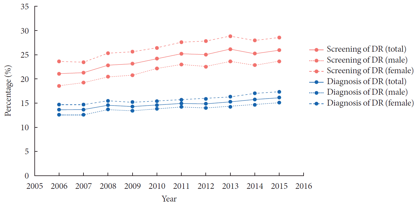
We performed a retrospective cohort study including people diagnosed with diabetes from 2006 to 2015 according to the Korean National Health Insurance Service-National Sample Cohort database, to analyze the changes in the prevalence, screening rate, and treatment patterns for diabetic retinopathy (DR) over 10 years. The proportion of people who underwent fundus screening for DR steadily increased over the past decade. The prevalence of DR increased from 13.4% in 2006 to 15.9% in 2015, while that of proliferative DR steadily decreased from 1.29% in 2006 to 1.16% in 2015. The proportion of patients undergoing retinal photocoagulation constantly decreased. The prevalence of DR increased over the past decade, while its severity seemed to have improved, with a decreased rate of proliferative DR and retinal photocoagulation. A higher proportion of patients underwent ophthalmic screening using fundus examination, but still less than 30% of patients with diabetes underwent comprehensive examination in 2015.
The prevalence of diabetes has increased annually, increasing from 8.6% in 2001 to 14.4% among adults ≥30 years in 2016 [123]. Based on the Diabetes Fact Sheet in Korea, 2018, there are still 37.4% of patients with diabetes who are not aware of their condition, with 9.2% of diagnosed patients remaining untreated [2].
Diabetic retinopathy (DR) is a major microvascular complication of diabetes, which occurs in approximately one-third of patients with diabetes worldwide, with an increased risk when associated with longer disease duration, elevated glycosylated hemoglobin (HbA1c), and the presence of hypertension [4]. Fundus examination is recommended at the time of diagnosis for patients with type 2 diabetes mellitus [5]. However, reportedly, only one-third of patients undergo fundus examinations [678].
Laser photocoagulation decreases the risk of moderate visual loss by 50% but is not effective to improve already impaired vision [9]. New treatment modalities such as intravitreal injections of an anti-vascular endothelial growth factor (VEGF) agent or corticosteroid implantation, have emerged in recent years [10]. Therefore, the aim of this study was to investigate the 10-year trend of DR and related clinical practice using the sample cohort data from the Korean Health Insurance Database.
We used the National Health Insurance Service-National Sample Cohort (NHIS-NSC) data from 2006 to 2015. The study protocol was reviewed and approved by the Institutional Review Board (IRB) of Ajou University Hospital, Suwon, Korea (IRB No. AJIRB-MED-EXP-19-347). Because the data in this database were de-identified, the requirement for informed consent was waived.
The list of variables with the corresponding International Classification of Diseases, 10th revision (ICD-10 codes), procedure codes, and Anatomical Therapeutic Chemical code is provided in Supplementary Table 1.
We identified patients with diabetes each year using the following criteria: (1) the presence of diagnostic codes for diabetes detected more than twice, or (2) a history of clinic visit and simultaneous prescription of glucose-lowering medications (for more than 30 days) and/or insulin. The occurrence of DR was defined based on the ICD-10 diagnostic codes. The crude prevalence rate was calculated as the number of cases per 100 people with diabetes. The age-adjusted prevalence rate was standardised against the weighted distribution of the people with diabetes studied in 2011 across sexes and 5-year age groups from 30 to 34 to 85 years and above.
The progression to proliferative DR (PDR) was defined as follows: (1) the presence of diagnostic codes of PDR; (2) the presence of diagnostic codes for DR with concomitant procedure codes for retinal laser photocoagulation; and (3) the presence of diagnostic codes for DR with concomitant operation codes for vitrectomy.
The screening for DR was defined by procedure codes for fundus examination, fundus photography, and/or fundus fluorescein angiography in that year. Laser treatment for DR included panretinal photocoagulation and focal/grid laser photocoagulation. Intravitreal and subconjunctival injections were also investigated with the following pharmacological agents: triamcinolone, dexamethasone implant, ranibizumab, and aflibercept. Intravitreal injection of bevacizumab was not investigated because of its off-label use in Korea.
All analyses were performed with SAS software version 9.4 (SAS Institute Inc., Cary, NC, USA). The prevalence of DR was calculated as the number of people with DR divided by the number of people diagnosed with diabetes in that year. The rates of ocular treatments were calculated as the number of persons receiving treatment by the number of those with DR.
The rate of DR screening and prevalence of DR steadily increased over time (Fig. 1). The mean screening rate increased from 20.6% in 2006 to 26.1% in 2015 but still remained below 30% (Supplementary Fig. 1A). The prevalence of DR increased from 13.4% in 2006 to 15.9% in 2015. Consistently more women presented with DR throughout the study period (Fig. 1). The maximum age was in the 60s until 2013 (15.9%), while age in 70s showed the highest prevalence from 2014 (18.0%) (Supplementary Fig. 1B). The rate of PDR among people with diabetes steadily decreased over time with consistently more men, from 1.29% in 2006 to 1.16% in 2015 (Supplementary Fig. 2).
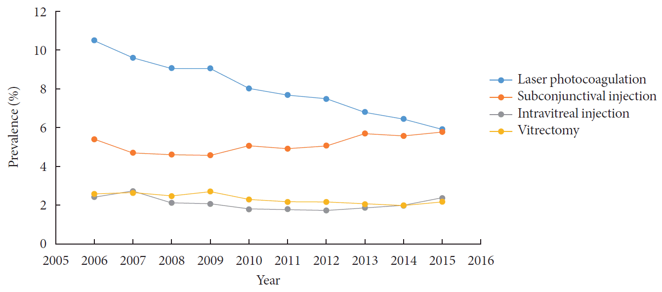
The proportion of patients who underwent laser treatment among those with DR constantly decreased over time (Fig. 2). The proportion of patients receiving subconjunctival injections slightly increased, while that of patients receiving intravitreal injections or vitrectomy was steady. None of the patients received ranibizumab or aflibercept among those who received anti-VEGF agents.
This retrospective study using NHIS-NSC of Korea showed that the prevalence of DR increased with a growing number of people undergoing fundus screening. Among patients with diabetes, only 26.1% underwent fundus examination, and the prevalence of DR was 15.9% in 2015.
A population-based study in Korea revealed a prevalence of 20% for DR in the 5th (2011, 2012) Korea National Health and Nutrition Examination Survey (KNHANES) [11]. In 2013, a Korean cohort study revealed that only 30% of patients with diabetes underwent comprehensive ocular examination [6]. Our results in the sample cohort were similar to those of Song et al. [6], who reported that less than 30% of patients with diabetes underwent DR screening in the nationwide cohort data. Fundus examination has not been included in the national health screening examination required by NHIS so far, and the inclusion would benefit in enhancing the screening of DR, which remains below 30%.
The severity of DR may have decreased over the past decade, along with a decreased rate of PDR. This may be partly because of the increased screening rate, i.e., earlier diagnosis of DR leading to proper treatment, as well as better glycemic control with newer antiglycemic agents. Approximately 36.7% of patients with diabetes reached the target of HbA1c < 7% in 2006 while 52.6% of patients reached the same level in 2013 to 2016 [21213]. This suggests that a better glycemic control has been reached over time, which has resulted in less aggravation of DR [14]. Another possible factor is the introduction of intravitreal anti-VEGF injections, reporting a low rate of DR worsening in patients treated with anti-VEGF agents [1516171819]. Bevacizumab is a globally used anti-VEGF agent for diabetic macular edema or complications of PDR [5]; however, intravitreal bevacizumab injections were not identified in this analysis, as this procedure is not covered by NHIS because of its off-label use in Korea. Moreover, the use of ranibizumab and aflibercept were not identified in this study, as they have been covered by NHIS since 2015 [20]. The effect of anti-VEGF agents in DR progression needs to be evaluated in further studies.
The decreased rate of laser photocoagulation may be associated with this lower severity of DR, which was similar to the results of Song et al. [6], who reported a decreased rate of laser treatment. This can be related to a decreased severity of DR while the introduction of intravitreal injections can also be responsible.
In conclusion, the proportion of patients undergoing ophthalmic screening was higher, but less than 30% of patients with diabetes underwent comprehensive examination in 2015. The inclusion of fundus examination in the national health screening protocol and/or provision of mandatory consultation to patients with diabetes by ophthalmologists might be helpful in promoting early detection of DR and/or undiagnosed diabetes.
ACKNOWLEDGMENTS
This study used NHIS Sample Cohort data (NHIS-2020-2-050) made by National Health Insurance Service (NHIS) of Korea. The authors declare no conflict of interest with NHIS.
Some of the data was released in October 2019 in the form of ‘Diabetes & Complications in Korea’ from the Korean Diabetes Association. We would like to thank the Committee of Public Relation, Korean Diabetes Association for helping with the work.
References
2. Kim BY, Won JC, Lee JH, Kim HS, Park JH, Ha KH, Won KC, Kim DJ, Park KS. Diabetes fact sheets in Korea, 2018: an appraisal of current status. Diabetes Metab J. 2019; 43:487–494.

3. Ko SH, Han K, Lee YH, Noh J, Park CY, Kim DJ, Jung CH, Lee KU, Ko KS. TaskForce Team for the Diabetes Fact Sheet of the Korean Diabetes Association. Past and current status of adult type 2 diabetes mellitus management in Korea: a National Health Insurance Service Database analysis. Diabetes Metab J. 2018; 42:93–100.

4. Yau JW, Rogers SL, Kawasaki R, Lamoureux EL, Kowalski JW, Bek T, Chen SJ, Dekker JM, Fletcher A, Grauslund J, Haffner S, Hamman RF, Ikram MK, Kayama T, Klein BE, Klein R, Krishnaiah S, Mayurasakorn K, O'Hare JP, Orchard TJ, Porta M, Rema M, Roy MS, Sharma T, Shaw J, Taylor H, Tielsch JM, Varma R, Wang JJ, Wang N, West S, Xu L, Yasuda M, Zhang X, Mitchell P, Wong TY. Meta-Analysis for Eye Disease (META-EYE) Study Group. Global prevalence and major risk factors of diabetic retinopathy. Diabetes Care. 2012; 35:556–564.

5. Stewart MW. Treatment of diabetic retinopathy: recent advances and unresolved challenges. World J Diabetes. 2016; 7:333–341.

6. Song SJ, Han K, Choi KS, Ko SH, Rhee EJ, Park CY, Park JY, Lee KU, Ko KS. Task Force Team for Diabetes Fact Sheet of the Korean Diabetes Association. Trends in diabetic retinopathy and related medical practices among type 2 diabetes patients: results from the National Insurance Service Survey 2006-2013. J Diabetes Investig. 2018; 9:173–178.

7. Lopez IM, Diez A, Velilla S, Rueda A, Alvarez A, Pastor CJ. Prevalence of diabetic retinopathy and eye care in a rural area of Spain. Ophthalmic Epidemiol. 2002; 9:205–214.
8. Sivaprasad S, Gupta B, Crosby-Nwaobi R, Evans J. Prevalence of diabetic retinopathy in various ethnic groups: a worldwide perspective. Surv Ophthalmol. 2012; 57:347–370.

9. Early Treatment Diabetic Retinopathy Study Research Group. Treatment techniques and clinical guidelines for photocoagulation of diabetic macular edema. Early Treatment Diabetic Retinopathy Study Report Number 2. Ophthalmology. 1987; 94:761–774.
10. Stitt AW, Curtis TM, Chen M, Medina RJ, McKay GJ, Jenkins A, Gardiner TA, Lyons TJ, Hammes HP, Simo R, Lois N. The progress in understanding and treatment of diabetic retinopathy. Prog Retin Eye Res. 2016; 51:156–186.

11. Lee WJ, Sobrin L, Lee MJ, Kang MH, Seong M, Cho H. The relationship between diabetic retinopathy and diabetic nephropathy in a population-based study in Korea (KNHANES V-2, 3). Invest Ophthalmol Vis Sci. 2014; 55:6547–6553.

12. Lim S, Kim DJ, Jeong IK, Son HS, Chung CH, Koh G, Lee DH, Won KC, Park JH, Park TS, Ahn J, Kim J, Park KG, Ko SH, Ahn YB, Lee I. A nationwide survey about the current status of glycemic control and complications in diabetic patients in 2006: the Committee of the Korean Diabetes Association on the Epidemiology of Diabetes Mellitus. Korean Diabetes J. 2009; 33:48–57.
13. Kim JH, Kim DJ, Jang HC, Choi SH. Epidemiology of micro- and macrovascular complications of type 2 diabetes in Korea. Diabetes Metab J. 2011; 35:571–577.

14. Chew EY, Davis MD, Danis RP, Lovato JF, Perdue LH, Greven C, Genuth S, Goff DC, Leiter LA, Ismail-Beigi F, Ambrosius WT. Action to Control Cardiovascular Risk in Diabetes Eye Study Research Group. The effects of medical management on the progression of diabetic retinopathy in persons with type 2 diabetes: the Action to Control Cardiovascular Risk in Diabetes (ACCORD) Eye Study. Ophthalmology. 2014; 121:2443–2451.
15. Bressler SB, Liu D, Glassman AR, Blodi BA, Castellarin AA, Jampol LM, Kaufman PL, Melia M, Singh H, Wells JA. Diabetic Retinopathy Clinical Research Network. Change in diabetic retinopathy through 2 years: secondary analysis of a randomized clinical trial comparing aflibercept, bevacizumab, and ranibizumab. JAMA Ophthalmol. 2017; 135:558–568.
16. Ip MS, Domalpally A, Hopkins JJ, Wong P, Ehrlich JS. Long-term effects of ranibizumab on diabetic retinopathy severity and progression. Arch Ophthalmol. 2012; 130:1145–1152.

17. Singh RP, Elman MJ, Singh SK, Fung AE, Stoilov I. Advances in the treatment of diabetic retinopathy. J Diabetes Complications. 2019; 33:107417.

18. Wykoff CC, Eichenbaum DA, Roth DB, Hill L, Fung AE, Haskova Z. Ranibizumab induces regression of diabetic retinopathy in most patients at high risk of progression to proliferative diabetic retinopathy. Ophthalmol Retina. 2018; 2:997–1009.

19. Mitchell P, McAllister I, Larsen M, Staurenghi G, Korobelnik JF, Boyer DS, Do DV, Brown DM, Katz TA, Berliner A, Vitti R, Zeitz O, Metzig C, Lu C, Holz FG. Evaluating the impact of intravitreal aflibercept on diabetic retinopathy progression in the VIVID-DME and VISTA-DME studies. Ophthalmol Retina. 2018; 2:988–996.

SUPPLEMENTARY MATERIALS
Supplementary materials related to this article can be found online at https://doi.org/10.4093/dmj.2020.0096.
Supplementary Table 1
List of diagnoses, treatments, procedures, and their corresponding codes
Supplementary Fig. 1
Screening rate (A) and prevalence (B) of diabetic retinopathy (DR) among patients with diabetes stratified by the age group in 2006 to 2015.
Supplementary Fig. 2
Prevalence of proliferative diabetic retinopathy (DR) stratified by sex in 2006 to 2015.


 PDF
PDF Citation
 Print
Print


 XML Download