

 PDF
PDF Citation
 Print
Print


서론
석회힘줄염(calcific tendinitis)은 건과 연부 조직에 석회가 침착되어 통증이 유발되는 질환으로 일반적으로 단일 관절을 침범한다. 특히 30세에서 60세 사이에서 잘 발생되고 특별한 외상 없이 젊은 여자에서 갑자기 발생하는 경우가 좀더 흔하며, 가장 흔하게 침범되는 부위는 견관절의 회전근개(rotator cuff)이지만 다른 부위에서도 발생된다고 보고되고 있다[1,2]. 이 석회화 과정은 독특하고 퇴행성 관절 질환과는 다르며 무증상인 경우가 많다. 석회힘줄염에서 발생하는 석회 침착의 과정은 칼슘의 형성과 용해의 진전 상태에 따라 석회화 전 단계(precalcific), 석회화 단계(calcific) 및 석회화 후 단계(postcalcific)의 세 단계로 분류된다. 석화화 단계는 세부적으로 형성기(formative phase), 휴지기(resting phase) 및 용해기(resorptive phase)로 나뉘며 독특한 형태의 방사선학적 및 병리학적 형태를 갖는데, 임상적인 증상과 연관되는 경우도 있다. 형성기에는 섬유연골세포 간질(fibrocartilagenous matrix)에 석회 결정이 침착하며, 이 시기의 석회는 분필 가루와 같은(chalk-like) 모습을 띠게 된다. 석회 침착의 반응이 없는 휴지기를 지나면 탐식 세포 및 다핵 거대세포들이 석회를 흡수하는 용해기 단계를 거치는데, 이 때의 석회 성상은 마치 치약과 같은(toothpaste-like) 형태를 보이며 통증이 심해지고 운동 범위가 줄어들 수 있다[1,3].
주관절 주위의 석회힘줄염에 대한 보고는 매우 드물며[4], 수술에 대한 결과를 보고한 경우는 거의 없다. 석회힘줄염의 치료에 대해서는 보존적 치료를 주로 하지만, 보존적 요법에 실패했을 경우에 수술적 치료가 필요하다고 하였다[5,6]. 저자들은 제주대학교병원 정형외과에서 2011년 11월부터 2018년 7월까지 주관절 주위의 통증으로 내원하여 방사선 사진 및 초음파 검사상 석회성 병변이 확인되고 보존적 치료 후 증상이 지속되어 수술적 치료를 시행하였던 환자에 대하여 결과를 분석하고 문헌 고찰과 함께 보고하고자 한다.
Go to : 
대상 및 방법
이 연구는 제주대학교병원 의학연구윤리심의위원회(Institutional Review Board)의 승인을 받아 후향적으로 시행되었고(No. 2020-09-009), 환자로부터의 연구동의서는 면제되었다. 2011년 11월부터 2018년 7월까지 주관절 주위의 통증으로 내원하여 방사선 사진 및 초음파 검사상 석회성 병변이 확인되고 수술적 치료를 시행하였던 7예를 대상으로 후향적으로 연구하였다. 모든 예가 여자였으며, 평균 연령은 40세(범위, 26–49세)였고, 석회 병변의 위치는 우측 및 외측이 6예로 가장 많았다. 추시 기간은 최단 2년에서 최장 8년 9개월로 평균 5년 10개월이었다(Table 1). 석회성 병변의 원인은 환자의 병력, 혈액 검사 및 영상 검사에서 특별히 찾을 수 없어 모든 예에서 특발성으로 판단하였다. 주관절 주위의 석회힘줄염을 진단받은 경우 먼저 생활양식의 변화, 비스테로이드성 소염제 사용을 통해 통증 조절 및 석회성 병변의 감소 등을 유도하는 보존적 치료를 시행하였다. 초기에 보존적 치료에도 불구하고 통증이 지속될 경우 통증 부위의 국소 주사, 체외 충격파 치료(extracorporeal shockwave therapy) 등의 보다 적극적인 치료를 시행하여 통증 감소 및 기능 향상을 유도하였다. 구체적으로 스테로이드 국소 주사는 한 차례만 우선적으로 시행하였다. 스테로이드는 트리암시놀론(triamcinolone acetonide, 40 mg/mL)을 리도카인(lidocaine, 0.1 mL)과 혼합하여 주사하였고, 한 번의 주사에도 통증의 호전이 없는 경우, 추가적인 치료를 원하는 경우, 그외 필요한 경우에 4–5회 정도를 1세트로 하는 체외 충격파 치료를 1주일에 1회씩 4-5회 정도를 1세트로 하여 치료하였다. 스테로이드 국소 주사는 최소 3개월에서 4개월의 간격을 두고 시행하여 합병증 발생을 줄이려고 노력하였으며, 체외 충격파 치료는 세트 사이에 1개월 정도의 간격을 두어 근육의 약화를 방지하려고 하였다. 적극적인 보존적 치료에도 반응하지 않고 통증이 지속되며 방사선 사진상 석회성 병변이 소실되지 않을 경우 수술적 치료를 시행하였으며, 석회힘줄염을 진단받은 후 수술까지의 시간은 평균 6.5개월(범위, 4–9개월)이었다(Table 1).
Table 1.
Overall data of the present study
![]()
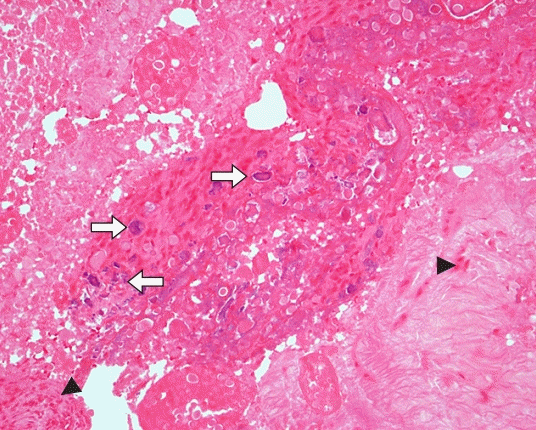
수술 방법은 상박신경총 차단 마취 하에 상완골 외상과(또는 내상과) 약 1 cm 원위부에서 피부선을 따라 약 5 cm의 종 절개를 가하여 노출시킨 후, 공통 수근 신전 건(또는 공통 수근 굴곡 건) 근막 절개술을 시행하였다. 모든 증례에서 수술 소견상 공통 수근 신전 건(또는 공통 수근 굴곡 건)에 부분 파열 소견과 파열된 건의 안쪽에 석회 성분으로 보이는 백색 물질이 축적되어 있었다(Fig. 1). 석회 침착물을 제거하여 조직 검사를 하였으며, 파열된 건의 변연부를 절제하고 부분 파열된 공통 수근 신전 건(또는 공통 수근 굴곡 건)을 비흡수성 봉합사(Prolene; Ethicon, Cincinnati, OH, USA)를 이용하여 단단히 봉합하였다. 수술 후 전완부를 중립 위치로 장상지 석고 부목(splint)을 2주간 유지하였다가 제거하고 수동 운동 기계(continuous passive movement)를 이용한 관절 운동을 시작하였으며, 주관절의 관절 운동 제한이 회복된 3주째부터 물리치료실에서 능동적 보조 운동 및 점진적 저항 운동을 허용하여 근력을 강화시켰다. 스포츠 활동 및 작업 등은 약 3개월째부터 허용하였다. 모든 예에서 조직 검사상 석회힘줄염을 확진하였다(Fig. 2).
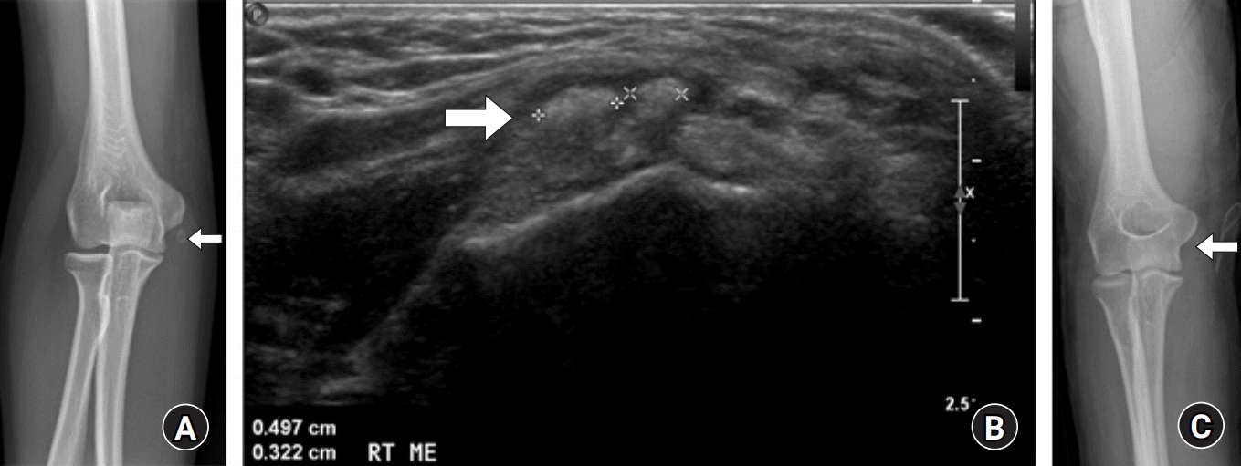
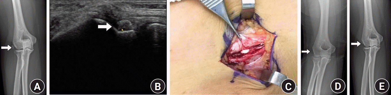 | Fig. 1.A 36-year-old female patient with calcific tendinitis in the lateral epicondylar area on the right elbow. (A) Preoperative anteroposterior radiograph shows small rounded calcific deposit at the insertion of the common extensor tendon (arrow). (B) Sonographic image revealed swelling and calcification at the insertion of the common extensor tendon (arrow). (C) Intraoperative photograph indicates toothpaste-like calcium deposits evident around the insertion site of the common extensor tendon. (D, E) Anteroposterior radiographs on immediate postoperative and the last follow-up period show complete resolution of calcific deposits without recurrence at the insertion site of the common extensor tendon (arrow).
|
수술 후 추시에서 주관절은 통증 없이 정상 관절 운동이 가능하였고 일상생활 및 스포츠 활동 등에 제한이 없었으며, 증상의 재발은 없었다(Figs. 3–5).
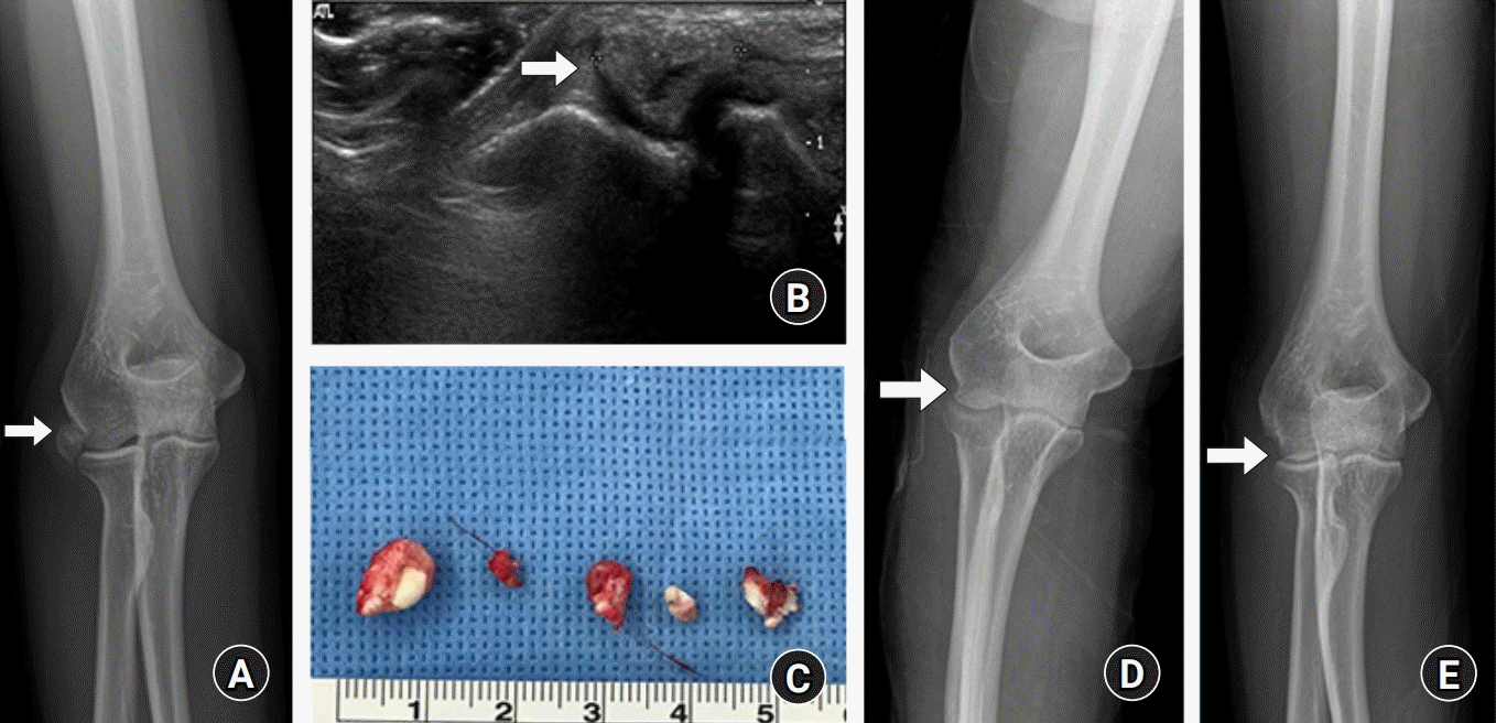 | Fig. 3.A 43-year-old female patient with calcific tendinitis on the lateral aspect of right elbow joint. (A) Preoperative anteroposterior radiograph shows a large rounded calcific deposit at the deep portion of the insertion of the common extensor tendon (arrow). (B) Sonographic image shows calcification at the insertion of the common extensor tendon and in contact with humeral cortex (arrow). (C) Intraoperative image indicates multiple calcific deposits after excision. (D, E) Anteroposterior radiographs on immediate postoperative and the last follow-up period show complete removal of calcific deposits without recurrence at the insertion of the common extensor tendon (arrow).
|
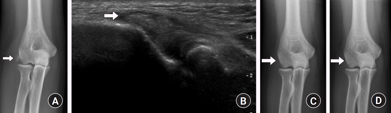 | Fig. 4.A 26-year-old female patient with calcific tendinitis in the lateral epicondylar area on the right elbow. (A) Preoperative anteroposterior radiograph shows small rounded calcific deposit at the insertion of the common extensor tendon (arrow). (B) Sonographic image revealed swelling and calcification at the insertion of the common extensor tendon (arrow). (C, D) Anteroposterior radiographs on immediate postoperative and the last follow-up period show complete resolution of calcific deposits without recurrence at the insertion site of the common extensor tendon (arrow).
|
 | Fig. 5.A 48-year-old female patient with calcific tendinitis under the medial epicondylar area on the right elbow. (A) Preoperative anteroposterior radiograph shows small rounded calcific deposit at the insertion of the common flexor tendon (arrow). (B) Sonographic image revealed swelling and calcification at the insertion of the common flexor tendon (arrow). (C) Anteroposterior radiograph on immediate postoperative day shows complete resolution of calcific deposits at the insertion site of the common extensor tendon (arrow).
|
수술의 결과는 환자의 주관적인 만족도를 5단계(매우 만족, 만족, 보통, 불만족, 매우 불만족)로 나누어 평가하였고, 수술 후 주관절 관절 운동, 안전성 및 손과 팔 등의 기능적 평가를 위해 Mayo Elbow Performance Index (MEPI)와 Disabilities of the Arm, Shoulder and Hand (DASH)를 조사하였다. MEPI 점수는 통증(45점), 운동 범위(20점), 안정성(10점), 일상 기능(25점)을 종합한 점수를 산출하여 매우 만족(excellent, 90–100점), 양호(good, 75–89점), 보통(fair, 60–74점), 불량(poor, <60점)으로 나누어 주관절의 기능을 평가하는 방법이다[7].
통계적 분석은 IBM SPSS Statistics, ver. 24.0 프로그램(IBM Corp., Armonk, NY, USA)을 이용하여 수술 전과 최종 추시 MEPI 점수, 수술 전과 최종 추시 DASH 점수를 Wilcoxon 부호-순위 검정(Wilcoxon signed-rank test)을 시행하여 p<0.05일 때 통계적으로 유의한 것으로 평가하였다.
Go to :
결과
수술 후 건 봉합 부위의 재파열이 있던 경우는 한 예도 없었으며, 감염이나 수술 부위 감각 이상 등의 합병증도 없었다.
환자의 주관적인 만족도는 3예에서 매우 만족이었으며, 4예에서는 만족의 결과로 환자들은 수술 후 전반적으로 만족한다고 하였다(Table 1). 수술 전 MEPI 점수는 중앙값 45점(범위, 30–65점; 불량 또는 보통)이었으나, 수술 후 최종 추시 시의 MEPI 점수는 중앙값 85점(범위, 85–100점; 양호 또는 매우 만족)으로 증가된 소견을 보였다. 수술 전 DASH 점수는 중앙값 88.8점(범위, 81.9–94.8점)이었으나, 수술 후 최종 추시 시의 DASH 점수는 중앙값 36.2점(범위, 31.9–38.8점)으로 호전된 소견을 보였다. 수술 전과 최종 추시 MEPI 점수와 DASH 점수를 Wilcoxon 부호-순위 검정한 결과, 유의수준 5% 미만으로 수술 전후의 차이가 있음을 확인할 수 있어 통계적으로 유의한 것으로 평가하였다(Table 2).
Go to :
고찰
석회힘줄염은 흔한 질환으로 수산화 인회석 칼슘 결정(calcium hydroxyapatite crystal)이 건 조직에 병적으로 침착되어 통증이 유발되는 질환으로 견관절의 회전근개(rotator cuff)에서 가장 많이 발생하며, 고관절, 수근 관절, 주관절, 슬관절, 족부 및 경부에서도 발생한다고 보고되고 있다[8]. 석회 침착의 기전은 아직 명확하게 규명되지 않았으나, 건과 연부 조직 내에 국소적인 저산소증 후에 화생(metaplasia)이 발생하며 섬유 연골이 형성되고, 염증성 세포에 둘러싸인 이영양성 석회 침착이 발생된다고 하였다[9]. 석회성 병변은 국소 스테로이드 주사로 인해 발생할 수도 있는데, 스테로이드 주사로 인한 석회화는 방사선 소견상 표재성의 석회화와 소금(salt grains)을 뿌려 놓은 것 같은 양상을 보이며 주사 바늘의 길(track)을 따라 나타나고, 주로 불용해성(insoluble) 또는 저용해성(low solubility)의 스테로이드에서 발생한다고 하였고, 조직학적으로 이영양성 석회 침착, 반흔 형성 및 만성 육아종성 반응을 보인다고 하였다[10]. 본 연구에서는 단순 방사선 사진상 주관절 외상과 또는 내상과의 공통 수근 신전 건이나 공통 굴곡 건 부착 부위에 방사선 비투과성 석회성 병변의 경계가 비교적 명확하고 균일하며, 조직 검사상 염증세포 침윤 및 미세 석회화만 있는 것으로 확인되어 스테로이드 국소 주사로 인한 석회화는 배제하였다. 석회힘줄염의 특징적인 증상은 통증, 국소 압통, 부종 및 발적 등과 같이 급성 염증성 반응으로 인한 것이지만, 혈액 검사에서 급성 염증 반응을 보이는 수치(백혈구, 적혈구 침강 속도, C 반응 단백)가 정상 소견을 보이므로 다른 염증성 질환들과 감별할 수 있다고 하였다[11].
석회힘줄염은 임상 증상과 함께 방사선 검사로 진단이 가능한데, 단순 방사선 사진은 가장 실용적인 평가 방법으로 석회 병변의 유무를 파악할 수 있을 뿐 아니라 석회 침착물의 양, 범위 및 밀도 등을 평가하는 데 있어서도 유용하며 비용 효과적이다[12,13]. 컴퓨터단층촬영(computed tomography)은 골성 침범을 평가하는 데 가장 좋은 방법이며, 특히 골성 미란(osseous erosion)이 있을 때 유용하다[14]. 비정상적인 위치의 석회화를 알아내는 데 도움을 주어 건 내 석회화를 진단할 수 있으며, 석회 침착물의 밀도 평가에도 가장 정확한 방법이다[15,16]. 초음파는 석회힘줄염의 진단에 좋은 방법이며, 실시간으로 보여지는 특성 때문에 치료적인 방법에서도 이용할 수 있다[15]. 초음파로는 석회 침착의 과정을 구분하기는 어렵기 때문에, 방사선 사진과 함께 이용한다면 석회힘줄염의 진단에 유용한 방법이 될 수 있다고 하였다[17]. 자기공명영상(magnetic resonance imaging) 검사는 연부 조직 이상의 정도를 파악할 수 있고 다른 관절 통증의 원인을 배제할 수 있기 때문에 석회힘줄염을 진단하는 데 중요하다[18]. 본 연구에서는 단순 방사선 사진상 주관절 외상과 또는 내상과의 공통 수근 신전 건이나 공통 굴곡 건 부착 부위의 방사선 비투과성 석회화 병변이 경계가 비교적 명확하며 균일한 소견이고 골성 침범 소견은 뚜렷하지 않아 추가 검사로 초음파를 시행하였고, 이를 통해 주관절 주위의 건 상태와 석회 침착물의 성상 및 모양을 확인하여 석회힘줄염을 진단할 수 있었다.
석회힘줄염에 대한 보존적 치료는 휴식, 비스테로이드성 소염제, 물리치료, 스테로이드 국소 주사 등이 있다. 치료 방법은 질환의 임상 증상 정도에 따라서 결정되며, 석회 침착물은 보통 단순 방사선 사진상 1, 2주가 지나면 자연스럽게 흡수된다는 보고도 있지만 증상의 발현 후 4주에서 8개월 사이에 소실된다는 보고도 있어 석회 침착물의 위치나 특성에 따라 다른 것으로 판단된다[19,20]. Uhthoff와 Loehr [1]는 석회함쥴염의 특성상 석회 침착물은 저절로 흡수될 수도 있는 것이라고 보존적 치료를 권하였다. Brinsden과 Wilson [6]은 족부의 장비골근(peroneus longus)에 발생한 석회힘줄염에서 비스테로이드성 소염제와 휴식으로 조기에 임상적인 증상의 완화를 보였다고 하였고, Cox와 Paterson [8]은 석회성 병변 주위에 스테로이드 국소 주사를 이용하여 즉각적인 증상의 소실을 보였다고 하였다. 스테로이드 국소 주사가 석회힘줄염에 효과적일 수도 있지만, 건 파열, 주사 후 통증, 피하 위축(subcutaneous atrophy), 피부 탈색(skin pigmentation) 등의 부작용이 나타날 수 있기 때문에 반복적인 주사는 피해야 한다. 다른 보존적 치료 방법으로는 체외 충격파 치료와 바늘 흡인(needle aspiration) 등의 방법이 있으며, 치료 효과는 다양하게 보고된다[15,21]. 석회힘줄염은 일반적으로 보존적 치료에 잘 반응하는 것으로 알려져 있기는 하지만, 본 연구에서 석회힘줄염을 진단받은 환자의 경우에는 적극적인 보존적 치료에도 불구하고 증상이 지속되거나 심해져 수술을 시행하게 되었다. 보존적 치료를 시작하고 시간이 지나면서 석회 침착물이 흡수되는 과정이 본 연구에서는 다른 증례처럼 나타나지 않았는데, 보통 스테로이드 국소 주사나 체외 충격파 치료를 시행하면 석회성 병변이 작아지면서 흡수되는 과정이 그 동안의 보고였지만 본 연구에서는 병변이 그대로 유지되고 통증 또한 비슷하게 지속되었다. 이러한 이유는 주관절 부위의 석회성 병변의 밀도가 다른 관절보다 높고 주관절 부위에 병변이 발생할 경우 다른 관절보다 흡수될 공간이 협소하여 보존적 치료에 잘 반응하지 않는 것으로 저자들은 판단하였다. Kim 등[22]은 석회힘줄염의 보존적 치료의 효과적인 기간에 대해서 적어도 6개월 정도의 치료 기간을 시도하는 것을 추천한다고 하였다. 일반적으로 보존적 치료 방법으로 증상의 호전을 볼 수 있지만, 2–3회의 스테로이드 주사 치료, 체외 충격파 및 적극적인 운동 치료를 포함한 보존적 치료에도 불구하고 증상이 심해지거나 통증이 일상생활을 장기간 방해할 때는 건 내의 석회 침착물의 수술적 제거를 고려할 수 있다.
석회힘줄염에서 석회 침착물에 대한 수술적 제거의 필요성에 대해서는 여러 이견이 있으며, 문헌상에서도 아직 최선의 치료에 대한 명확한 기준 및 보고가 없다. 하지만 보존적 치료는 수술적 치료에 비해 치료 기간이 길고 재발이 발생할 수 있다는 단점이 있으며, 보존적 치료를 먼저 시행하여 이에 반응이 없는 환자에 한하여 선택적으로 수술을 시행하는 경우에도 수술을 받을 때까지 환자의 통증을 조절하는 보존적 치료의 연장 및 운동 제한이 발생하는 문제가 나타날 수 있다[23]. 이에 최근의 연구에서는 조기에 적극적인 치료를 시행하여 일상으로 빨리 복귀할 수 있는 방법을 권장하고 있다. 견관절에서의 석회힘줄염의 치료에 대한 보고이기는 하지만 Kim 등[24]은 4주 이상의 보존적 치료에도 통증이 지속 및 악화되어 일상생활에 지장을 초래하는 경우에 조기의 수술적 치료로 석회 침착물을 제거하면, 통증이 빠르게 감소하고 기능 회복이 우수하여 환자의 만족도가 높고 일상생활로 빠르게 복귀할 수 있었다고 하였다. 본 연구에서도 적어도 4개월 이상의 적극적인 보존적 치료에도 불구하고 통증이 지속되고 관절 운동 제한으로 인해 일상생활에서의 기능이 심하게 제한된다고 판단되어 수술적 치료를 통하여 좋은 결과를 얻을 수 있었다.
본 연구는 증례의 수가 적고 후향적 연구라는 점, 수술한 그룹과 비교할 수 있는 대조군이 없어 결과를 비교할 수 없었던 점이 제한점이라고 할 수 있다.
Go to :
 XML Download