

 PDF
PDF Citation
 Print
Print


서론
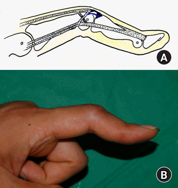
수부 신전건 중 중앙 건(central slip) 손상은 치료를 요하는 수부 외상의 2% 정도로 알려져 있다[1]. 이러한 중앙 건의 부착 부분인 중위지골 배측 기저 부분이 약화 혹은 손상되면, 근위 지관절(proximal interphalangeal joint)은 신전 지연(extension lag) 혹은 제한이 되고, 원위 지관절(distal interphalangeal joint)은 과신전을 보이는 ‘단추구멍 수지 변형(boutonniere or buttonhole deformity)’이 발생할 수 있다. 최초 손상 시에는 근위 지관절의 신전 제한 소견만 보이지만, 시간이 경과함에 따라 측부대(lateral band)가 근위 지관절의 수장 측으로 전위되어 중앙 건과 측부대의 길이 균형이 깨지게 되고, 전방으로 전위된 측부인대의 변형된 당김 기전으로 원위 지관절이 과신전되면서 단추구멍 변형이 초래된다[2,3] (Fig. 1).
이러한 변형은 주로 앞서 설명한 중앙 건의 직접적인 열상 혹은 폐쇄성 외상 이후 시간이 경과하면서 발생할 수 있다. 수상 당시 건손상을 간과하였거나, 봉합을 시행했음에도 적절한 봉합이 이루어지지 않아 변형이 나타나게 되는 경우가 많다. 이러한 외상성 중앙 건 손상 외에도 류마티스 관절염, 골관절염, Dupuytren 구축, 활차 손상, 화상 등에 의해서도 발생할 수 있다[4].
단추구멍 변형은 수부외과 의사가 경험할 수 있는 증례가 제한적이고, 현재까지도 치료에 대한 명확한 지침이 없어, 치료하는 의사의 지식과 경험에 의존해 치료가 결정되는 경향이 있다. 본 종설에서는 단추구멍 변형의 치료를 결정할 때 고려해야 할 요소들을 논의하고, 비수술적 혹은 수술적 치료에 대해 고찰하고자 한다.
Go to : 
단추구멍 변형의 치료에서 고려 사항
1. 급성 혹은 만성 변형
중앙 건의 단독 손상만으로는 단추구멍 변형이 발생하지 않는다[5,6]. Grau 등[6]은 18구의 사체 실험 연구를 통해 단순 중앙 건의 손상은 근위 지관절의 신전 지연만 보일 뿐이며, 삼각인대(triangular ligament)와 가로 및 사선 망상 인대(transverse and oblique retinaculum ligament)까지 절제해야 측부대가 수장 측으로 전위되면서 단추구멍 변형이 발생함을 증명하였다. Grundberg와 Reagan [5]은 만성 추지 변형에서 근위 지관절 배측에서 중앙 건을 절제하였을 때, 원위 지관절의 추지 변형은 호전되고, 근위 지관절은 단추구멍 변형 없이 2도 미만의 신전 지연만이 있었다고 보고했다. 최초 중앙 건에 손상이 발생하면 배측에 측부대를 지지해 주는 구조물에 장력이 커지고, 점차적으로 삼각인대나 사선 망상 인대가 기능을 상실한다. 그리하여 Clavero 등[7]은 중앙 건 손상 이후 7일에서 14일까지는 단추구멍 변형이 뚜렷하지 않을 수 있다고 기술하였다.
이렇게 중앙 건 손상 이후 단추구멍 변형의 발생 시점이 명확하지 않음에도 급성과 만성 변형으로 나누는 이유는 그 치료와 예후가 다르기 때문이다. 급성과 만성을 구분하는 시점에 대한 명확한 기준은 없으나, Posner와 Green [8]은 수상 이후 2주까지를 급성, 2주에서 8주까지는 아급성, 8주 이상을 만성으로 분류하였다. 급성 변형의 경우, 창상이 있거나 중위 지골 배측 기저부 골편을 동반한 폐쇄성 견열 골절인 경우를 제외하고는 근위 지관절 최대 신전 위치에서 4, 5주간 고정하는 보존적 치료를 권고하였고, 이 경우는 예후가 양호하다[9-11]. 대부분의 아급성 변형은 수동적 정복이 가능하며 관절 자체는 유연하다. 이러한 아급성 변형의 치료도 급성과 유사하나, 8주 미만의 손상이라 하더라도 관절이 수동적으로 정복이 되지 않고 구축되었을 경우 만성에 준해서 치료한다. 대체로 좋은 치료 결과를 보이는 급성 및 아급성과 다르게 만성의 경우 중앙 건 외에 다른 해부학적 요소들을 고려해야 하며, 치료 방법과 결과 역시 다양하다[4]. 만성의 경우 삼각인대의 기능이 소실되어 측부대가 수장 측으로 전위되어 있으며, 이차적으로 가로 망상 인대와 사선 망상 인대가 구축에 의해 고정되어 있다[8]. 이러한 만성 단추구멍 변형은 다음에 소개할 변형의 단계에 따라 치료 방향을 달리한다.
2. 변형의 단계
만성 단추구멍 변형에서 Burton은 3개의 단계를 제시했고, Green’s Operative Hand Surgery 7판에서는 근위 지관절 관절염이 있는 경우까지 총 4단계로 분류하였다[10] (Table 1).
Table 1.
Stages of boutonniere deformity by Burton [10]
![]()
만성 단추구멍 변형일지라도 Burton stage I, II 혹은 심지어 stage III 역시 보존적 치료를 먼저 권한다. 근위 지관절을 최대 신전 위치에서 6주에서 12주 가량 casting, splinting, 혹은 dynamic splint를 이용하여 고정시키는데, 이때 원위 지관절은 고정하지 않고 능동적 혹은 수동적 운동을 허용한다[10-12]. 근위 지관절의 수동적 신전이 제한되는 3단계의 경우 단순한 신전 기전 회복을 위한 건 수술로는 한계가 있으며, 수술 전 보존적 치료를 통한 근위 지관절 수동 관절 범위를 회복한 뒤 건 수술을 시행하거나, 근위 지관절 관절 유리 수술을 건 수술과 함께 시행해야 한다[10,12]. 만약 퇴행성 관절염이 동반된 4단계라면, 건의 신전 기전 재조정(rebalancing) 수술과 동시에 관절에 대하여 관절 치환술 혹은 관절 유합술을 함께 시행하는 것을 고려해야 한다.
Bellemère [11]는 Burton의 분류에 더해 Strauch에 의해 수정된 단추구멍 변형 단계를 제안하였고, 각 단계에 따른 치료 방법을 제시하였다(Table 2). 각각의 단계에서 근위 지관절의 신전 제한 기준을 30도 이하인 경우 A로, 이상의 경우 B로 나누었으며, 단계에 따른 다른 수술적 치료를 권고하였다.
Table 2.
Stage of boutonniere deformity and recommended surgical treatment with failed conservative management
PIP, proximal interphalangeal joint; DIP, distal interphalangeal joint; CS, central slip; LB, lateral band.
Adapted from Bellemère [11].
![]()
3. 변형에 따른 기능적 장애 및 환자 요소
단추구멍 변형은 손가락 굴곡에 영향을 미칠 정도의 변형이 발생하지 않는 한, 근위 지관절 신전 제한으로 육안으로 보이는 변형 이외에는 기능적 제한 혹은 통증과 같은 임상적으로 유의한 증상은 크지 않다[12]. 그래서 경도의 변형은 단순 외형적인 이유로 병원을 찾게 된다[4]. 기능적 장애를 호소하는 단추구멍 변형에서도, 근위 지관절 구축에도 불구하고 많은 환자들은 오히려 원위 지관절을 구부릴 수 없어 불편감을 호소한다[10]. 이러한 원위 지관절의 굴곡을 해결하기 위해 만성 단추구멍 변형에서 종말 건(terminal slip) 절제술이 수술적 치료의 한 가지 방법이 된다[13,14].
대체로 단추구멍 변형에서는 보존적 치료를 권고하며, 근위 지관절의 30도 미만 신전 장애는 수술적 치료로 교정하기 힘들다[10]. 수술적 치료를 시행한 이후에도 관절 고정 및 재활 운동에 대한 수술 후 관리가 중요하며, 변형의 재발 혹은 근위 지관절 굴곡 제한 등 좋지 않은 결과를 가져올 수 있다. 따라서 수술적 치료 결정 전에 충분한 설명을 통해 환자 스스로 자신의 상태에 대하여 이해할 수 있게 하고, 특히 수술 결과 및 수술 후 발생 가능한 합병증에 대해서 명확히 인지시켜야 한다. 수술 이후 치료 종결 전까지 재활을 포함한 환자의 적극적인 협조가 중요하다[12].
4. 외상 외 원인에 의한 변형
외상에 의한 중앙 건 손상으로 발생하는 변형 외에도 류마티스 관절염, 골관절염, Dupuytren 구축, 활차 손상, 화상 등에 의해서도 변형이 발생할 수 있기 때문에, 치료를 선택하기 전에 정확한 변형의 원인을 파악하는 것 또한 치료 방향 결정 및 예후를 예측하는 데 중요하다.
먼저 외관적으로 비슷해 보이는 가성 단추구멍 변형(pseudo boutonniere deformity)과 감별이 필요하다. 가성 단추구멍 변형은 주로 근위 지관절의 과신전 손상 이후, 근위 지관절의 수장판의 섬유화로 인한 관절 굴곡 구축으로 발생한다. 이러한 가성 변형은 신전 기전이나 삼각인대에 손상은 없으며, 원위 지관절 굴곡은 가능하다. 근위 지관절 과신전에 의해 수장판에 의한 중위지골 수장측 견열 골편이 동반되기도 한다(Fig. 2). 진성 변형과 감별을 위한 이학적 검사로, Elson 검사와 Boyes 검사가 있다. 임상에서 더 많이 알려지고 신뢰성을 가지는 Elson 검사는 근위 지관절을 90도 굴곡한 상태에서 원위 지관절 과신전의 가능 여부로 판단된다[15]. 진성의 경우 측부대가 전방으로 전위되고 근위 지관절을 굴곡하더라도 파열된 중앙 건에 의해 측부대가 전진을 하지 못하여, 결국 원위 지관절 과신전 기전으로 작용이 가능하여 검사 양성 소견을 보인다. 정상 수지나 가성 변형에서는 중앙 건이 근위 지관절 90도 굴곡 시에 늘어나고 긴장되므로, 인위적으로 원위 지관절을 신전시키려 해도 긴장된 중앙 건에 의해 측부대의 신전력이 약해져 원위 지관절에 전달되지 못한다.
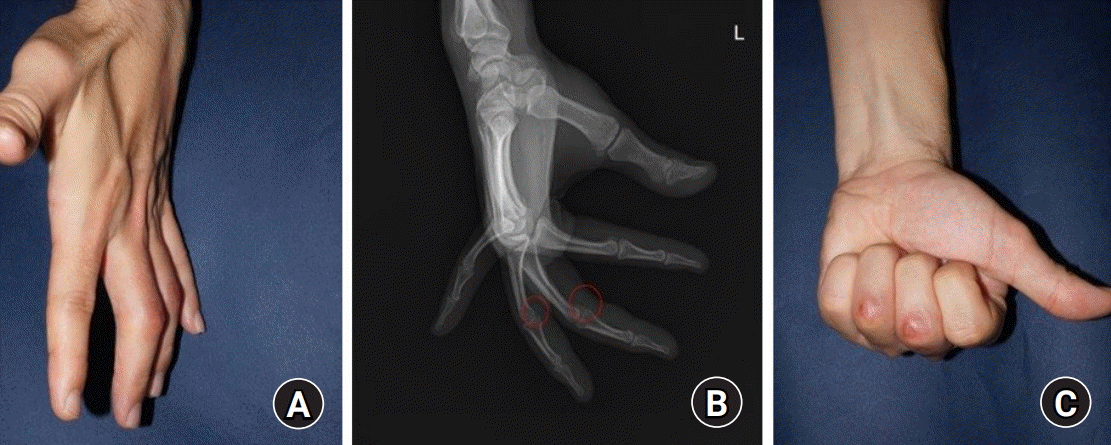 | Fig. 2.Pseudo boutonniere deformity. (A) A 36-year-old male patients presented proximal interphalangeal joint flexion contracture on left 3rd and 4th finger 3 months after hyperextension injury in affected finger joint. The plain X-ray presented volar small bony fragments marked with red circles (B) and the finger distal interphalangeal joint flexion was intact (C).
|
10년 이상의 류마티스 관절염 이환력이 있는 환자의 24%에서 단추구멍 변형을 보인다고 보고되었다[16]. 류마티스 특유의 활액낭염이 근위 지관절 배측에서 발생하면서 관절에 부착하는 중앙 건의 기능을 손상시켜 근위 지관절 신전 지연을 초래하면서 변형이 시작된다[17]. 원인은 외상이 아닌 활액막염이지만, 이후 변형 완성까지의 삼각인대 파열 등 기존에 설명한 경과를 거치게 된다. 류마티스에 의한 변형 역시 변형의 단계에 따른 비수술적 혹은 다양한 수술적 치료가 소개되고 있지만, 류마티스에서 동반될 수 있는 관상면 상 부정 정렬, 피부 상태 불량, 건의 기능 저하, 인접 관절의 활액막염 등은 타 원인에 의한 변형에 비해 나쁜 예후를 보일 수 있다[18]. 특히나 류마티스 환자에서 단추구멍 변형은 단순 연부 조직 수술만으로는 변형을 해결하기 어려울 수 있으며, 장기적인 결과 역시 예측이 어렵고 재발 내지는 변형의 지속을 보이는 좋지 않은 결과를 가져올 수 있다[19,20].
심한 Dupuytren 변형에선 주로 사상대(spiral cord)와 중앙대(central cord)에 의한 근위 지관절 굴곡 구축이 발생하며, 근위 지관절의 지속적인 굴곡 구축은 원위 지관절 수장판의 신장과 횡형 망상 인대의 구축으로 인한 측부대의 수장측 전위를 발생시킨다[21,22]. 최종적으로 Dupuytren 질환은 진성 혹은 가성 단추구멍 변형을 유발할 수 있다[23]. Dupuytren 변형에서 발생한 단추구멍 변형의 증례는 더욱 드물기 때문에, 정립된 방법 없이 일부 저자의 경험에 따른 치료 방법이 소개되고 있다. 본 저자들은 Dupuytren 질환과 동반하여 발생한 단추구멍 변형의 환자에서, 먼저 통상적인 Dupytren 구축 치료에 준해서 수장측에서 근막 절제술을 시행하여 근위 지관절 굴곡 구축을 해결한 뒤, 추가적으로 원위 지관절 과신전 및 굴곡 제한에 대해 종말 건 절제술을 시행하여 단추구멍 변형을 마저 해결한 경험이 있다(Fig. 3).
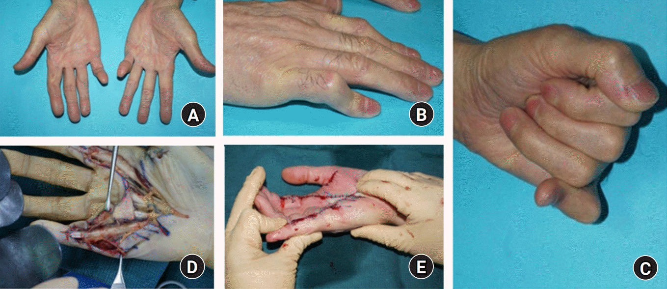 | Fig. 3.Boutonniere deformity in Dupuytren contracture disease. (A) A 65-year-old male patient presenting severe Dupuytren contracture on both hand. (B, C) The left 5th finger is also accompanied by boutonniere deformity. (D, E) With aggressive palmar fasciectomy and dorsal terminal tendon tenotomy, the deformity could be corrected.
|
화상에 의한 이차적인 수지 변형으로도 단추구멍 변형이 발생 가능하다. 일반적인 단추구멍 변형과 같이 근위 지관절 배측에 화상 자체 혹은 화상 이후 감염에 의한 중앙 건 손상으로 변형이 발생할 수 있으나, 화상으로 인한 주변 연부 조직 손상 및 구축 같은 여러 복합적인 요소들이 작용하며, 그 수술적 치료 예후 역시 좋지 않은 경우가 많다[4]. 이 경우 마찬가지로 정립되고 일반적인 수술적 방법을 적용하기 어려우며, 수부 화상 치료에 경험이 많은 의사에 의해 손상된 구조물 전반에 대한 접근이 이루어져야 한다. 따라서 이에 대해 발표된 연구도 경험 많은 의사에 의한 증례 보고에 한정된다[4].
Go to :
단추구멍 변형의 치료
근위 지관절과 원위 지관절 운동은 해부학, 생역학적으로 서로 연관되어 영향을 미치는 복잡한 구조이며, 섬세한 근위 지관절의 신전을 회복하고자 하는 단추구멍 변형의 치료에 있어 수술 후 치료 결과가 만족스럽지 못한 때가 많아, 수술적 치료의 결정과 세부적인 수술 방법을 신중하게 선택해야 한다.
1. 수술적 치료의 적응증
Green’s Operative Hand Surgery 7판에서는 급성 변형은 비수술적 치료를 권하며, 만성의 경우에도 일차적으로 보존적 치료를 시도하고 실패 시에 수술적 치료를 권하고 있다. 하지만 급성의 경우에도 외부 창상이 동반되어 중앙 건 파열이 있는 경우나 중위지골 배측에 전위된 골편이 동반되어 있을 경우, 수술적으로 파열된 중앙 건을 봉합하거나 골편의 정복 고정을 시도할 수 있다[7]. 또한 보존적 치료를 위한 보조기 착용이 어려운 급성 단추구멍 변형 환자에서 근위 지관절을 신전 위치에서 금속핀(Kirschner wire, K-wire)으로 관절 고정 수술 후 4–6주 뒤에 관절 범위 운동을 시작할 수도 있다[12].
만성 변형에서는 Fox와 Chang [9]이 종설을 통해 수술적 치료 의 적응증으로 (1) 보존적 치료 실패, (2) 변형이 진행하는 고정된 구축, (3) 근위 지관절에 수동적 운동이 가능한 경우, (4) 큰 사물을 잡을 수 없을 경우의 네 가지를 제시하였고, 금기증으로 (1) 근위 지관절의 심한 관절염, (2) 내과적 치료가 제대로 이루어지지 않은 류마티스 관절염, (3) 근위 지관절 수동적 운동이 제한될 때, (4) 최대 굴곡이 가능하며 정상적인 악력을 보일 경우를 제시하였다.
2. 보존적 치료
근위 지관절 최대 신전 위치에서 4–6주 고정하여 중앙 건의 자연 치유를 유도한다. 이 때, 원위 지관절의 굴곡 운동은 허용하여, 측부대를 중위지골의 배측인 원래의 해부학적 위치로 유도한다. 또한 원위 지관절 굴곡 운동은 구축된 측부대를 스트레칭 시켜, 이차적으로 근위 지관절의 신전 기전을 근위로 이동시킬 수 있다. 근위 지관절 신전 상태로 4–6주 고정 이후에도 4–6주 동안 간헐적 혹은 야간 부목 고정을 추가로 시행한다[10].
3. 수술적 치료
수술적 치료의 원칙은 원위지 관절의 과잉된 신전력을 근위 지관절의 신전력으로 전환시키는 것이다[24]. 이때 근위 지관절의 굴곡 기능에 장애를 유발하지 않는 범위에서 신전을 회복해야 하며, 지나친 근위 지관절 신전은 근위 지관절 굴곡의 제한을 일으켜 단추구멍 변형에 비해 더 큰 기능적 악화를 가져올 수 있다[12].
중앙 건의 개방성 파열의 경우 파열된 건을 봉합하는 수술을 요한다. 때때로 중앙 건의 건열 골절의 경우에선 골편이 작거나 전위가 있지 않다면 보존적 치료를 시행하며, 2 mm 이상의 골편은 K-wire를 통해 고정하는 수술을 요한다.
급성 변형의 수술적 치료와 달리, 만성 변형은 다양한 수술적 방법이 소개되고 있다. 이는 만성 변형 내에서도 변형의 단계를 포함한 다양한 요소들이 작용하기 때문에 일률적인 수술적 방법을 적용하는 것이 제한되기 때문이다. 또한 수부외과 의사일지라도 상대적으로 만성 변형의 수술적 치료 경험은 적기 때문에 치료 방법의 비교 연구를 수행하기 어렵다. 결국, 만성 단추구멍 변형은 정립된 수술적 치료 방법 없이, 경험 있는 수부외과 의사들의 증례 연구를 통해 다양한 수술적 치료 방법이 보고되고 있다[25,26]. 수술적 치료 방법은 수술적 대상과 방법에 따라 크게 종말 건 절제술, 측부대 수술법, 중앙 건 수술법, 건 이전술 및 건 이식술, 단계적 신전 기전 재조정 수술, 관절 고정술로 나눌 수 있다.
1) 종말 건 절제술
단추구멍 변형에 있어, 간단한 신전 기전 재조정 수술로는 종말 건 절제술(terminal tendon tenotomy; distal Fowler)이 있다[27]. Dolphin [27]이 처음으로 두 개의 증례에 대해 보고하여 Dolphin tenotomy라고도 한다. 이론적으로 종말 건을 절제하여 과도한 원위 지관절의 신전 기전을 약화시키고, 절제된 종말 건과 중앙 건이 근위부로 같이 후퇴하면서 근위 지관절 신전 기전을 상대적으로 강화시켜 최종적으로 근위 지관절의 신전 기전을 회복할 수 있다[10]. 그러나 이 수술은 원위 지관절의 굴곡은 향상시킬 수 있으나 근위 지관절의 신전 회복에 대해서는 이견이 있다[13,14]. 따라서 종말 건 절제술은 주로 30도 미만의 근위 지관절 신전 지연을 보이는 만성 변형에서 주로 원위 지관절의 굴곡 향상을 위해 시행할 수 있다.
2) 측부대 수술법
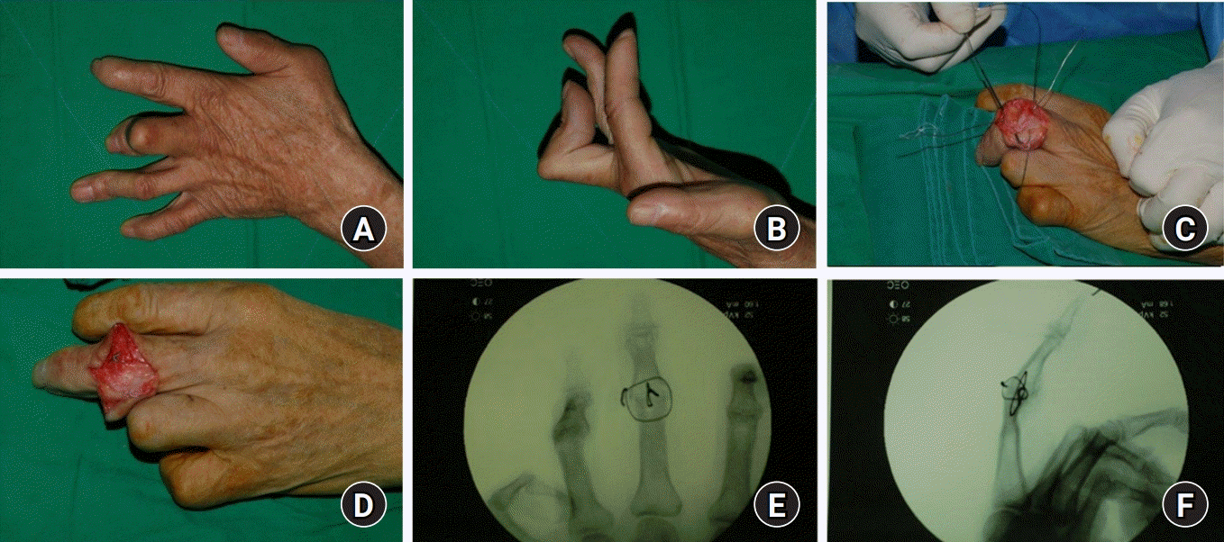
전방으로 전위된 측부대를 배측으로 이동시켜 근위 지관절의 신전을 향상시키는 방법이다. Littler와 Eaton [24]은 배측 절개를 통해 측부대를 주변 조직과 박리하여 배측으로 이전시킨 뒤 양측 측부대를 중위지골 중앙에서 같이 봉합하였다. 이 술기를 통해 내재적(intrinsic), 그리고 외재적(extrinsic) 신전력을 근위 지관절과 원위 지관절 모두에 효과적으로 전달할 수 있다(Fig. 4). 다만, 지나친 원위 지관절 신전으로 인해 굴곡이 제한되는 단점이 있으며[24,28], 비해부학적 건 재조정 수술로 그 결과 역시 일정하지 않다[26].
 | Fig. 4.Lateral band dorsal translocation and sutured together. (A) A 61-year-old female patient presented over 90° extension lag in left 3rd finger. (B, C) With dorsal approach, the central tendon almost lost function and bilateral lateral bands were released and suture together at the center of middle phalanx. (D, E) The patient recovered 0° of extension in proximal interphalangeal joint but also presented distal interphalangeal joint flexion deficit.
|
3) 중앙 건 수술법
중앙 건 술식은 손상된 조직이 반흔 조직으로 변하면서 근위 지관절 신전 지연이 발생된 중앙 건에 대한 술식이다. 배측 접근을 통해 반흔 조직을 절제한 이후 절제된 만큼 중앙 건을 전진 봉합하여 손상 전의 중앙 건의 길이를 회복하는 방법으로, 여기에 신전력을 더하기 위해 일부의 저자들은 측부대를 재부착하기도 한다. 종말 건 절제술이나 측부대 수술법과 달리 해부학적 복원술에 해당한다.
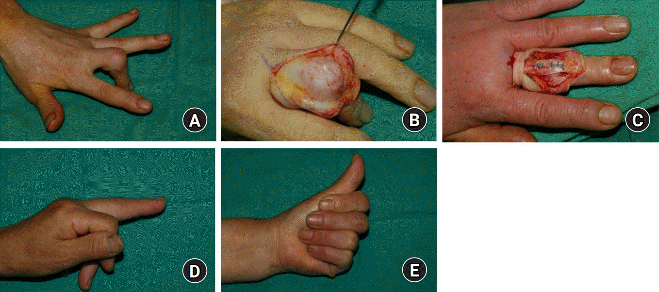
저자들 역시 만성 단추구멍 변형에서 수술적 치료의 적응증에 해당하는 Burton stage I형 혹은 II형에서 해당 술식을 먼저 고려한다. 근위 지관절 배측을 통해 접근하여, 중앙 건의 과거 손상으로 인해 늘어지고 반흔 조직으로 채워진 부분을 확인한다. 먼저 중앙 건의 양측을 종으로 절개하여 측부대와 분리시킨 뒤, 측부대를 박리한다. 확대경(surgical loupes)을 이용하여 중앙 건의 실질 및 반흔 조직을 구분하고, 중앙 건의 원위 부분에서 가능한 한 반흔 조직 부분을 절제한다. 이때 절제하는 길이는 절제 이후 남은 근위부와 원위부를 봉합했을 때 근위 지관절 신전 0도를 목표로 하여 과신전 및 굴곡 제한이 생기지 않게 주의한다. 중앙 건 단열 말단을 봉합하기 전, 근위 지관절에 굴곡 구축이 있을 경우 수장대 및 측부 인대를 박리하여 수동적 근위 지관절 신전을 회복할 수 있게 하여야 한다. Loop suture method로 건 봉합을 하는데, 봉합 강도를 높이고 봉합 부분에 가해지는 전단력을 줄이기 위해 봉합 나사(suture anchor)를 중앙 건 닿는 부분에 삽입 후 남겨진 실을 이용하여 추가 봉합한다. 봉합된 건의 상태를 고려하여 필요시 근위 지관절 0도에서 관절 고정 금속핀(K-wire)을 삽입 후 4주 뒤에 외래에서 제거한다. 수술 이후 근위 지관절만 4주에서 6주 고정하며 원위 지관절은 수술 후 통증이 줄면 관절 운동을 허용한다(Fig. 5).
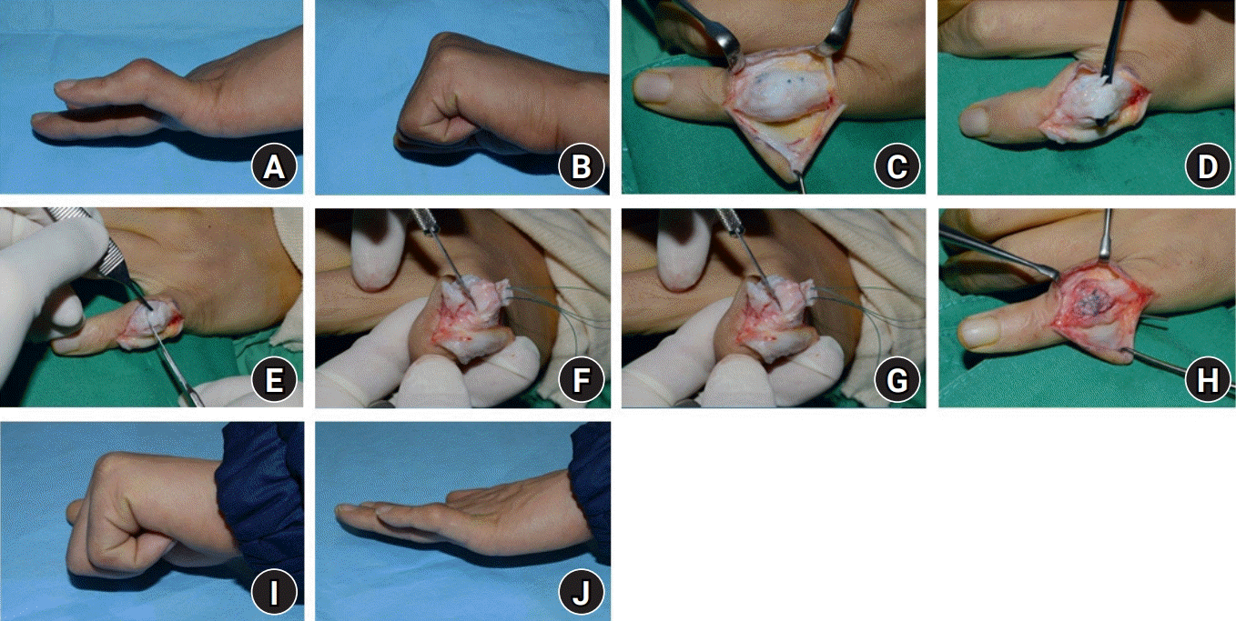 | Fig. 5.Central slip surgery. (A, B) A 35-year-old man with a 40° extensor lag of the proximal interphalangeal (PIP) joint and 20° hyperextension of distal interphalangeal joint after 10 months of initial tendon repair surgery at outer hospital in left 5th finger. (C) With a lazy S-shaped incision over the PIP joint, the elongated scar tissue is identified within the underlying joint capsule. (D) The bilateral sides of the central tendon are incised and released for advancement. (E) The proper length of scar tissue preventing the PIP full extension is resected, including the underlying joint capsule. (F) The volar plate and collateral ligament were release through exposed joint. (G, H) The proximal stump is advanced and the end repair between the distal stump containing the joint capsule using 4-strand core sutures at 0° PIP extension is performed. An additional Mitek Micro QuickAnchor (DePuy Mitek Inc., Raynham, MA, USA) is inserted and sutured to the proximal stump to enhance suture stability. (I, J) At the final clinical follow-up, the range of motion is recorded.
|
이러한 해부학적 중앙 건 복원 방법은 과거 여러 저자들에 의해 보고되었는데, 세부적인 부분에서 차이를 보인다[25,29,30]. Pardini 등[29]은 Burton stage I형의 단추구멍 변형에서, 중앙 건의 원위 1 cm 근위부에서 반흔 조직을 제거한 뒤 양측 절단단을 봉합하고, 측부대는 중앙 건에 측면 봉합하였다. 평균 33일을 근위 지관절 최대 신전위에서 K-wire로 고정한 뒤 관절 운동을 하여 수술 후의 기능적 결과를 측정하는 Souter’s 기준상[31], 59%가 우수(good) 혹은 보통(fair)의 결과를, 41%가 불량(poor)한 결과를 보였다.
Grundberg [30] 역시 비슷한 술기를 사용하였는데, 차이점은 절제하는 반흔 조직 중앙 건을 3 mm로 정하여 같은 길이를 제거하고자 노력하였다는 점이다. 수술 이후 근위 지관절 관절 운동 범위는 7도에서 89도로, 원위 지관절은 7도에서 51도로 향상된 소견을 보고하였다.
4) 건 이전술 혹은 건 이식술
일부 저자들이 신전 기전의 회복을 위해 천수지 굴건, 장장건 등을 이용한 건 이전술 혹은 이식술을 보고하였다. 이러한 증례 보고는 저자들마다 서로 다른 술식으로 적은 증례를 대상으로 시행하였고, 다양한 결과를 보고해 왔다[32-34]. 건 이전술 혹은 건 이식술의 명확한 적응증을 기존 연구 역시 제시하지 않으며, 경험이 많은 수술자가 아닌 이상 앞서 소개한 건 재조정 수술에 비해 먼저 고려되어야 할 술기로 보기 어렵다. Chung 등[35]은 중앙 건에 대한 수술이 건 이전술이나 건 이식술에 비해 좀더 나은 관절 운동 범위를 보이며, 환부 창이 적으므로 중앙 건 수술이 좀 더 선호된다고 하였다.
5) 단계적 신전 재조정술(Curtis procedure)
1983년 Curtis 등[36]은 외상성 단추구멍 변형에서 단계적인 치료법을 제시하였다. 이들은 근위 지관절 수장측 관절낭의 구축으로 수동적 근위 지관절 신전이 제한될 경우, 부목 고정이나 필요 시 선행적 수술 치료를 통해서라도 수동적 신전 제한을 회복한 이후에 단계적 술식을 시행하였다. 수술의 시작은 환자의 능동적 운동 확인을 위해 국소 마취 하에 진행하되, 4단계 수술까지 진행할 경우를 대비하여 전신마취나 액와신경총 마취를 같이 준비하였다. 환자 1단계는 근위 지관절 배측 관절낭의 위에 신전건 박리술과, 측부대를 전방으로 전위시키는 횡형 망상 인대를 유리한다. 2단계에서는 횡형 망상 인대를 수장측에서 절제한다. 3단계에서는 앞서 소개한 Fowler의 술식을 변형하여 중위지골 위치에서 측부대를 단계적으로 절제하여 연장한다. 4단계에서는 중앙 건의 반흔 조직을 절제하고 재전진 봉합시킨다. 1단계 수술 이후 2단계 수술 진행 여부는 압박대를 푼 뒤 환자에게 해당 수지를 신전하도록 하여, 최대 신전 회복 시 수술적 치료를 중단하고 아닐 경우 다음 술기로 진행한다. 2단계 수술 이후 마찬가지로 압박대를 풀고, 능동적 신전을 확인한다. 근위 지관절의 능동적 신전 각도가 20도 미만이면 바로 4단계 술식으로, 20도 미만인 경우 3단계 술식으로 진행하게 된다. 이러한 술식은 비교적 간단한 술기부터 적용하여 교정 정도를 보면서 추가 술기 진행 여부를 선택할 수 있다는 장점이 있다.
6) 관절 고정술
Burton stage IV와 같이 관절염까지 진행하였거나, 관상면의 변형이 동반된 경우, 근위 지관절 굴곡 구축에 따른 신전 제한으로 기능의 장애가 크며 고령인 경우 등에서 고려해 볼 수 있다(Fig. 6.)
 | Fig. 6.Arthrodesis of proximal interphalangeal joint. (A, B) A-65-year old male patient with severe boutonniere deformity with fixed 100° proximal interphalangeal joint flexion contracture with coronal deformation in left 3rd finger. (C-F) The arthrodesis using transosseous two wirings was performed.
|
정리하면, 만성 단추구멍 변형의 수술적 방법들에 절대적인 적응증은 없으며, 어떤 치료도 최선의 결과를 보장하지 못한다. Kaplan [37]은 시간이 경과하여 확립된 단추구멍 변형에서 신전 기전을 회복시키는 수술은 수부 영역에서 가장 힘든 수술이라고 기술하였고, Burton은 단추구멍 변형 수술은 수부 수술에 경험이 많은 외과의에 의해 수행되어야 한다고 주장하였다[10]. 경험이 많은 수부 외과의라 할지라도 그 결과가 일정하지 않거나, 수술 전보다 좋지 않은 결과를 가져올 수 있다[12]. 특히나 Steichen 등[38]은 근위 지관절의 수동적 신전이 되지 않는 경우, 수 개월 이상 경과한 폐쇄성 파열의 경우, 재수술인 경우, 그리고 45세 이하에 비해 45세 이상에서 수술적 결과가 좋지 않음을 보고하였다.
Go to :
 XML Download