

 PDF
PDF Citation
 Print
Print


INTRODUCTION
Extramammary Paget disease (EMPD) is a rare malignant neoplasm of the skin mainly involving the genital region, including the vulva, penis, perianal and periurethral areas [1]. Perianal Paget disease (PPD) accounts for 20% of EMPD [2].
Currently, wide local excision is the standard treatment modality for PPD. To reduce the risks of recurrence, excision with a sufficient safety margin is necessary, which results in wide defects of the skin and soft tissue surrounding the anus. Reconstruction of these defects is challenging because the perianal area can be easily contaminated and is difficult to be kept clean and bacteria-free. Creating an ileostomy in order to protect the wound from fecal contamination has often been suggested [3]; however, this could induce additional morbidity and reduce the patient’s postoperative quality of life.
The superior gluteal artery perforator (SGAP) flap has been utilized as a versatile option for reconstruction of the gluteal area, including sacral pressure sore reconstruction [4]. Owing to the vicinity of the donor site to the defect, this flap could be used for reconstruction of the perianal region. However, there have been few reports regarding the use of SGAP flaps for reconstruction of perianal defects [5]. We present a case in which reconstruction of a perianal defect was performed using a well-tailored SGAP flap following wide excision of tissue affected by PPD.
Go to : 
CASE REPORT
A 77-year old woman was referred from the department of colorectal cancer with a mass encircling the anus, which was diagnosed as EMPD on a punch biopsy. Surgery was planned to be performed by a combined general surgery (GS) plastic surgery team.
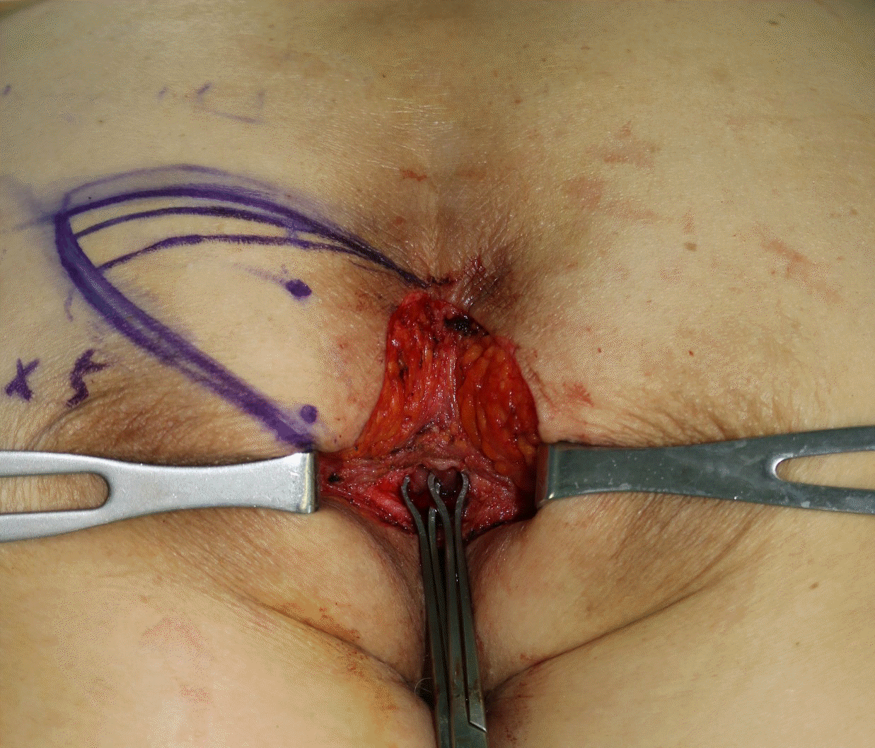
The patient was placed in a jack knife position under general anesthesia. Wide excision was performed by the GS team with cancer-free resection margin, leading to an 8×5 cm circumferential perianal defect (Fig. 1). Two perforators were identified in the adjacent tissue using an audible pencil doppler and a 10×5 cm SGAP flap was designed on her left buttock (Fig. 2). The flap was elevated along the suprafascial, dissecting two perforators with sufficient length for implantation. Primary defatting of the flap was done using Metzenbaum scissors to match the contour of the anal cleft and to avoid bulkiness. An opening was made within the flap at the proper location of the anus, with the skin and soft tissue trimmed to prevent tissue crowding. The anal mucosa tagged beforehand by a GS surgeon was pulled through the hole and sutured to the flap (Fig. 3). A temporary diverting colostomy was not needed. A drain was inserted into the left buttock donor site.
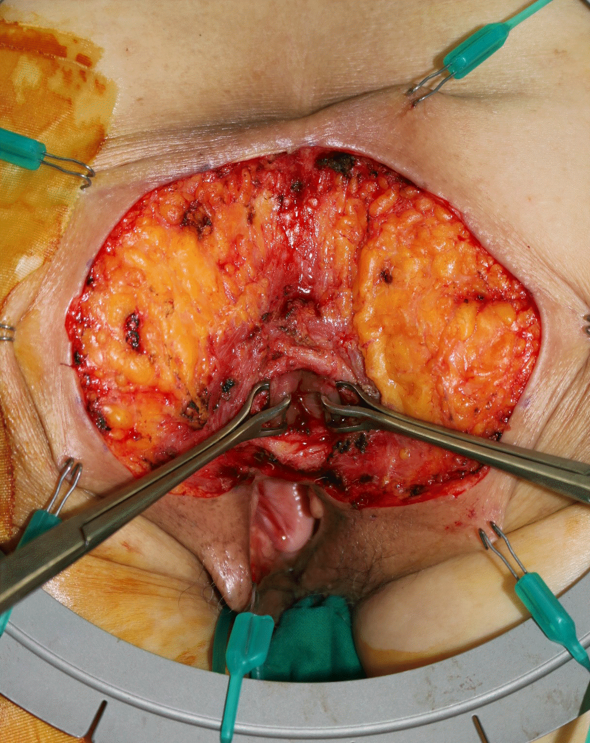 | Fig. 1.Tissue defect encircling the anus, sized 10 × 5 cm. Written informed consent for publication of their clinical images was obtained from the patient.
|

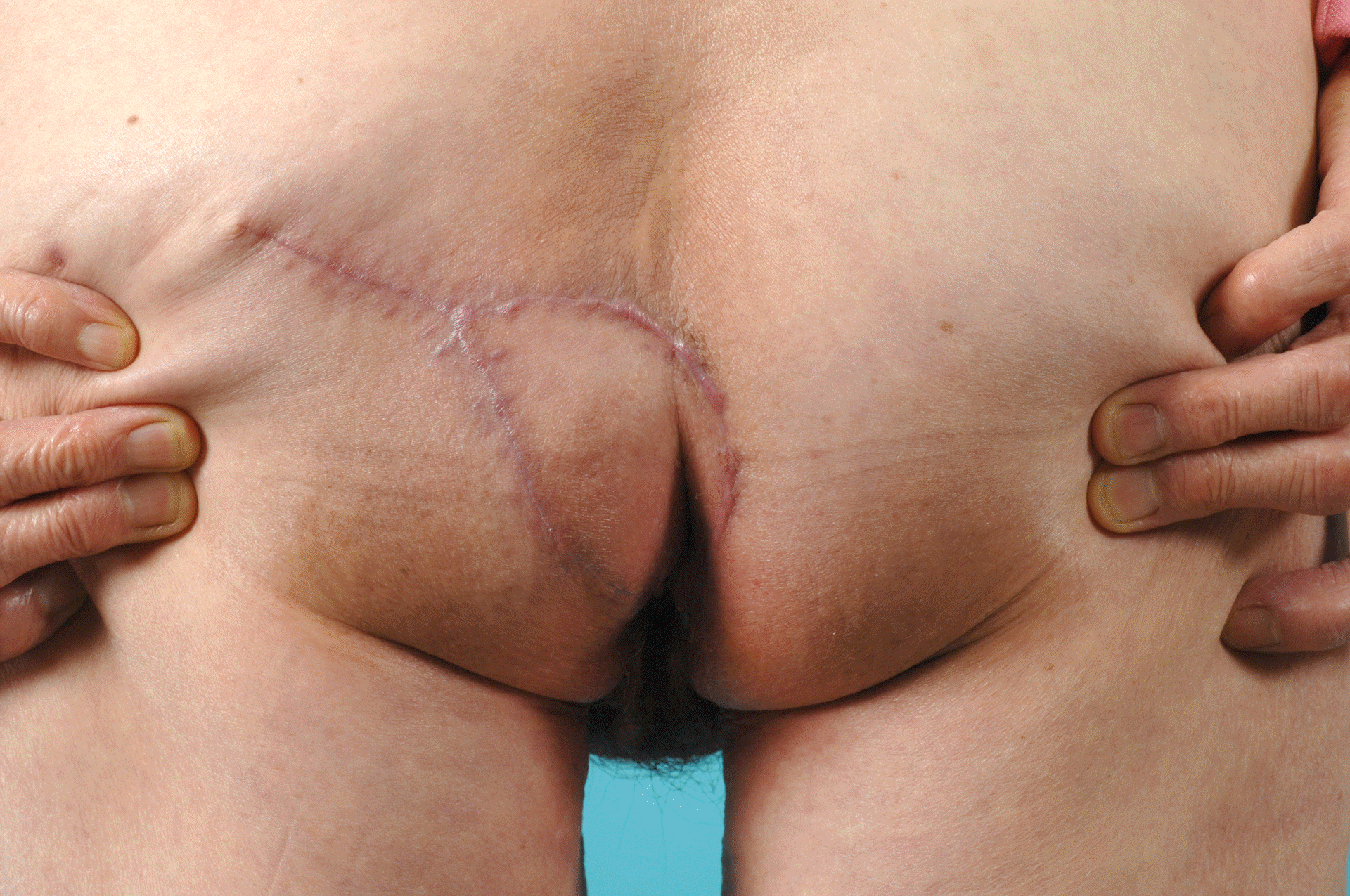
The patient was kept nil per os for 14 days postoperatively to prevent wound contamination by stool. The wound successfully healed without any complications. Laxatives were prescribed to protect the perianal wound from pressure imparted when passing stool for postoperative one month. In the follow up visit, preservation of postoperative anal functions including soft defecation and sensation as well as satisfactory tissue contouring were observed (Fig. 4).
This study was approved by the Institutional Review Board of the Samsung Medical Center (IRB No: 2020-10-041). All authors declare that written informed consent was obtained from the patient for publication of this case report and accompanying images.
Go to :
DISCUSSION
The perianal skin is a common site for EMPD, Bowen disease, and squamous cell cancer, with wide local excision as the treatment of choice [6]. In the literature, reports on reconstruction of perianal soft tissue have mainly described surgeons’ experiences with skin grafts and, in some cases, multiple V-Y advancement flaps.
Skin grafts are susceptible to bacterial infection, which leads to prolonged wound healing or graft failure. Postoperative care is challenging as the patient is required to remain in the prone position during the recovery period. Furthermore, even when the wound is fully healed there is a risk of compromised anal function due to contraction of the grafted skin. The V-Y advancement flap is a popular method that provides reliable outcomes in PPD patients. It enables the anatomical reconstruction of the natal cleft and anus if the flap is appropriately designed [6]. To provide adequate tissue coverage and preserve anatomical function, bilateral or multidirectional flaps are often required. This results in prolonged operation time and increased donor site morbidity. Tension from the bilateral flaps can result in widening of the anus. In case of a local recurrence of PPD after reconstruction with a bilateral flap, further excision and reconstruction can be challenging. Flap bulkiness is another shortcoming of the V-Y advancement flap. Defatting or thorough tailoring of the V-Y flap can be difficult. Furthermore, tripoint formation is inevitable in V-Y advancement flaps, which can result in scar contracture that can hinder anal function.
Our approach using the SGAP flap has three strengths. First, the flap has reliable perfusion, which enables not only fast wound healing but also the stability to withstand formation of a hole within the flap. Subsequently, there was no scar contracture due to poor blood supply. Second, the flap could be tailored in accordance with the adjacent tissue, and the flap thickness was adjusted using primary defatting. Defatting could be performed safely because the perforators were directly visualized and could be easily avoided. Suprafacial plexus is formed in multiple directions; therefore, defatting and hole fabrication wouldn’t compromise flap perfusion. Third, SGAP flap reduces donor site morbidity compared to the bilateral V-Y advancement flap. However, there are limitations to the SGAP flap; particularly, meticulous dissection is required to avoid any tension on perforators on the flap. Adequate defatting is necessary to obtain ideal clinical outcomes.
Reconstructive procedures to repair perianal defects following the wide excision of PPD should provide tissue coverage that is resistant to infection and pressure from postural changes, without impairing anal function. We used an SGAP flap and produced an opening in the flap for the anus. Primary defatting was done to achieve an optimal contour with the adjacent tissue. Satisfactory tissue contouring and defecation were observed. Therefore, well-tailored SGAP flaps may be a good option for the reconstruction of perianal defects encircling the anus.
Go to :
 XML Download