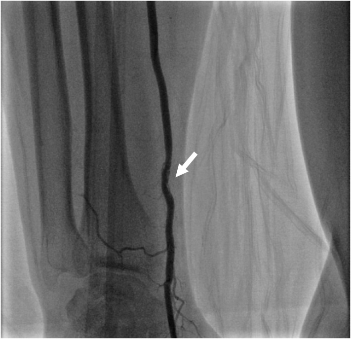
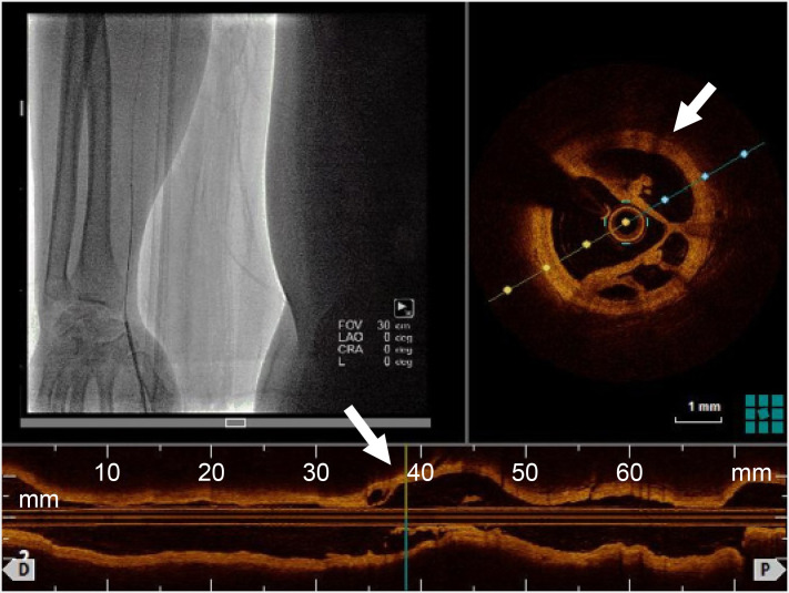
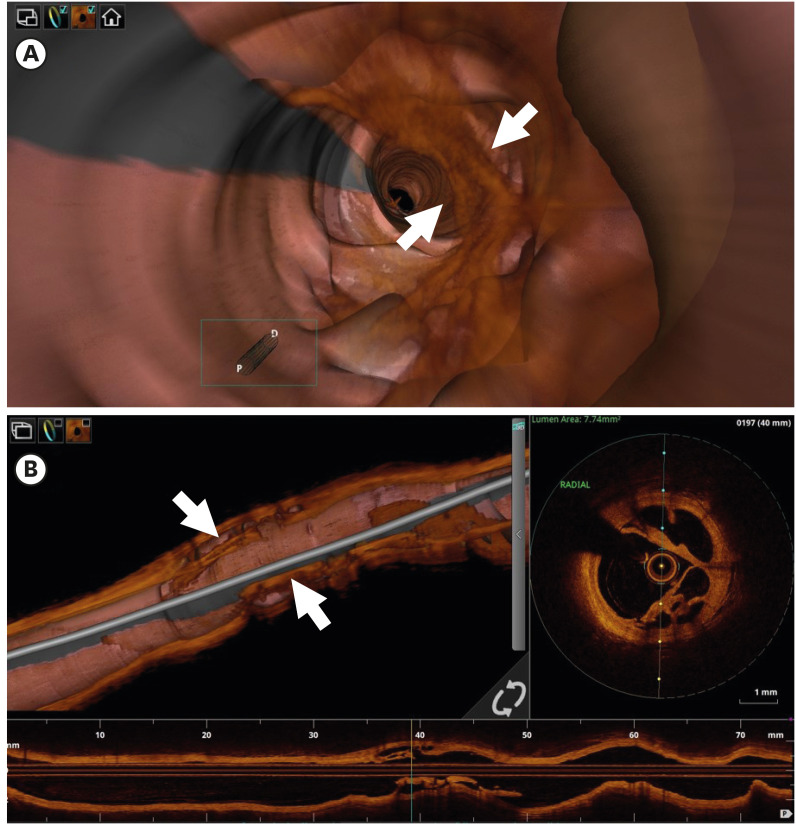
 | Figure 1Radial arterial angiography via sheath at right anatomical snuffbox revealed intraluminal hazziness at previous radial puncture site for transrdial intervention 10 years ago. |
A 52-year-old man who underwent stent implantation in the coronary artery via right transradial access (TRA) with 6F sheath 10 years ago was admitted to the emergency room due to chest pain. Coronary artery angiography was performed via distal radial access at anatomical snuffbox because of prior history of radial access and chronic kidney disease.1)2) When the wire and catheter went up to the radial artery, the operator felt some resistance. After successful primary distal transrdial intervention for acute myocardial infarction, angiography at right radial artery via sheath at anatomical snuffbox revealed intraluminal hazziness at radial artery which had been punctured for previous intervention 10 years ago (Figure 1). The lesion was examined with optical coherence tomography (OCT; Dragonfly™ OPTIS™ Imaging Catheter; Abbott, Abbott Park, IL, USA, pullback speed: survery mode 36 mm/sec, maximal contrast dose: 8 mL at 4 mL/sec). Because of arterial dissection issue, we reduced contrast volume which purge blood from the lumen. The OCT showed a lotus root-like appearance at the lesion3)4)(Figures 2 and 3). We quessed this appearance came from stenosis, thrombosis, occlusion reopen at previous puncture site. We believe this findings would be the very first OCT image of spontaneous recanalization of radial arterial occlusion via distal TRA and provide some evidences of occlusion or stenosis caused by puncture trauma and their spontaneous healing process. Two years later, the follow-up ultrasound image showed a patent radial artery at previous radial puncture site (Supplementary Video 1). As solution of this event, balloon angioplasty may be considered via distal radial access.5)
References
1. Kim Y, Ahn Y, Kim I, et al. Feasibility of coronary angiography and percutaneous coronary intervention via left snuffbox approach. Korean Circ J. 2018; 48:1120–1130. PMID: 30088362.

2. Kim Y, Lee JW, Lee SY, et al. Feasibility of primary percutaneous coronary intervention via the distal radial approach in patients with ST-elevation myocardial infarction. Korean J Intern Med. 2020.

3. Kang SJ, Nakano M, Virmani R, et al. OCT findings in patients with recanalization of organized thrombi in coronary arteries. JACC Cardiovasc Imaging. 2012; 5:725–732. PMID: 22789941.

4. Kadowaki H, Taguchi E, Kotono Y, et al. A lotus root-like appearance in both the left anterior descending and right coronary arteries. Heart Vessels. 2016; 31:124–128. PMID: 25142445.

5. Bae DH, Lee SY, Lee DI, Kim SM, Bae JW, Hwang KK. Percutaneous angioplasty at previous radial puncture site via distal radial access of anatomical snuffbox. Cardiol J. 2019; 26:610–611. PMID: 31701513.

Go to : 
SUPPLEMENTARY MATERIAL
Supplementary Video 1
The 2 year follow-up ultra-sound image showed a patent radial artery at previous radial puncture site. There were no intraluminal haziness, stenosis and occlusion in ultrasound scan.


 PDF
PDF Citation
 Print
Print


 XML Download