

 PDF
PDF Citation
 Print
Print


INTRODUCTION
Serrated polyposis syndrome (SPS), also known as hyperplastic syndrome, is a rare disease that is characterized by multiple serrated or hyperplastic polyps (HPs), mainly in the proximal colon. The World Health Organization clinical diagnostic criteria for SPS are as follows: 1) >5 serrated lesions/polyps proximal to the rectum, all being >5 mm in size, with >2 being 10 mm in size; 2) >20 serrated lesions/polyps of any size distributed throughout the large bowel, with >5 being proximal to the rectum.1
HPs were traditionally included in the non-neoplastic category. Unlike patients with sporadic small and distal HPs, patients with SPS have an increased risk of colorectal cancer. Edelstein et al.2 reported that the standard incidence ratio for colorectal cancer was 18.72 in patients with serrated polyposis. The reasons for the increased incidence of colorectal cancer are as follows. A diagnosis of SPS is often missed because flat lesions are difficult to recognize. Another problem with SPS is that there are so many polyps; the physicians cannot remove them all in an endoscopic procedure. This paper reports a case of SPS with a synchronous colon adenocarcinoma treated with an endoscopic mucosal resection (EMR).
Go to : 
CASE REPORT
A 61-year-old man visited the Kangwon National University Hospital for a follow-up colonoscopy. He had received an endoscopic resection for a polyp in the sigmoid colon in a private clinic one month earlier. The polyp was identified as a well-differentiated intramucosal adenocarcinoma (pTisNx) (Fig. 1). The cancer lesion was 1.2 cm in size with a polypoid (Ip Paris classification) type and was located at the proximal sigmoid with lamina propria invasion. No lymphovascular or perineural invasion was observed. Other parts of the polyp showed serrated adenoma on a pathology review. Many polyps were noted at other sites, but they were not removed.
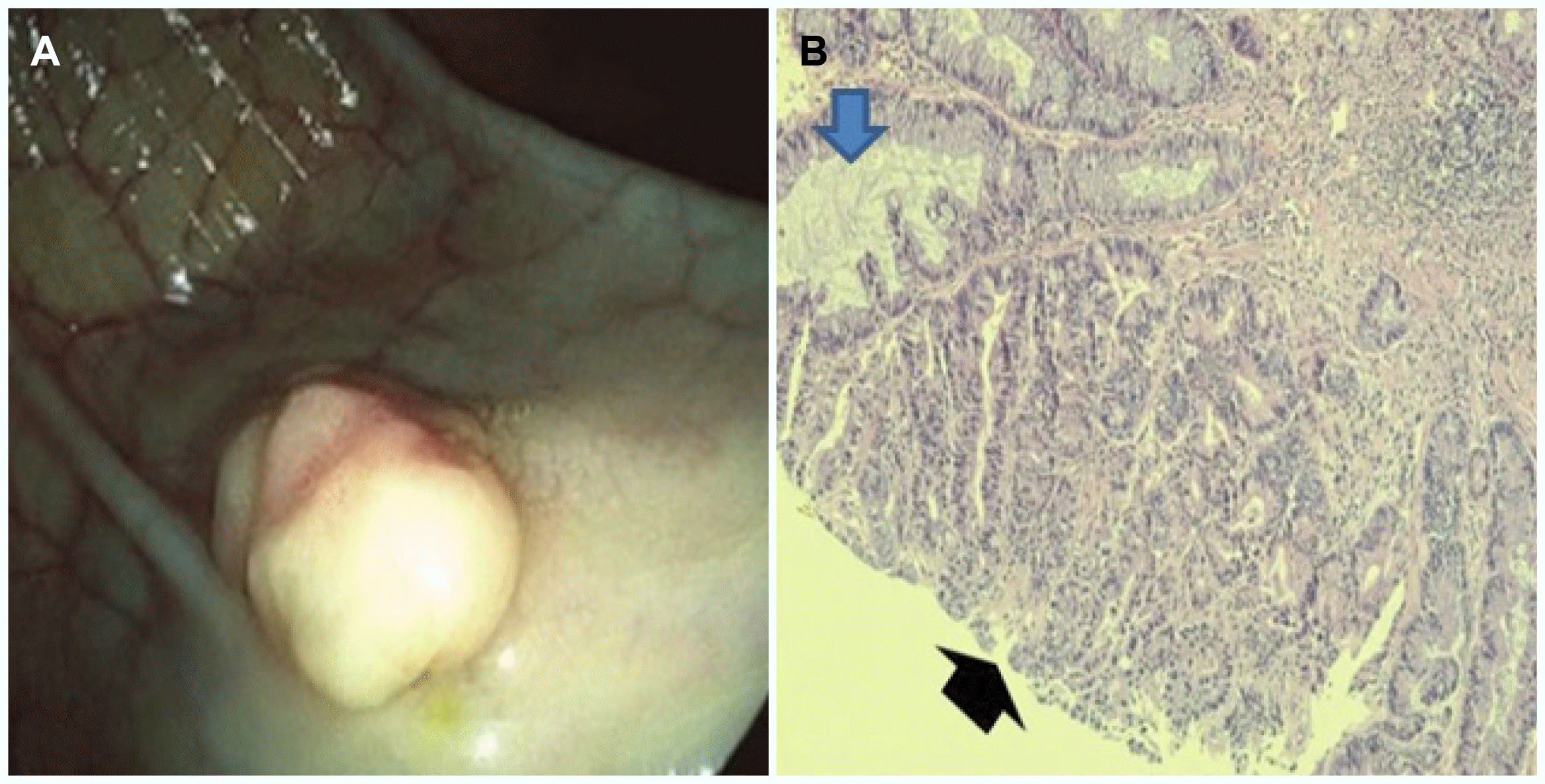 | Fig. 1(A) Colonoscopic finding showing a polyp in the sigmoid colon identified as well-differentiated adenocarcinoma. (B) Histology examination showing focal high-grade dysplasia (black arrow), suggesting well-differentiated adenocarcinoma and epithelial serration with extension to the crypt bases (blue arrow), suggestive of a sessile serrated adenoma (H&E, ×100).
|
The patient had a 20-year history of diabetes mellitus. He did not complain of any gastrointestinal symptoms and had no family history of colorectal cancer or other malignancies. He had a 40 pack-year smoking history and social drinking history (one bottle×15 days/month for 40 years).
The physical examination showed that the abdomen was soft and flat, and the bowel sounds were normo-active. No tenderness or rebound tenderness was noted. The chest and abdominal X-ray results were normal. The complete blood count profile showed 7,100 white blood cells/mcL, hemoglobin level of 13.7 g/mcL, and a platelet count of 272 K/mcL. The other laboratory findings were also within the normal ranges, except for his fasting serum glucose level was elevated (135 mg/dL). Carcinoembryonic antigen and other tumor markers were not checked. No specific findings were observed in abdominal CT.
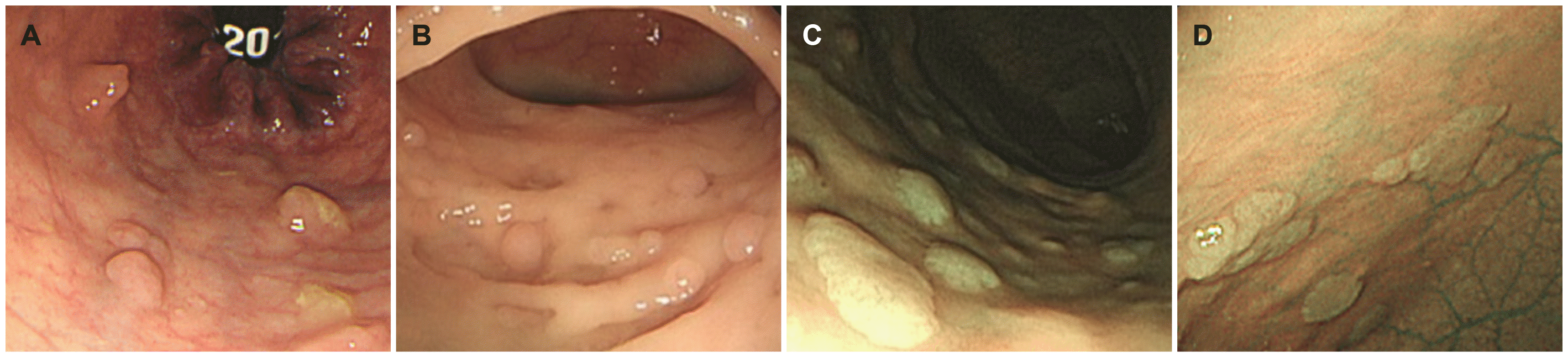
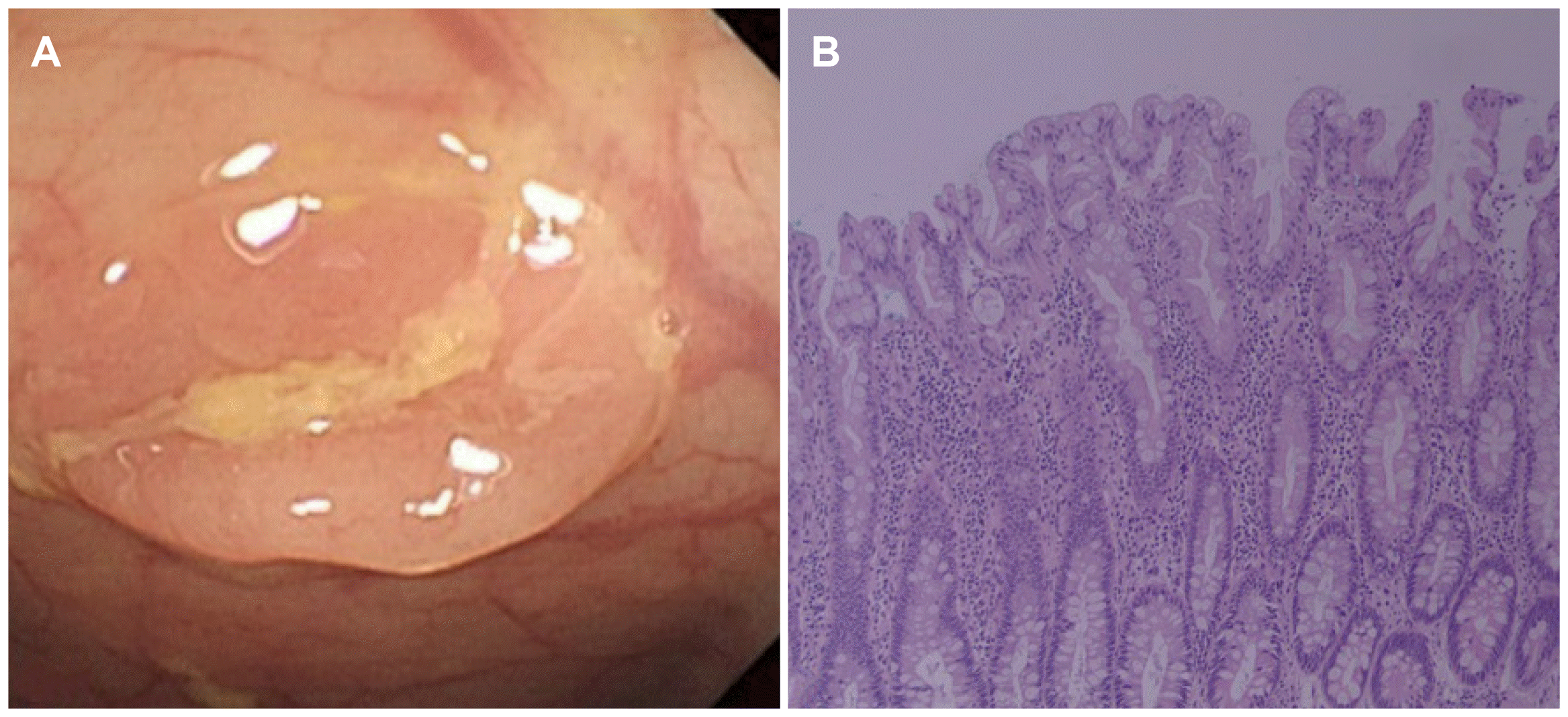
A colonoscopy performed in the Kangwon National University Hospital revealed multiple polyps (more than 40) of variable sizes throughout the entire colon (Fig. 2). They were distributed mainly from the descending colon to the rectum. Thirteen polyps, 1.0 to 2.0 cm in size, were removed by EMR. Histologically, one polyp in the descending colon was a tubular adenoma, while most of them were confirmed histologically as HPs. Some were identified as serrated adenoma (Fig. 3). Two of the HPs might have been sessile serrated polyps. On the other hand, the L- or T-shaped parts of the crypts were too small to be diagnosed as serrated polyps in the histology examination.
Go to :
DISCUSSION
Although multiple sporadic HPs resemble multiple adenomatous polyps endoscopically, they do not show any association with the development of adenocarcinoma. On the other hand, the progression of mixed hyperplastic adenomatous polyps and sessile serrated adenomas (SSA) to adenocarcinoma has been reported. In particular, SPS showed a high risk of developing adenocarcinoma (Table 1).3-5 The pathological findings for typical HP have serrations in the upper portion of the crypts with otherwise normal architectures and a normal proliferative zone at the base of the crypt. SSA have serrations at the base of the crypt with branching and dilation. Traditional serrated adenomas (TSAs) have serrated crypts with cytologic dysplasia.6 SSA and TSAs have additional pathological findings, such as the presence of horizontally oriented crypts, large areas without endocrine cells, focal mucus overproduction, and frequent (focal or diffuse) eosinophilia of the cytoplasm.7 In the present case, the patient had more than 50 polyps in the entire colon distributed mainly from the descending colon to the rectum. Most of them were confirmed histologically as HPs, and some were identified as serrated adenoma. The patient was diagnosed with SPS based on the World Health Organization criteria (II) mentioned above.
Table 1
Summary of Histologic Findings and Treatment in Three Patients Reported as Serrated Adenoma with Adenocarcinoma in the Previous Reports
| Study | Sex/age | Histology | Treatment |
|---|---|---|---|
| Yamauchi et al. (2002)3 | F/76 | Serrated adenoma/Adenocarcinoma | Transverse colectomy |
| Pyleris et al. (2014)4 | M/58 | Serrated adenoma/Adenocarcinoma | Total colectomy |
| Chino et al. (2016)5 | F/77 | Serrated adenoma/Adenocarcinoma | Surgery |
![]()
In the epidemiology of patients with SPS, there is no sex predominance. The mean age at the time of diagnosis is 55 years, and 10% to 50% of the SPS patients have a family history of colorectal cancer.8 Smoking and obesity have been identified as potential risk factors of SPS.9
The pathogenesis of CRC in SPS involves the serrated pathway instead of the adenoma-carcinoma sequence. The serrated pathway is divided into the sessile serrated pathway and the traditional serrated pathway. This might be associated partially with the presence of conventional adenoma. A recent genetic study on the origins of CRC arising in SPS patients showed that these tumors have diverse molecular profiles.10 SSA are characterized by a high rate of BRAF mutations. They are likely to be precursor lesions of most CRCs evolving through the serrated neoplastic pathway in SPS.11 The sessile serrated pathway develops SSA into dysplasia or microsatellite instability/stability cancer through a BRAF mutation, CpG island methylator phenotype, and MLH-1 promoter hypermethylation. The traditional serrated pathway develops TSA with high-grade dysplasia and MSS cancer through a pathway related to KRAS/BRAF mutations.12 In the present case, the endoscopic finding was a polypoid lesion-like adenoma. A mucosal pit pattern was not identified. Molecular genetics assays were not performed. On the other hand, this focal adenocarcinoma might have developed through the serrated pathway based on the histological findings.
Endoscopically, serrated polyps appear sessile, flat, and similar to the surrounding mucosa and are usually covered by mucus. These special features make their detection more difficult.13 Because of the malignant potential of these lesions, particularly in the context of SPS, early endoscopic detection becomes more important. In this regard, new endoscopic techniques, such as a magnifying videoscope with a chromoendoscopy, narrow-band imaging, and confocal laser endomicroscopy, are useful. Two recent retrospective studies on SPS were published in Korean. One study reported that the prevalence of SPS was 0.06%, whereas the other study estimated the prevalence at 0.025%.14,15 In western studies, the prevalence of SPS ranged from 0.08% to 0.66%.16-18 The prevalence of SPS was reported to be 8.4% in Japan.19 Magnifying video endoscopy with chromoendoscopy is used for the initial colonoscopies in Japan, which can explain the difference between Japan and other countries. The results of the study in Japan suggest that the prevalence of SPS might be higher than what is known.
Until recently, neither a screening method nor an optimal therapeutic strategy for SPS had been established. Immunological fecal occult blood testing has a high diagnostic accuracy for the detection of colorectal cancer.20 On the other hand, fecal occult blood tests could be less suitable for screening because serrated adenomas are less likely to bleed than conventional adenomas. Large polyps in SPS should be removed and followed up periodically. In general, polyps with an abnormal shape or larger than 5 mm in size should be removed, and repeat colonoscopy should be performed routinely in three years or less. SPS appears to be stricter compared to other conditions.21 Bleijenberg et al.22 recommended 2-year surveillance rather than 1-year surveillance to reduce the colonoscopy burden without increasing the risk of advanced neoplasia.
This paper reports a case of SPS that showed both well-differentiated adenocarcinoma and serrated adenoma from the same lesion by EMR. Distinguishing it completely as an adenomatous polyp was impossible by the gross findings on colonoscopy. Patients with SPS should undergo regular screening colonoscopies to remove potential premalignant lesions.
Go to :
 XML Download