

 PDF
PDF Citation
 Print
Print


서 론
담낭의 용종성 병변(gallbladder polypoid lesions)은 최근 다양한 영상진단법의 발전과 건강검진의 활성화로 발견 빈도가 증가하고 있고, 복부 초음파를 받는 성인의 4-7% 정도에서 발견되는 매우 흔한 질환이다.1-3 담낭의 용종성 병변은 담낭용종(gallbladder polyp), 선근종증(adenomyomatosis) 등 다양한 질환을 포함하지만, 일차적으로 가장 흔한 담석(gallstone)과의 감별이 필요하다. 담낭용종은 담낭 내부 점막의 융기성 병변으로 복부 초음파 검사에서 체위 변동에 따른 움직임이 없고, 담낭벽과 유사하거나 고에코(hyperechogenicity)를 보이는 질환으로, 대부분은 비신생물용종(non-neoplastic polyp)이기 때문에 추후 악성화 가능성이 거의 없지만, 3-8% 정도는 신생물 용종(neoplastic polyp)으로 담낭암으로 진행할 수 있다.1 신생물/비신생물용종의 감별 진단을 위해 복부 초음파(transabdominal ultrasonography, TAUS), 전산화단층촬영, 초음파 내시경(EUS) 등의 다양한 영상 검사가 이용되지만 두 질환을 감별하기는 매우 어렵다. 일반적으로 담낭용종의 크기가 10 mm 이상 또는 복통 등의 증상을 동반한 담낭용종은 악성화 가능성이 높아 담낭 절제술을 권유한다. 그렇지만 담낭용종은 대다수가 무증상이고, 신생물용종이 의심되어 수술을 시행하였음에도 비신생물용종으로 최종 진단되는 경우가 적지 않기 때문에, 수술적 치료의 결정은 환자의 유익(benefit)과 위해(risk)를 면밀히 고려하여 결정되어야 한다. 본고에서는 가장 최근의 2017년 유럽 가이드라인4을 소개하고, 국내 실정에 맞는 담낭용종의 치료 및 추적 관찰의 최신 지견에 대해 살펴보고자 한다.
Go to : 
본 론
1. 담낭용종의 병리 및 분류
2010년 세계보건기구 종양 분류 기준에 따르면, 담낭의 상피성 종양 중 전암성 병변은 선종(adenoma), 담관상피내종양(biliary intraepithelial neoplasm), 담낭내유두상종양(intracholecystic papillary neoplasm, ICPN), 점액낭종양(mucinous cystic neoplasm, MCN)으로 구분된다.5 담낭암의 발암 기전은 선종-암종 변환(adenoma-carcinoma sequence)과 이형성증-암종 변환(dysplasia-carcinoma sequence) 두 가지 발암 단계가 연관되는 것으로 추정된다. 담석 및 염증으로 유발된 담관상피내종양은 이형성증-암종 변환의 중요 소견으로 담낭암의 주 발암기전으로 생각되지만, 담낭 선종의 경우도 크기가 10 mm 이상의 담낭 선종은 약 20-55% 내외에서 담낭암으로 진행할 수 있기 때문에 선종-암종 변환도 또 다른 중요한 발암기전으로 생각된다.
담낭용종은 영상학적인 진단으로 콜레스테롤 용종이 가장 흔하지만, 일부는 신생물용종인 선종과 ICPN 등으로 진단된다. 과거에는 신생물 담낭용종을 선종으로 통칭하였지만, 내강 내 돌출성 성장을 하는 유두상 형태의 ICPN은 췌담관계 유두상 종양(intrapapillary mucinous neoplasm)의 담낭 내 발병 형태로 생각된다. 현재까지도 담낭용종에 대한 병리 진단 분류는 모호하고, 최근 세계보건기구에서는 ICPN과 선종 모두를 담낭의 상피성 종양 전암성 병변의 진단 분류에 남겨두었기 때문에 기존의 유두상 선종(papillary adenoma)과 ICPN은 병리학적으로 진단이 겹치는 문제가 발생할 수 있다(Table 1).
Table 1
Histology Classification of Polypoid Lesions of the Gallbladder and Extrahepatic Bile Ducts
![]()
담낭 선종은 대개 직경 1-2 cm의 단발성 유경성 또는 무경성 형태로 내부에는 혈관 및 결체 조직으로 구성되고, 외부는 이형성을 동반한 상피 증식 조직이 관찰되는 병변이다(Fig. 1). 이 중 일부는 암성 변화가 발생하여 담낭암으로 진단될 수 있다. 이에 비하여 콜레스테롤 용종은 콜레스테롤증(cholesterolosis)의 한 형태로 지질 포말양 대식세포(lipid-laden foamy macrophage)가 점막 내 고유층에 침착된 용종이다(Fig. 1). 콜레스테롤증은 미만형으로 담낭벽 전체에 걸쳐 나타나지만, 약 30%에서는 용종 양상으로 나타나며 다발성으로 발견되는 경우가 많고 얇은 목을 가지는 유경성 모양이 흔하다. 콜레스테롤 용종은 추후 악성 변화 가능성이 없는 비신생물용종이지만, 드물게 용종이 점막에서 떨어져 나오면서 췌장염, 담관염 등을 일으키기도 한다. 또다른 양성 질환인 담낭 선근종증(adenomyomatosis)은 염증으로 인해 점막이 비정상적으로 증식되고 점막층이 두꺼워지며 하부의 근층으로 함몰이 발생하여 생기는 병변으로, 미만형, 분절형 또는 국소형으로 발생한다. 특징적인 병리학 소견은 과증식된 점막층이 근층으로 함몰되어 낭상 구조를 띄는 Rokitansky-Aschoff sinus가 있으며, 이는 EUS 등의 검사에서 미세낭종(microcyst) 양상으로 관찰된다. 담낭 선근종증의 경우 만성 담관염의 아형이고 비종양성 용종이기 때문에 수술적 치료가 반드시 필요하지는 않지만, 담석을 동반한 경우에는 추후 악성화 위험도가 증가하여 수술 여부를 결정한다.
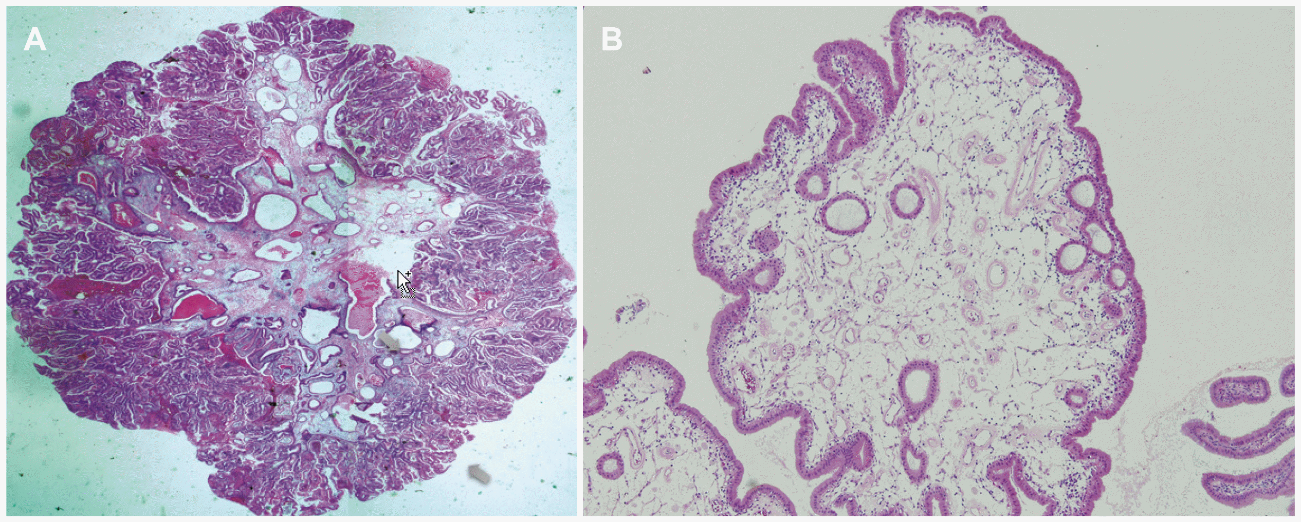 | Fig. 1(A) microscopic feature of an adenomatous polyp. The polyp was composed of a glandular structure with a tall columnar epithelium and stroma. In the center, a dilated gland and vessels surrounded by loose connective tissue were noted (H&E staining, ×40), (B) microscopic features of a cholesterol polyp. Lipid laden macrophage containing a polyp was observed (H&E staining, ×40).
|
2. 담낭 신생물용종의 위험인자
대표적인 담낭용종의 악성화 위험인자는 크기, 개수, 모양, 연령 등으로 알려져 있다. 크기는 가장 중요한 치료 결정인자로 10 mm 이상 담낭용종은 악성화의 위험도가 37-88%까지 증가되기 때문에 담낭 절제술이 필요하다. 하지만 10 mm 이하의 담낭용종도 이미 담낭암으로 발전하거나 또는 악성 변화의 가능성이 있는 선종이 발견되기 때문에 여러 악성화 위험인자를 고려하여 치료 계획을 수립하는 것이 필요하다. 일반적으로 단일 용종, 무경성 용종, 50세 이상의 나이, 담석, 원발성 경화성 담관염(primary sclerosing cholangitis)은 악성 변화의 위험인자로 간주되지만,4 이에 대한 근거 연구는 부족하다. Bhatt 등8의 메타분석 연구에서 10 mm 이하 담낭용종 중 단일(solitary) 용종은 악성화 위험도가 4.3%로 다발성 경우보다 높았지만, 다른 연구에서는 용종의 개수가 담낭암 발생과 연관이 없었다.9,10 담낭용종의 형태학적 분류와 관련해서는 Kwon 등9의 연구에서 무경성 담낭용종의 악성화 위험도는 OR 7.70 (95% CI 2.48-23.95)으로 높았고, 메타분석 연구에서도 10 mm 이하 무경성 담낭용종의 악성 예측도는 13.9%로 높게 보고되어,8 무경성 용종이 관찰되는 경우는 악성 변화의 가능성을 항상 염두해 두어야 한다. 담석은 아직 근거가 불충분한데, Aldouri 등11은 담석이 악성 변화 위험도를 증가(HR 3.2, 95% CI 1.42-7.22)시킨다고 보고하였지만, Park 등10은 담석과 담낭용종 악성 변화의 연관성을 찾지 못하였다. 담석은 증상이 있거나 합병증이 동반되는 자체만으로도 수술의 적응증이 되지만, 무증상 담석과 담낭용종이 동반된 경우에 과연 수술적 치료가 필요한지는 추가 연구가 필요하다. 또한 환자의 연령도 위험인자로 생각되는데, Park 등12의 연구에서는 50세를 기준으로 나누었을 때 신생물/비신생물용종 발생 빈도에 차이가 없었지만, Bhatt 등8의 메타분석 연구는 50세가 넘는 경우 10 mm 이하의 무경성 용종 악성 위험도가 20.7%로 높았고, 다른 국내 연구10에서도 57세를 기준으로 악성 변화의 위험도가 증가하는 것이 보고되었다. Kim 등13의 EUS 연구에서는 115명의 수술을 받은 담낭용종 환자에서 50세 이상 연령과 담석 동반 여부는 신생물용종의 위험인자가 아니었고, 유경성(OR 3.39, 95% CI 1.28-9.03)과 단일 용종(OR 3.68 95% CI 1.33-10.19)은 위험인자로 확인할 수 있었다. 이러한 다양한 연구 결과를 반영하여, 담낭용종 치료 계획은 검사 방법의 차이점과 다양한 위험인자를 복합적으로 고려하여 결정되어야 한다.
3. 담낭용종의 치료 전략
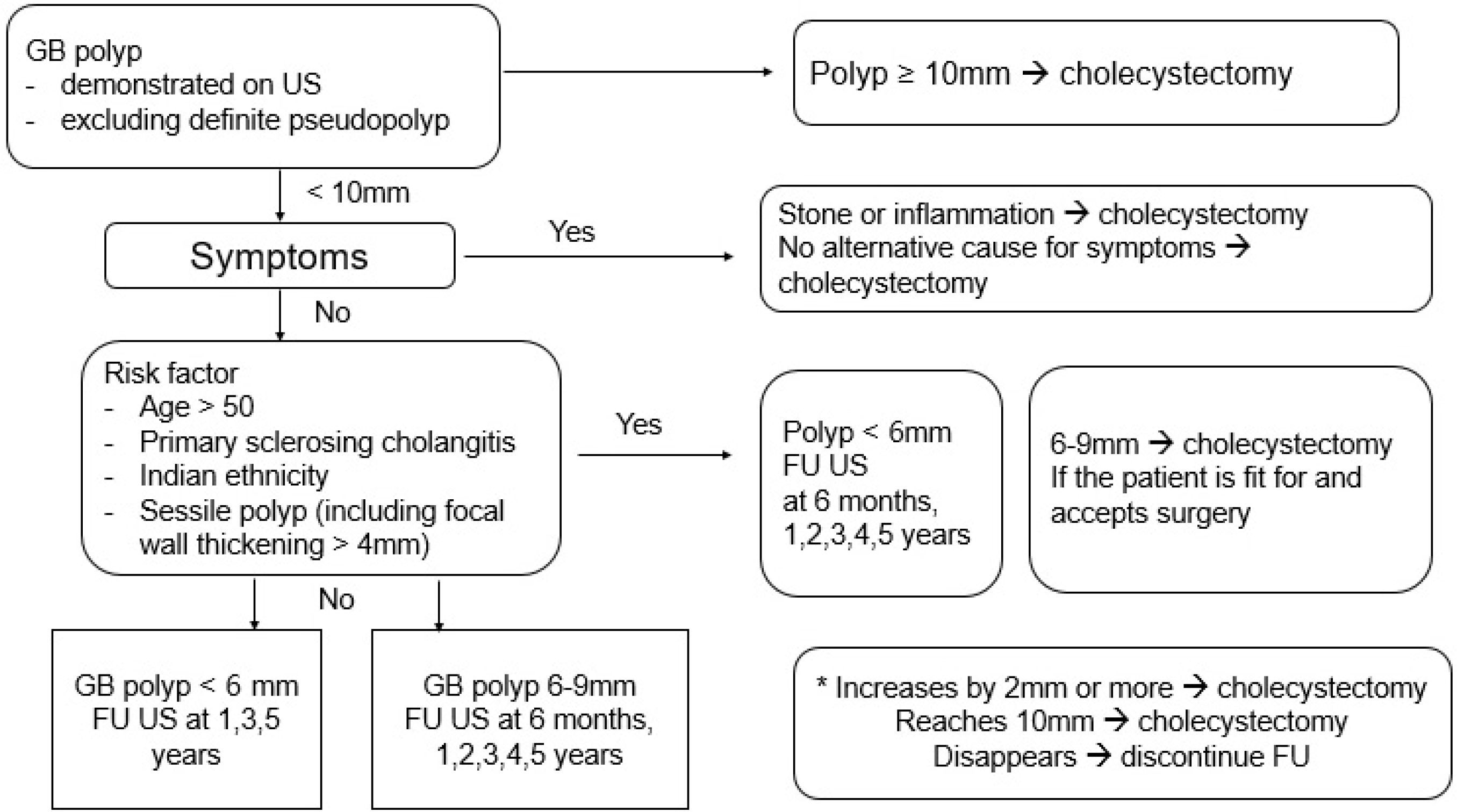
가장 최근 발표된 담낭용종의 치료지침은 2017년 유럽 영상의학과, 외과, 소화기내시경학회가 공동으로 제작한 “담낭용종의 치료 및 추적 관찰 유럽 진료지침”이다.4 유럽 치료지침에서는 담낭용종을 TAUS 상에서 내강 내로 돌출되어 있는 구조물로서 움직임과 posterior acoustic shadowing이 없는 병변으로 정의하였다. Comet tail 징후가 있는 경우는 콜레스테롤 용종, 염증성용종 또는 선근종증 등의 가성용종(pseudopolyp)으로 구분하였고 추가적인 검사 및 치료는 필요하지 않았다. 용종의 악성화를 예측할 수 있는 인자들 중 가장 중요한 인자는 용종의 크기였고, 치료지침에서는 담낭용종의 크기가 10 mm 이상인 경우 담낭 절제술을 추천하였고(moderate quality evidence, 89% agreement), 또한 연관 증상이 있으면서 다른 통증 원인을 배제할 수 없는 경우도 수술적 치료를 권고하였다(low quality evidence, 89% agreement). 담낭암의 위험인자는 50세 이상, 원발성 경화성 담관염(primary sclerosing cholangitis), 인디언 인종, 4 mm 이상의 담낭벽 비후를 포함한 무경성(sessile) 용종이었다. 용종 크기가 10 mm보다 작은 경우에도 이러한 위험인자가 있으면 다른 진단 및 치료적 접근을 시도하는데(low-moderate quality evidence, 78% agreement), 담낭암의 위험인자가 있는 6-9 mm 크기의 용종은 담낭 절제술을 고려하였다(low-moderate quality evidence, 78% agreement) (Fig. 2).
4. 추적 관찰 및 자연 경과
흥미롭게도 유럽 2017년 진료지침에서는 담낭용종의 추적 관찰과 크기 변화에 따른 접근 방법을 추가로 제시하였다. 담낭암의 위험인자가 없는 6-9 mm 크기의 용종 및 5 mm 크기 이하의 위험인자가 있는 용종은 6개월, 1년째 TAUS를 시행하고 이후로는 1년 간격으로 5년간 추적 관찰을 시행한다. 그 외 담낭암의 위험인자가 없는 5 mm 이하 크기의 용종은 진단 후 1년, 3년, 5년째 TAUS 추적 검사를 추천하였다(low quality evidence, 78% agreement). 또한 TAUS 추적 검사에서 연간 2 mm 이상 커지는 경우 또는 10 mm까지 커진 경우는 담낭 절제술을 권유하였고(moderate quality evidence 78% agreement, moderate quality evidence, 100% agreement), 용종이 없어지는 경우는 추적 검사를 종료하도록 하였다(moderate quality evidence, 100% agreement) (Fig 2).
사실 담낭용종의 자연 경과는 잘 알려져 있지 않고, 여러 연구에서 악성화를 예측할 수 있는 용종 성장 속도는 기존 연구에서 정확히 확인하지 못하였다. Park 등12의 보고에 따르면 1,558명을 추적 검사하였을 때 신생물용종은 33명에서 발생하였고, 진단 후 누적 발견율이 1년 1.7%, 5년 2.8%, 8년 4%로 증가하였고, 1년 이상 추적 관찰이 가능하였던 1,027명 중 36명(3.5%)에서 크기가 증가하고, 이 중 9명이 신생물용종으로 확인되었다. 이를 통해 추적 관찰 시 크기가 증가하는 것은 신생물용종의 중요한 예측인자로 간주할 수 있었고 악성화의 연간 누적 위험도는 5년 뒤에도 증가하기 때문에 정기적인 추적 검사는 최소한 5년 이상 지속하는 것을 권고하였다. 그러나 최근 보고된 미국의 후향적 코호트 연구에서 담낭용종의 유무가 담낭암의 발생률에 영향이 없었고, 연간 2 mm 이상 크기 증가도 담낭용종의 자연 경과로 담낭암의 발생과 큰 연관성이 없다는 상반된 결과를 보고하여 추가적인 연구가 필요하다.14
5. 새로운 시도와 발전 방향
상기 기술한 진료지침에서 진단과 추적 관찰의 방법으로 TAUS를 이용하였지만, 충분한 경험과 다양한 장비의 활용이 가용한 의료기관에서는 EUS 등의 정밀 검사를 이용하여 용종의 감별과 담낭암의 병기 설정할 수 있다. 특히 EUS는 보다 면밀하게 고해상도로 담낭용종을 관찰할 수 있기 때문에 다양한 추가 연구가 시도되었다. 특징적인 담낭용종 EUS 소견으로 hypoechoic foci, hyperechoic spot, 미세낭종(microcyst) 등이 이용된다. Hypoechoic foci는 용종의 변위부와 구분되는 중앙부의 상대적인 저음영 부위(hypoechoic foci)인데, 이는 조직학적으로 용종 주변부의 결체 조직과 대비된 중앙부의 혈관 밀집 조직으로 신생물용종의 진단에 도움이 된다.1 또한 용종 내부의 hyperechoic spot은 콜레스테롤 용종을 시사하고, 미세낭종은 담낭 선근종증의 특징적인 소견이다. 최근에는 CEH-EUS 및 도플러 측정을 이용한 담낭용종의 감별이 시도되고 있다. CEH-EUS는 용종 내부의 미세혈관을 조영하는 영상 검사로 담낭용종의 감별에 도움이 될 수 있지만,6,7 짧은 시간 내에 실시간으로 판정을 내리기 어렵기 때문에 제한이 많다. 담낭용종 내부 혈액의 흐름을 측정하는 도플러 측정법은 실시간 EUS를 시행하면서 내부의 혈관을 확인할 수 있는 검사법으로 간편하게 사용할 수 있는데, 저자의 전향적 연구에서 신생물용종은 강한 도플러가 관찰되어 진단 정확도가 73.9%로 측정되었다(Fig. 3).13 하지만 일부 콜레스테롤 용종에서도 내부에 약한 도플러 소견이 관찰되는 경우도 적지 않아 확진 방법보다는 보조적인 영상 진단법으로 활용이 가능할 것으로 생각된다.
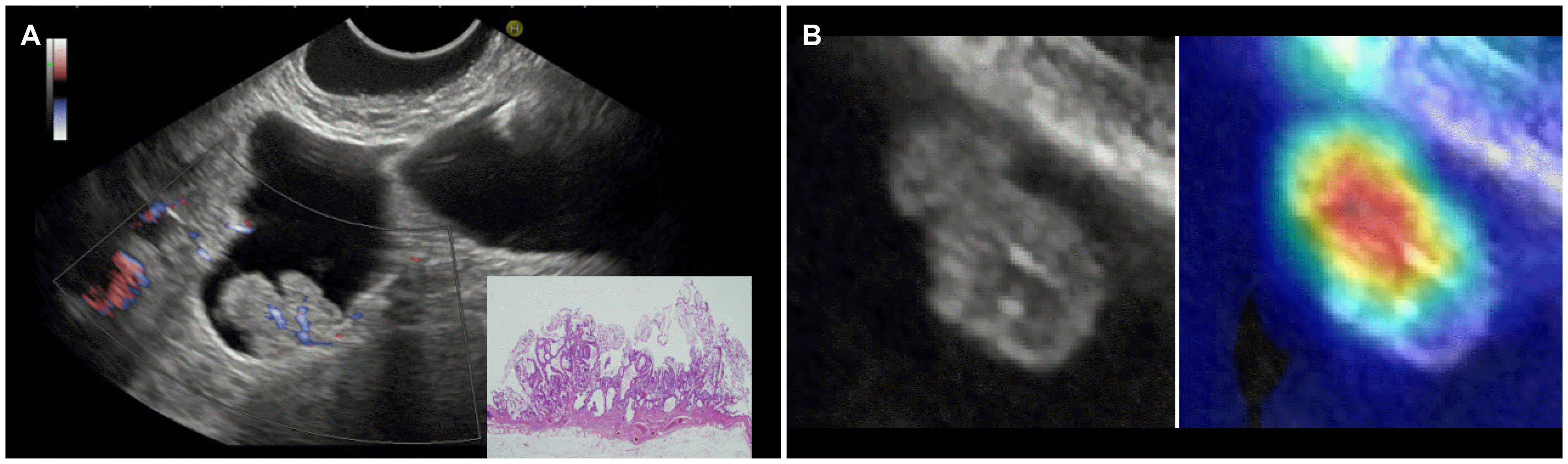 | Fig. 3(A) Color Doppler flow endoscopic ultrasonography image shows a strong continuous flow in an adenomatous polyp, which is compatible with prominent small feeding arterioles in gallbladder polyps. (B) Class activation map of a gallbladder polyp in the image classification through deep learning of the endoscopic ultrasonography-artificial intelligence system.
|
최근에는 의료 영상에 대한 인공지능의 도입으로 담낭용종에서도 deep learning 기법을 이용한 진단법이 실험되고 있다. 담낭용종의 감별에 있어 주관적인 TAUS 검사의 단점을 극복하기 위해 deep learning based decision support system을 도입한 연구에서는 인공지능 진단 보조 시스템이 TAUS 검사자 간의 진단 불일치율 및 가양성율(false positive rate)을 줄일 수 있는 것으로 보고하였다.15 저자들도 EUS 영상의 인공지능 deep learning 사전 연구를 시행하였고, 담낭의 융기성 병변 영상 1,039장을 학습(train), 내부 검증(internal validation), 시험(test)하여 EUS-artificial intelligence 시스템을 개발하고, 다기관 외부 검증을 시행하여 EUS-artificial intelligence 시스템의 담석과 용종의 감별 정확도는 89.8%, 신생물과 비신생물용종의 감별 정확도는 74.4%임을 확인하였다(Fig. 3). 이와 같이 주관적인 검사자의 판독 차이를 보정하고 객관화할 수 있는 인공지능의 활용은 담낭용종 감별에 보조적인 진단 기법으로 효과가 기대되지만, 다양한 기법 및 추가적인 검사 방법에 대해서는 아직도 심도 깊은 연구가 필요하다.
Go to :
결 론
담낭 질환의 추적 관찰은 임상 증상과 악성화 가능성을 예측하여 결정된다. 현재까지 증상 또는 합병증과 연관된 담낭 결석, 10 mm 이상 크기의 담낭용종, 악성 변화가 의심되는 담낭벽 비후가 담낭 절제술의 적응증으로 간주되지만, 최근 복강경 담낭 절제술이 보편화되고 수술 연관 합병증이 매우 감소하였기 때문에, 개복 담낭 절제술의 경험을 기반으로 제시된 과거의 담낭 절제술 적응증은 시대의 흐름에 맞추어 보다 확대될 가능성이 있다. 전술한 바와 같이 담낭용종은 크기 및 담낭암의 위험도를 고려하여 수술 여부를 결정하고, 추적 검사를 통해 장기간 악성 변화의 가능성을 배제해야 한다. 그러나 현재는 구체적인 추적 관찰 방법 및 기간 등에 대한 연구 자료가 부족하기 때문에 추가적인 연구가 필요하고, 한국인에 적합한 새로운 담낭용종의 진단 및 치료지침이 필요하다.
Go to :
 XML Download