

 PDF
PDF Citation
 Print
Print


INTRODUCTION
Epiploic appendages are small fatty pouches covered by visceral peritoneum and situated along the large intestine, particularly at the sigmoid colon. They are vulnerable to torsion, which leads to ischemic necrosis and subsequent inflammation. This entity is called primary epiploic appendagitis and can be misdiagnosed easily due to the lack of characteristic clinical and laboratory findings.1 Traditionally, they are diagnosed during exploratory laparotomy or diagnostic laparoscopy performed for other causes of right lower quadrant pain.2,3 In recent years, the diagnostic accuracy of abdominal ultrasound and CT has improved, and an increasing number of cases are diagnosed preoperatively.
On the other hand, there are still cases that are diagnosed during laparoscopy. The treatment of choice is conservative with or without the use of NSAIDs, while the use of antibiotics remains controversial.4,5 Laparoscopic excision should be considered in cases of an uncertain diagnosis, persistent symptoms, or recurrence.
This paper reports the case of a 42-year-old male who was misdiagnosed with acute appendicitis. A definite diagnosis was made by laparoscopy. The purpose of this review is to summarize all the existing data regarding primary epiploic appendagitis mimicking acute appendicitis and highlight the most important points for the differential diagnoses of epiploic appendagitis from acute appendicitis. Medline was investigated from 1960 to March 1, 2020. The keywords, epiploic appendagitis, torsion of an epiploic appendage, necrosis of an epiploic appendage, and infarct of an epiploic appendage, were selected to identify all reports possibly related to primary epiploic appendagitis. The reference lists of all relevant studies and reviews were scanned for additional studies.
Go to : 
CASE REPORT
A 42-year-old male visited the emergency department with a history of abdominal pain localized in the right lower abdomen in the previous eight hours. The pain was described as dull and constant, and it was associated with anorexia and nausea. No history of fever, vomiting, diarrhea, constipation, or urinary complaints was noted. The patient reported no prior medical or surgical history. An abdominal examination disclosed a soft, non-distended abdomen, with normal bowel sounds and deep tenderness at the Mc Burney point. Rebound tenderness in the right iliac fossa, Rovsing's sign, psoas sign, and obturator sign were all negative.
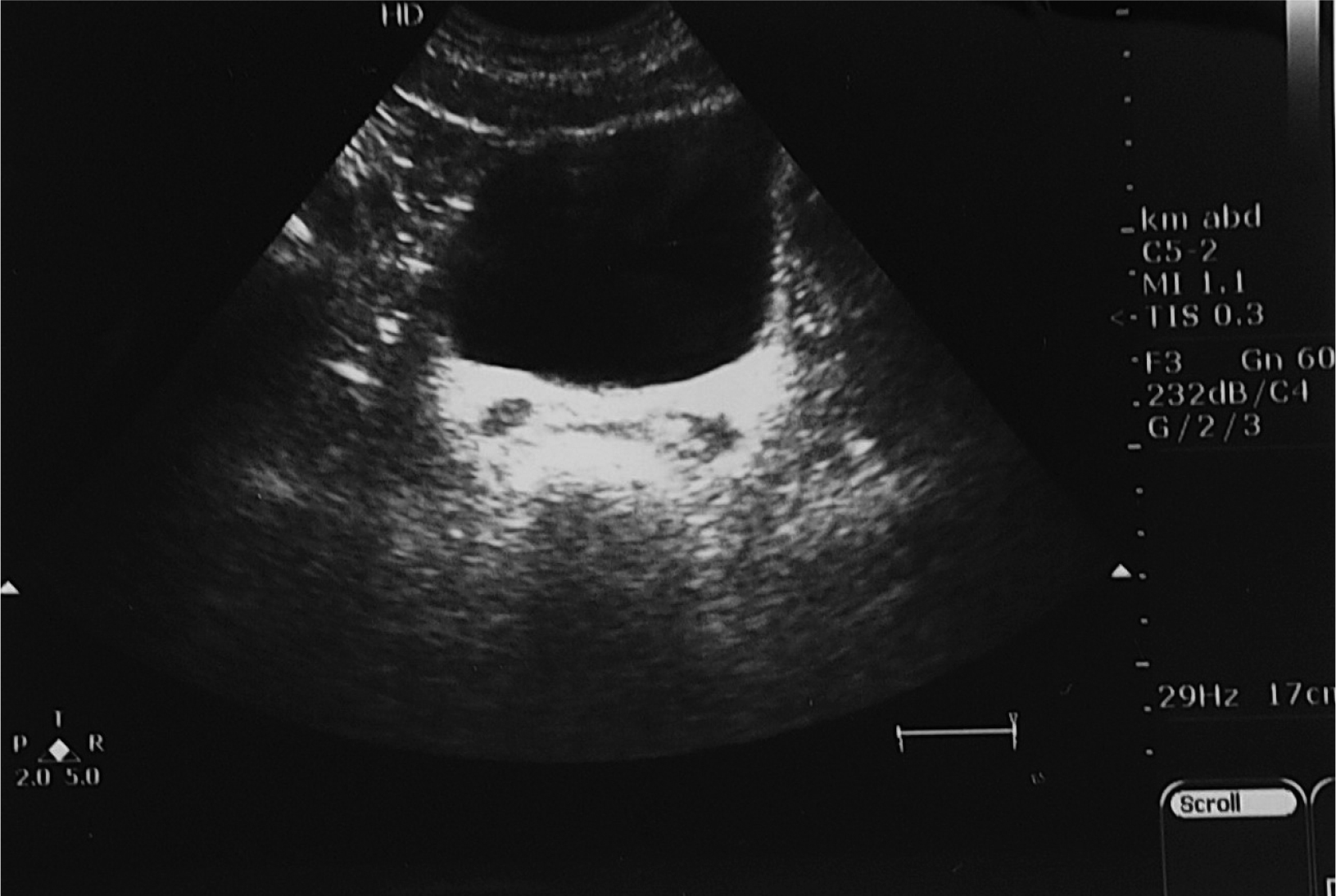
Laboratory studies revealed mild elevation of CRP (0.7 mg/dL) without any other inflammatory markers (white blood cell count 7.4 K/UL, neutrophils 53.5%, and erythrocyte sedimentation rate 8 mm/hour). Abdominal ultrasound showed no free fluid, while the abdominal organs were normal (Fig. 1). The appendix, however, was not visualized. The only finding was probe tenderness in the right iliac fossa.
The patient was admitted to the surgical department for observation, and antibiotics were commenced, but the pain did not settle. In the afternoon, the pain became intense, and rebound tenderness was found in the right iliac fossa on palpation. In the authors’ department, abdominal CT was not performed routinely for further evaluation of lower quadrant pain except for particular cases. Therefore, a decision was made to carry out diagnostic laparoscopy instead of an abdominal CT due to the high clinical suspicion of acute appendicitis.
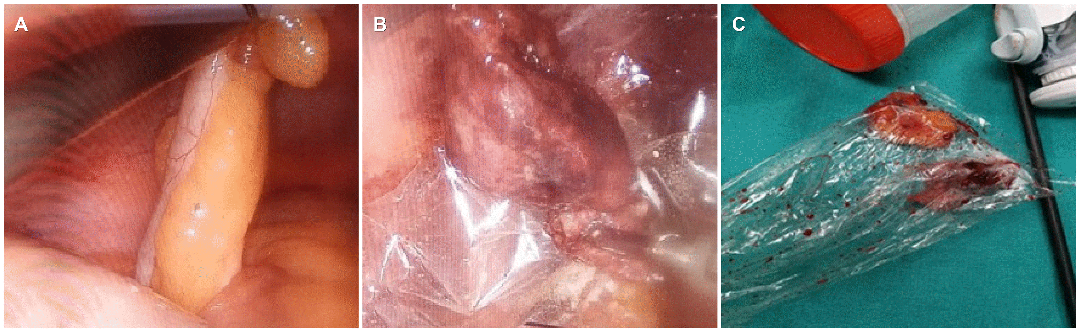
Nevertheless, the appendix was identified, exposed, and found to be normal. A twisted, necrotic epiploic appendage originating from the antimesenteric border of the mid ascending colon was noted (Fig. 2). There was no mesenteric lymphadenopathy or Meckel’s diverticulum. Laparoscopic appendectomy and resection of the necrotic epiploic appendix were performed using a harmonic scalpel (Ethicon Endo-Surgery, Johnson & Johnson, Cincinnati, USA). No antibiotics were administered postoperatively. The patient recovered uneventfully, and he was discharged after two days. Histology indicated an infarct of the epiploic appendage, hemorrhagic and necrotic adipose tissue, and infiltration by foamy macrophages, while the vermiform appendix was normal.
Go to :
DISCUSSION
The most common cause of acute right lower quadrant pain is acute appendicitis. Epiploic appendagitis is a frequent mimic of appendicitis clinically, which can be either primary or secondary. The primary form is caused by the torsion of an epiploic appendage or the spontaneous thrombosis of the central vein, which leads to ischemic or hemorrhagic infarction and subsequent inflammation. In contrast, secondary epiploic appendagitis is the inflammation of epiploic appendages due to the extension from adjacent inflamed organs, particularly in cases of diverticulitis, appendicitis, cholecystitis, or pancreatitis.6
The estimated frequency of primary epiploic appendagitis in patients with abdominal pain is 1.1-1.3%, and its incidence is 8.8 cases/million/year. Approximately 1% of patients with an initial diagnosis of acute appendicitis might have epiploic appendagitis.7,8 The condition usually affects adults in their fourth and fifth decades of life, and there is a male predominance. The commonest site is the sigmoid colon (50%) followed by the descending (26%), ascending colon and cecum (22%), while it is rarely encountered in the transverse colon (2%).1-4,7,9-14
Epiploic appendages, also known as epiploic appendices or appendices epiploicae, are small fatty pouches covered by visceral peritoneum. They are attached to the antimesenteric border of the colon via a stalk and protrude into the peritoneal cavity. Each is supplied by one or two end arteries originating from the vasa recta longa of the colon and drained by a single tortuous vein. Vesalius was the first to describe them in 1543, while Virchow suggested that loose intraperitoneal bodies could be detached epiploic appendages. Approximately 50-100 epiploic appendages measuring 2-5 cm in length and 1-2 cm in thickness can be found all over the colon.1 They are arranged in two rows: anteriorly along the taenia libera and posterolaterally along the taenia omentalis, except for the transverse colon, which contains only one row as the greater omentum attaches to the taenia omentalis. Occasionally they might be larger and reach up to 15 cm, particularly in obese people and in those who lose weight.1,2 Their function remains unclear. They might act as a protective cushion during peristalsis, fat storage, or contribute to intestinal immunity.1
The initial manifestation of primary epiploic appendagitis mimicking acute appendicitis is variable. The most frequent appearance is acute pain in the right iliac fossa. The pain is described as dull, constant, and non-exacerbating with physical movements. Other symptoms include postprandial fullness and early satiety, but fever, nausea, vomiting, diarrhea, and constipation are rarely present. A clinical examination reveals localized tenderness and occasionally rebound tenderness.1-4,7,9-14 In most cases, all routine laboratory parameters, such as inflammatory markers, liver transaminases, pancreatic amylase, lipase, and urinalysis, are within the normal limits. Occasionally, a mild leukocytosis and a mild elevation of CRP are observed.2,13 Table 1 lists the demographics and clinical presentations of patients with primary epiploic appendagitis.
Table 1
Demographics and Clinical Presentation of Patients with Primary Epiploic Appendagitis
| Author | Patients/sex ratio (M/F) | Mean age (years) | Right side epiploic appendagitis | Symptoms except for pain | Clinical findings |
|---|---|---|---|---|---|
| Van Breda Vriesman et al. (1999)7 | 40 (28/12) | 42 | 6 (15) | Nausea 5% | Focal tenderness 100% |
| Vomiting 5% | Rebound 25% | ||||
| Mass 10% | |||||
| Zissin et al. (2002)9 | 33 (24/9) | 44.6 | 9 (27.3) | None | Focal tenderness 84.9% |
| Son et al. (2002)3 | 8 (8/0) | 34.9 | 1 (12.5) | Fever 12.5% | Focal tenderness 100% |
| Rebound 25% | |||||
| Mass 25% | |||||
| Legome et al. (2002)12 | 19 (10/9) | 37.8 | 5 (26) | Anorexia 32% | Focal tenderness 100% |
| Nausea 36% | Guarding 47% | ||||
| Vomiting 21% | |||||
| Constipation/diarrhea 10% | |||||
| Fever >38℃ 15% | |||||
| Sand et al. (2007)1 | 10 (7/3) | 44.6 | 2 (20) | Nausea 4.7% | Focal tenderness 100% |
| LLQ 80% | Vomiting 4.7% | Rebound 10% | |||
| RLQ 20% | Diarrhea 14.2% | ||||
| Ozdemir et al. (2010)13 | 12 (9/3) | 40 | 4 (33) | Nausea 16.6% | Focal tenderness 100% |
| Vomiting 16.6% | Rebound 24.9% | ||||
| Urinary tract symptoms 8.3% | Rigidity 16.6% | ||||
| Chen et al. (2011)11 | 21 (15/6) | 40 | 2 (9.5) | Nausea 4.7% | |
| Vomiting 4.7% | |||||
| Diarrhea 14.2% | |||||
| Choi et al. (2011)2 | 31 (22/9) | 40 | 13 (41.9) | Focal tenderness 100% | |
| Rebound 25.8% | |||||
| Nadida et al. (2016)4 | 12 (4/8) | 51.7 | 6 (50) | Nausea 25% | |
| Vomiting 8.3% | |||||
| Ergelen et al. (2017)14 | 45 (23/22) | 42.3 | 16 (35) | Fever 6.6% | Focal tenderness 100% |
| Rebound 13.3% |
![]()
Distinguishing primary epiploic appendagitis from acute appendicitis preoperatively is very challenging. In contrast, primary epiploic appendagitis needs to be distinguished preoperatively from acute appendicitis because it is managed conservatively without surgical operation. One clinical attribute of primary epiploic appendagitis is focal, non-migratory pain situated in the right iliac fossa, which is accompanied by an absence of inflammatory markers. On abdominal ultrasonography, it is depicted as a hyperechoic ovoid non-compressible mass surrounded by a subtle hypoechoic rim. The mass is situated adjacent to the colonic wall, while the adjacent fat is hyperechoic due to inflammation.4,5,8,13,15 Ultrasound can reveal the inflamed epiploic appendage to the anterior abdominal wall.5,8 On Doppler images, a specific feature is the absence of blood flow in the mass, while there might be increased blood flow in the adjacent inflamed fat.4,8,15 On the other hand, abdominal ultrasound is an operator- dependent examination, and primary epiploic appendagitis is not always visible.
Currently, abdominal CT is the gold standard for a diagnosis of primary epiploic appendagitis. Despite the location of primary epiploic appendagitis, the imaging criteria encompass the following five special features: an ovoid mass with fat attenuation measuring 1-3.5 cm in the greatest diameter; hyperdense ring sign, which is a hyperdense ring that encircles the ovoid mass and corresponds to the inflammatory visceral peritoneum; central dot sign, which is the presence of a hyperdense dot centrally that illustrates the thrombosed vein; fat stranding sign, which is the severe mesentery inflammation compared to minimal asymmetric thickening of the adjacent bowel wall; thickening of the parietal peritoneum due to the attachment of the inflamed epiploic appendage. The fat stranding sign indicates that the primary site of the inflammation is the epiploic appendage rather than the bowel wall. All the CT findings are summarized in Table 2.4-11,14-18 Abdominal MRI can be an alternative to abdominal CT, particularly in children and pregnancies. The imaging findings include an ovoid mass with high signal in T1 and T2 images, while rim enhancement can be observed in contrast agent (gadolinium) enhanced T1 images.19
Table 2
Computed Tomography Findings of Patients with Primary Epiploic Appendagitis
| Author | Ovoid mass of fat density | Hyperdense ring sign | Central dot sign | Fat stranding sign | Parietal peritoneal thickening |
|---|---|---|---|---|---|
| Rioux et al. (1994)5 | 100 | 100 | 28.5 | 93 | |
| Mollà et al. (1998)8 | 100 | 100 | 57 | 100 | 57 |
| Van Breda Vriesman et al. (1999)7 | 100 | 59 | 41 | 38 | |
| Zissin et al. (2002)9 | 100 | 84.8 | |||
| Ng et al. (2006)16 | 100 | 100 | 42.8 | 100 | |
| Jalaguier et al. (2009)6 | 100 | 100 | 77 | 100 | 93 |
| Chen et al. (2011)11 | 100 | 100 | 33.3 | 100 | |
| Hasbahceci et al. (2012)15 | 100 | 100 | 75 | 100 | 60 |
| Hwang et al. (2013)10 | 100 | 89.3 | 17.9 | 14.3 | |
| Saad et al. (2014)20 | 100 | 100 | 61 | ||
| Nadida et al. (2016)4 | 100 | 100 | 16.7 | ||
| Nugent et al. (2017)17 | 100 | 100 | 79 | 74 | |
| Ergelen et al. (2017)14 | 100 | 100 | 26.6 | 100 | |
| Almeida et al. (2018)18 | 100 | 100 | 51 | 100 | 81 |
![]()
In the authors’ hospital as well as in others all over the world, the initial assessment of right lower quadrant pain included an abdominal ultrasound. CT of the abdomen is obtained only in special circumstances. Sharp, non-migratory pain in the right iliac fossa accompanied by the absence of inflammatory markers and a normal abdominal ultrasound might be an indication for CT to avoid unnecessary laparoscopy or laparotomy.
The treatment of choice for primary epiploic appendagitis is conservative with or without the use of NSAIDs because it is considered a self-limiting disease.4,20 The symptoms are usually resolved within two weeks, while the CT findings require more time to subside.10 Antibiotics are administered widely, 1,3,10,11,16,18 even though their therapeutic benefit is not well-documented.5-7 Most studies report a low rate of recurrence with conservative treatment.1,3,5,7,8,10,13,14 Nevertheless, a few studies revealed a recurrence rate of 5-17%.12,15,18 Sand et al.1 reported that there is a tendency of recurrence in conservatively treated patients. Four out of ten patients in this study (40%) had the same pain at the same locality, for two days, on average, four weeks before their appearance to the emergency department. The indications of surgery include an uncertain diagnosis, no improvement in symptoms despite conservative management, and relapse. Many authors recommend a laparoscopic resection of the affected epiploic appendages coupled with prophylactic appendectomy in such cases.1 In the authors’ hospital as well as in other institutions around the world, prophylactic appendectomy is carried out in cases of diagnostic laparoscopy or exploratory laparotomy for right lower quadrant pain, even if the appendix is normal.
Go to :
 XML Download