

 PDF
PDF Citation
 Print
Print


INTRODUCTION
The incidence of rectal foreign body insertion is not known precisely, but it is uncommon. On the other hand, the incidence has been increasing.1,2 Various types of foreign bodies and household objects, including plastic or glass bottles, vegetables, such as carrots and cucumbers, and adult objects, such as vibrators and dildos, have been reported.2-4 Proper treatment for removal depends on the type, size, shape, and location of the foreign body in the rectum; hence, various methods have been used.2,4-6 Transanal endoscopic removal is difficult because the risk of complications increases with larger and sharper foreign bodies, such as perforation and peritonitis. This paper reports the successful endoscopy- assisted removal of a large rectal foreign body using the Valsalva maneuver without anesthesia.
Go to : 
CASE REPORT
A 53-year-old male presented at the emergency department complaining of lower abdominal pain three days after inserting an empty plastic bottle in his rectum for sexual gratification. His vital signs, physical examination, and laboratory tests were normal without signs of peritonitis. A plain abdominal radiograph revealed a huge oval object lying longitudinally in the rectosigmoid region and an absence of free air (Fig. 1). The rectal foreign body was not palpable by a digital rectal examination (DRE), and the anal sphincter tone was normal. A manual attempt at transanal extraction of the foreign body failed. Therefore, endoscopic removal without anesthesia was attempted. On endoscopy (GIF-H260; Olympus Medical Systems, Tokyo, Japan), the large foreign body was embedded tightly at the just proximal to the middle rectal fold (Fig. 2). Several attempts to grasp it with regular endoscopy accessories were unsuccessful because of the large diameter of the foreign body and the hard but smooth material. Tenaculum forceps with sharp hooks in the tips of the forceps were then used. The tenaculum forceps (23.0 cm, straight) was inserted into the rectum under endoscopic assistance (Fig. 3A). The hard end of the foreign body was grasped successfully with the hooks of the tenaculum forceps (Fig. 3B), but there was insufficient space in the rectum because the foreign body was large and tightly embedded in the rectal wall. Forceful removal of the foreign body could have resulted in serious damage to the rectum. On the other hand, the foreign body moved very slightly on its own when the patient exerted force as if defecating. Under endoscopic guidance, a small amount of tension was applied to the forceps to remove the distal end of the foreign body from the rectal fold, and the patient was instructed to perform the Valsalva maneuver while in the left lateral position. As he performed a powerful Valsalva maneuver, tension was applied to the tenaculum forceps, and the foreign body was slowly pulled out through the anal canal and removed successfully. The foreign body was 17×6×3 cm in size, with a capacity of 520 mL (Fig. 4A). After extraction, the patient’s anal sphincter remained intact and contracted normally. Multiple linear, shallow, active ulcers with diffuse edematous mucosa without perforation 10 cm to 25 cm from anal verge were noted on the follow-up endoscopy (Fig. 4B). The patient did not receive sedatives during the procedure, and he tolerated the procedure well. He was discharged two days later with normal bowel movements and no evidence of incontinence.
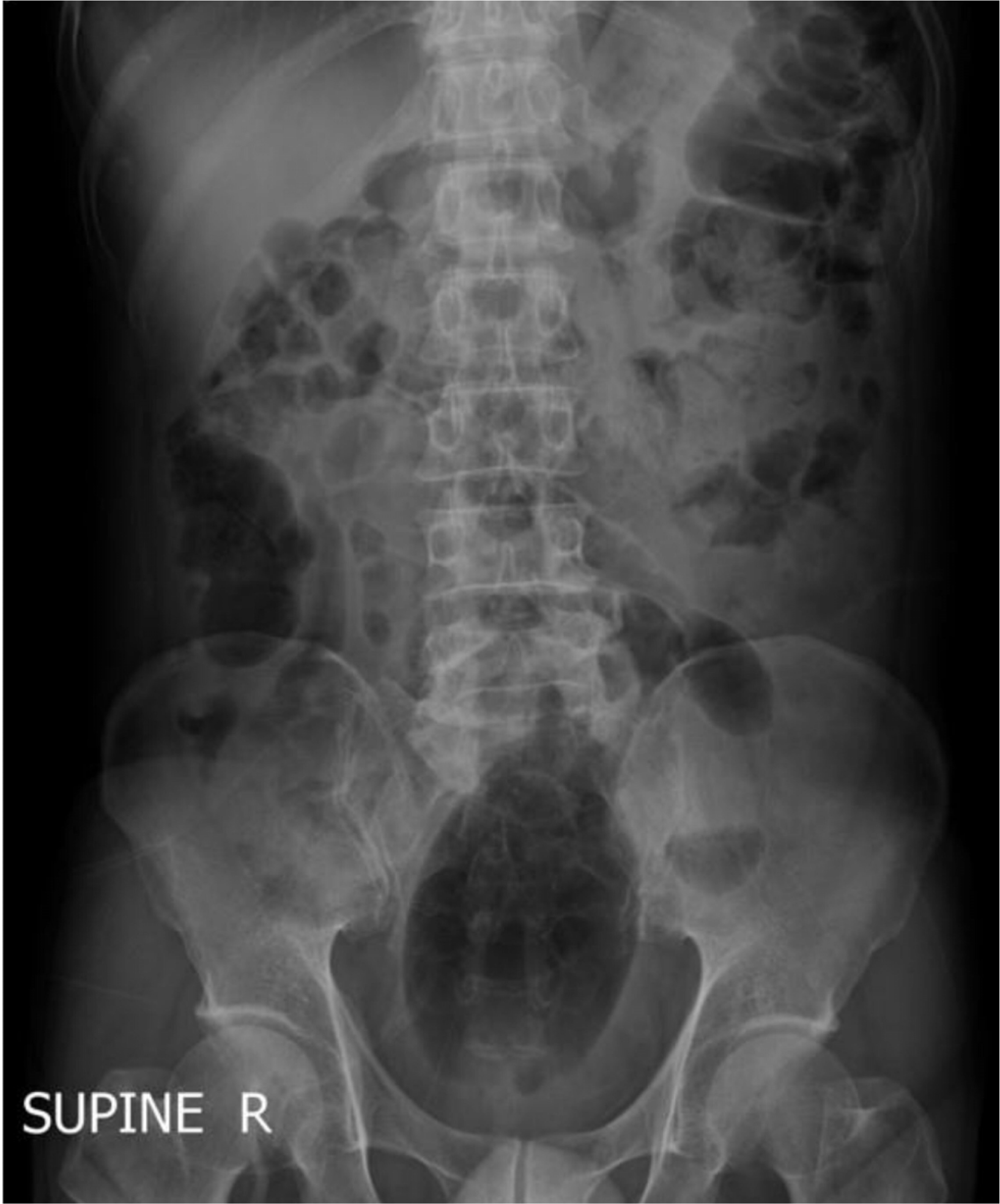 | Fig. 1Plain abdominal radiograph revealed a huge, oval object lying longitudinally in the rectosigmoid region.
|
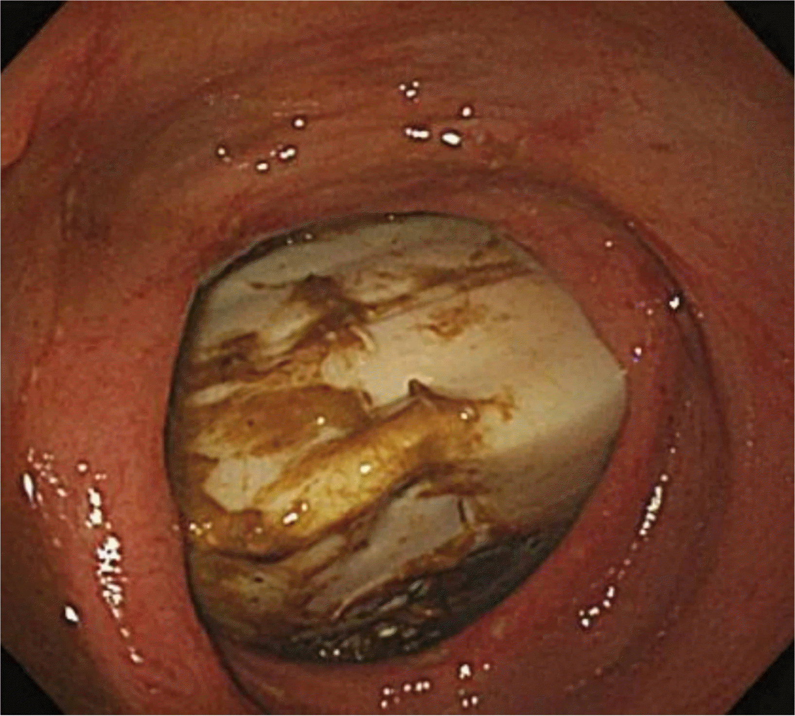 | Fig. 2Endoscopy demonstrated a large foreign body embedded tightly, just proximal to the middle rectal fold.
|
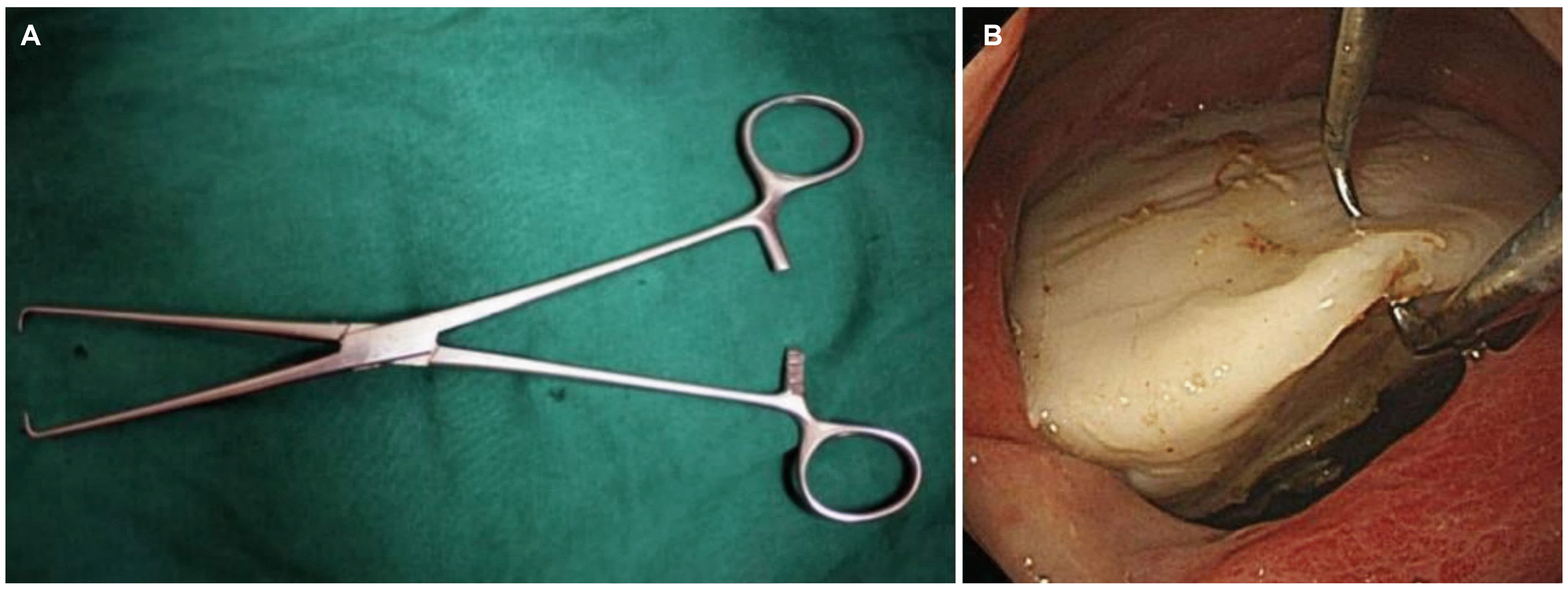 | Fig. 3Tenaculum forceps (23.0 cm, straight) (A) was inserted into the distal rectum under endoscopy, and (B) the rectal foreign body was grasped successfully with the hooks of the tenaculum forceps under endoscopy.
|
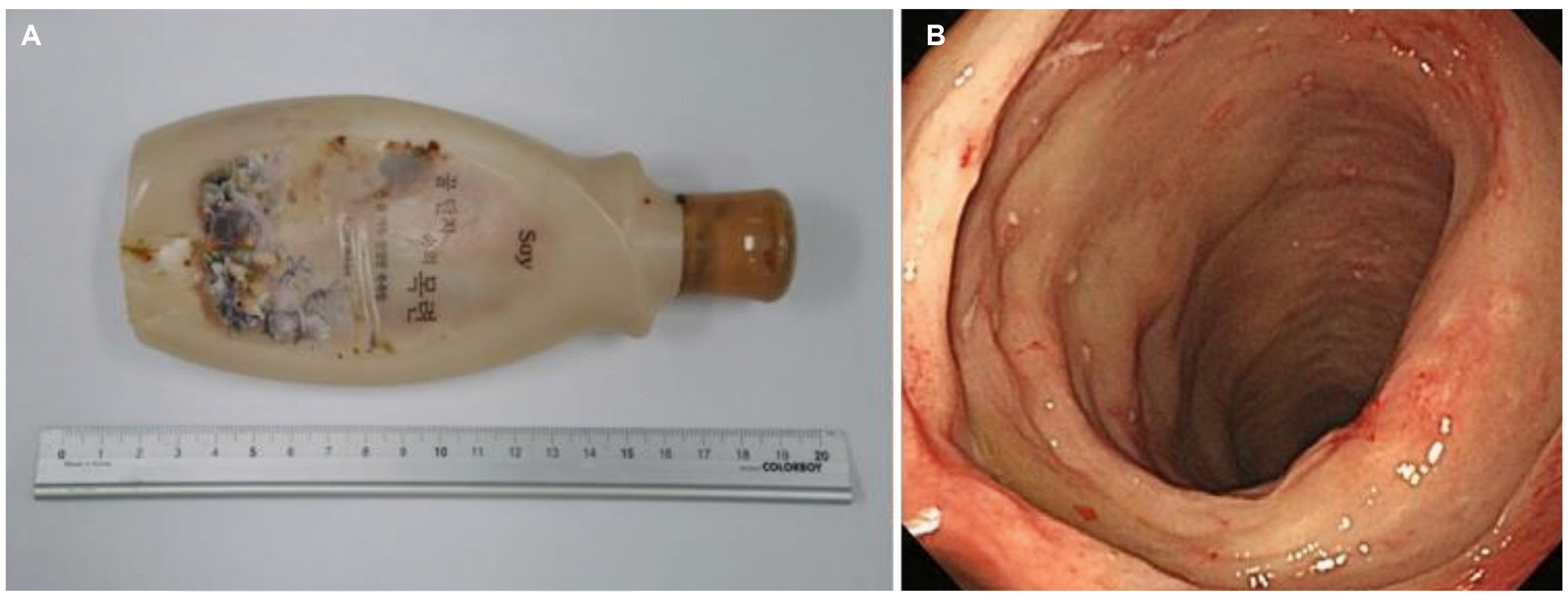 | Fig. 4Foreign body (A) removed successfully from the distal rectum measuring 17×6×3 cm with a capacity of 520 mL. (B) An endoscopic image obtained after extracting the rectal foreign body showed multiple linear shallow active ulcers with diffuse edematous mucosa and 10 cm to 25 cm in size from the anal verge.
|
Go to :
DISCUSSION
Patients with a foreign body in the upper gastrointestinal tract often visit the emergency department, but patients with a rectal foreign body are less common. The precise incidence of rectal foreign bodies is unknown, but it has been increasing.1,2 Most previously reported patients with a rectal foreign body were male with a mean age of 44 years.1,7 The main reason for rectal foreign body insertion was voluntary sexual purposes, rather than therapeutic purposes.2 Typically, patients are unwilling to provide details of the incident, so they come to the hospital late and delay treatment, sometimes resulting in serious complications.
Many types of rectal foreign bodies have been encountered. The most common type was a household object, such as a bottle or glass (42.2%). Other objects include the following: plastic bottles; foods, such as cucumbers and carrots; sports equipment; light bulbs; sex toys, such as vibrators and dildos; and cocaine packets.2,4,7
The most effective method for removing a rectal foreign body is determined by the shape, size, material, and location of the object. As there are various types of foreign bodies, various methods of removal have been reported.2,5,6 Therefore, careful history taking, physical examination, and abdominal X-ray or CT scans must be performed before planning treatment.2
The patient’s history can reveal the approximate type, size, and insertion period of the foreign body. Large and long foreign bodies can be embedded deeply in the rectum, and extended insertion periods can cause edema of the rectal mucosa and anal sphincter convulsion, hindering removal via a transanal approach.5 Surgical removal should be performed when a physical examination, abdominal X-ray, and CT scan indicate peritonitis or perforation.2,3 Further information can be obtained through DRE. Manual removal can be considered if a foreign body is palpable through DRE and located in the digital rectum. On the other hand, rigid or flexible proctosigmoidoscopy is required if the foreign object is not palpable through DRE.2,3 The status of the anal sphincter should be evaluated through the DRE. A decreased anal sphincter tone or visible injury is likely to be caused by insertion. An increased anal sphincter tone may indicate a secondary muscular spasm or a foreign body that has been inserted for a long time. If the anal sphincter tone is altered in either direction, removing the foreign body via the transanal approach will cause severe sphincter injury and should be avoided.2,3
Endoscopy is very effective in safely removing foreign bodies. Physicians can assess the location of the foreign body and the condition of the surrounding rectal mucosa with an endoscope. In addition, using endoscopic devices, such as snare and forceps, physicians can successfully remove a foreign body with minimal damage to the rectum and avoid surgery.2,3
In the present case, the patient’s diagnosis and treatment were delayed because he visited the hospital 3 days after inserting the object. The foreign body was large and long, and it was embedded deeply in the rectum, which was a serious concern for possible injury. Furthermore, the surface of the foreign body was smooth, and no protruding parts were visible under endoscopy, so conventional endoscopic devices could not grasp it. Tenaculum forceps, which are typically used in obstetrics, have long, sharp hooks that can be useful for catching smooth, round objects.8 Fortunately, the patient’s case was not complicated by perforation or peritonitis. His anal sphincter tone was normal, and no damage had occurred during insertion. Endoscopy confirmed that the foreign body moved slightly when the patient strained, as in defecating. The patient strained to evacuate the rectal body using the Valsalva maneuver. While performing the Valsalva maneuver, the patient’s defecation straining force increased, and the foreign body was removed without intervention according to the peristalsis of the rectum.
Previous reports have indicated that the Valsalva maneuver is helpful for removing a foreign body.2 When the Valsalva maneuver is performed during defecation, it raises the intra- abdominal pressure with a contraction of the chest, diaphragm, and abdominal muscles, and it increases the anorectal angle, making it almost straight.9,10 These processes increase the intrarectal pressure and activate rectal peristalsis to cause internal and external sphincter relaxation that facilitates defecation.9,10
The anal sphincter tone must be released to remove a foreign body by the transanal approach, and perianal nerve blocks, spinal anesthesia, and intravenous conscious sedation can be utilized. On the other hand, such procedures can result in serious neurological complications.3,8
This case had the advantage of minimizing rectal damage because the patient performed Valsalva maneuver voluntarily in a conscious state without anesthesia, and the procedure was conducted with endoscopic guidance.
To date, there are no clear standard treatment guidelines for removing large foreign bodies from the rectum. On the other hand, the first thing to consider is which method is least invasive and safest. If possible, patients should be treated with minimally invasive techniques, preferably while conscious.2,6 The present case showed that the removal of a large foreign body without force using the Valsalva maneuver under endoscopic guidance could help minimize the rectal damage.
Go to :
 XML Download