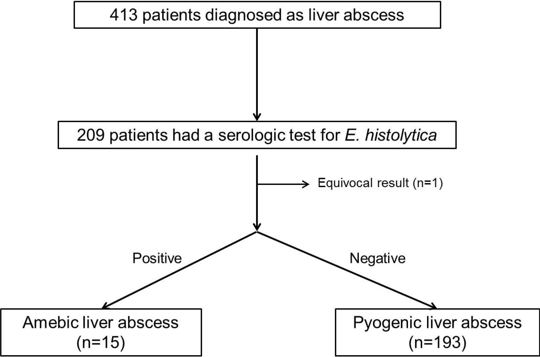
서 론
대상 및 방법
결 과
1. 아메바 및 화농성 간농양 환자의 기본 특징
Table 1
| Amebic liver abscess (n=15) | Pyogenic liver abscessd (n=193) | p-value | |
|---|---|---|---|
| Age (years) | 57 (49-74) | 66 (54-76) | 0.173 |
| Male | 12 (80.0) | 123 (63.7) | 0.267 |
| WBC (/mm3) | 13,780 (10,990-17,340) | 12,600 (8,785-17,135) | 0.937 |
| Hb (g/dL) | 12.4 (11.2-12.9) | 12.1 (10.9-13.4) | 0.625 |
| PLT (×103/mm3) | 221 (173-375) | 177 (117-245) | 0.024 |
| AST (U/L) | 45 (31-49) | 55 (36-100) | 0.432 |
| ALT (U/L) | 41 (32-78) | 57 (34-107) | 0.318 |
| ALPa (U/L) | 495 (326-1,029) | 467 (273-893) | 0.592 |
| GGT (U/L) | 147 (75-345) | 129 (64-246) | 0.521 |
| Total bilirubin (mg/dL) | 1.0 (0.6-1.9) | 1.0 (0.6-1.6) | 0.853 |
| Albumin (g/dL) | 3.0 (2.6-3.2) | 3.0 (2.6-3.3) | 0.774 |
| Creatinine (mg/dL) | 0.94 (0.85-1.08) | 1.06 (0.88-1.33) | 0.174 |
| Glucose (mg/dL) | 149 (123-236) | 137 (113-178) | 0.341 |
| PT INR | 1.25 (1.19-1.29) | 1.20 (1.13-1.29) | 0.797 |
| CRP (mg/dL) | 16.7 (11.1-21.3) | 18.0 (11.5-25.2) | 0.669 |
| Procalcitoninb (ng/mL) | 1.47 (0.32-2.44) | 3.07 (0.86-24.5) | <0.001 |
| HBsAg | 0 (0.0) | 8 (4.5) | 1.000 |
| Anti-HCV | 0 (0.0) | 0 (0.0) | NA |
| Anti-HIV | 1 (10.0) | 0 (0.0) | 0.063 |
| Diabetes mellitus | 8 (53.3) | 54 (28.1) | 0.047 |
| Immune suppressant use | 0 (0.0) | 1 (0.5) | 1.00 |
| Malignancyc | 1 (6.7) | 3 (1.6) | 0.260 |
WBC, white blood cell; Hb, Hemoglobin; PLT, platelet; AST, aspartate aminotransferase; ALT, alanine aminotransferase; GGT, gamma glutamyltranspeptidase; PT INR, prothrombin time international normalized ratio; CRP, C-reactive protein; HBsAg, hepatitis B s antigen; HCV, hepatitis C virus; HIV, human immunodeficiency virus.
aFor ALP, the reference range was 104-338 U/L before November 24, 2015 (phase I), and 40-129 U/L in male and 35-104 U/L in female since November 24, 2014 (phase II). All the patients with amebic abscess were included in period I. Among the patients with pyogenic abscess, 164 were included in period I, and the remainder were in period II. Therefore, only the patients in period I were analyzed.
bThe value was missing in four out of 15 patients with amebic abscess and 58 out of 193 patients with pyogenic abscess.
cThe malignancy was hepatocellular carcinoma in the amebic abscess group and gastric, duodenal, and colon cancer in the pyogenic liver abscess group each other.
dEight patients with pyogenic liver abscess were ultimately diagnosed as the one secondary to cholangitis. HBsAg was tested in the amebic group and in 178 of 193 among the pyogenic group. Anti-HCV was tested in all among the amebic group, and in 175 of 193 in the pyogenic group. Ani-HIV was tested in 10 out of 15 among the amebic group, and in 150 out of 193 among the pyogenic group.
Table 2
2. 아메바 및 화농성 간농양 환자의 방사선학적 특징 및 임상경과
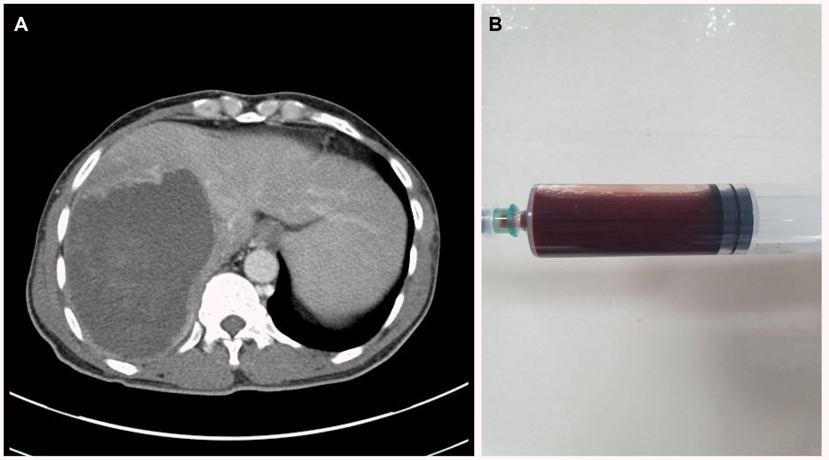
 | Fig. 2Computed tomographic findings of liver abscess. (A) Internal septation. (B) Peripheral rim enhancement (arrow). (C) Double target sign (arrowheads). (D) Hyperemia in adjacent hepatic parenchyma (arrow). (E) Cluster sign. (F) Gas formation; the image from (A-D) originated from the patients with amebic liver abscess, and the other (E, F) were from patients with a pyogenic liver abscess. |
Table 3
| Amebic liver abscess (n=15) | Pyogenic liver abscess (n=193) | p-value | |
|---|---|---|---|
| Number of abscess pockets | 0.501 | ||
| 1 | 11 (73.3) | 145 (75.1) | |
| 2 | 1 (6.7) | 23 (11.9) | |
| 3 | 2 (13.3) | 9 (4.7) | |
| ≥4 | 1 (6.7) | 16 (8.3) | |
| Maximal diameter of largest lesion (cm) | 6.5 (1.8-13.0) | 5.0 (1.0-13.8) | 0.262 |
| Dominant lobe | 0.057 | ||
| Right lobe | 13 (86.7) | 123 (63.7) | |
| Left lobe | - | 54 (28.0) | |
| Caudate lobe | - | - | |
| Both lobes | 2 (13.3) | 16 (8.3) | |
| Internal septationa | 5 (33.3) | 88 (46.1) | 0.340 |
| Peripheral rim enhancementa | 13 (86.7) | 111 (58.1) | 0.030 |
| Double target signa | 7 (46.7) | 48 (25.1) | 0.124 |
| Cluster signa | 0 (0.0) | 59 (30.9) | 0.007 |
| Hyperemia in adjacent hepatic parenchymaa | 11 (73.3) | 110 (57.6) | 0.233 |
| Gas formationa | 1 (6.7) | 12 (6.3) | 1.00 |
| Abutting to liver capsulea | 14 (93.3) | 164 (85.9) | 0.699 |
| Extrahepatic extension of the abscessb | 4 (26.7) | 35 (18.3) | 0.491 |
| Morphologic categroya | 0.508 | ||
| Multiple small lesions | 0 (0.0) | 13 (6.8) | |
| Multilocular confluent lesion | 5 (33.3) | 71 (37.2) | |
| Unilocular round lesion | 10 (66.7) | 107 (56.0) | |
| Percutaneous drainage use | 14 (93.3) | 133 (69.6) | 0.072 |
| Hospital stay (days) | 13.9 (0-61) | 15.0 (0-69) | 0.916 |
| Death during admission | 0 (0.0) | 9 (4.7) | 1.000 |
3. 아메바 및 화농성 간농양 환자의 동정균 분포
Table 4
| Amebic liver abscess (n=15) | Pyogenic liver abscessc (n=193) | |
|---|---|---|
| No growth | 11 (73.3) | 85 (44.0) |
| Klebsiellaa | 4 (26.7) | 89 (46.1)a |
| Streptococcusb | 11 (5.7) | |
| Escherichia coli | 7 (3.6) | |
| Enterococcus | 4 (2.1) | |
| Enterobacter | 3 (1.6) | |
| Proteus | 3 (1.6) | |
| Pseudomonas | 2 (1.6) | |
| Staphylococcus hominis | 2 (1.0) | |
| Candida albicans | 2 (1.0) | |
| Bacillus spp. | 2 (1.0) | |
| Clostridium | 1 (0.5) | |
| Corynebacterium | 1 (0.5) | |
| Gemella morbillorum | 1 (0.5) | |
| Fusobacterium | 1 (0.5) |
aAmong the amebic abscess group, all were Klebsiella pneumoniae, and among the pyogenic liver abscess group, 88 were Klebsiella pneumoniae and 1 Klebsiella oxytoca, respectively.


 PDF
PDF Citation
 Print
Print


 XML Download