

 PDF
PDF Citation
 Print
Print


INTRODUCTION
ERCP is an important procedure for the diagnosis and treatment of biliary and pancreatic disease, but successful biliary cannulation is not always achieved, even when performed by experienced endoscopists. In difficult cannulations, the procedure is prolonged, and repeated manipulation of the papilla can increase the risk of procedure-related complications.1 Several rescue techniques can be attempted after the failure of standard biliary cannulation. Common rescue methods include needle knife fistulotomy (NKF) or needle knife papillotomy.2 The double-guidewire technique (DGT) is also used, but it has been reported to increase the risk of post-ERCP-pancreatitis (PEP). Therefore, the European Society of Gastrointestinal Endoscopy guidelines recommended DGT as a backup technique.3
PEP is the most common complication associated with ERCP, occurring in 4-10% of patients. Prophylactic pancreatic duct (PD) stenting prevents PEP. Therefore, PD stenting can be considered in cases of difficult cannulation and inadvertent cannulation of the PD. This study evaluated the success and complication rates of NKF after unsuccessful cannulation using a standard method or DGT.
Go to : 
SUBJECTS AND METHODS
1. Patients and design
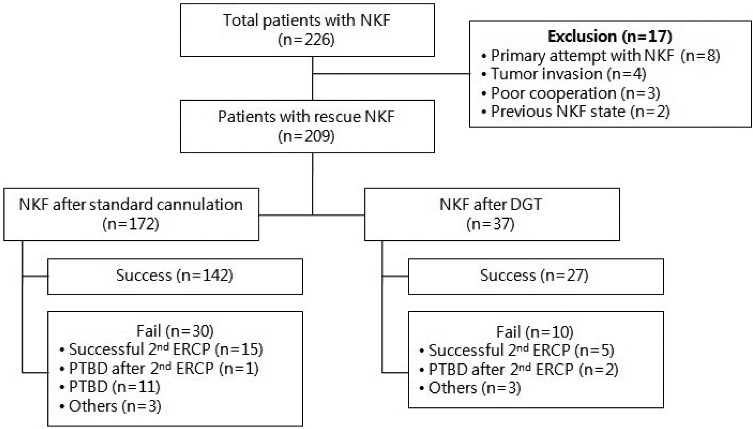
Between January 2009 and December 2016, the records of patients with pancreaticobiliary disease, who were admitted to Pusan National University Yangsan Hospital in Korea for diagnostic or therapeutic ERCP, were reviewed retrospectively. A total of 226 patients underwent ERCP with NKF. Of these, 17 were excluded because of a primary attempt with NKF (n=8), tumor invasion in the ampulla orifice (n=4), poor patient cooperation (n=3), or previous NKF (n=2). Therefore, 209 patients underwent ERCP with NKF as a rescue method after the initial failure of standard cannulation or DGT. In both groups, NKF was performed when selective cannulation was not achieved within 5 minutes. A PD stent was placed in the patients with 3 times more inadvertent guidewire insertions into the PD (Fig. 1).
All ERCP procedures were performed by two endoscopists (DH Kang and HS Nam) with more than 5 years of experience with therapeutic ERCP. The Ethics Committee of Pusan National University Yangsan Hospital approved this study (IRB No. 05-2018-037).
2. Definition of complications
Post-ERCP complications were defined as any procedure-related adverse events, including pancreatitis, bleeding, and perforation. PEP was defined as pancreatic abdominal pain occurring within 24 hours after the procedure with serum amylase or lipase levels that were at least 3 times higher than the normal range. The severity of pancreatitis was graded by consensus.4 Asymptomatic hyperamylasemia after ERCP was defined as a serum amylase level that increased 3 or more times in the first 24 hours following the procedure with no abdominal pain. Clinical data, such as vital signs, physical examination, and laboratory examination, were collected before ERCP and at 24 hours after ERCP. A decrease in the hemoglobin level of more than 3 g/dL, need for transfusion, or clinical evidence of bleeding (hematemesis, melena) were denoted as bleeding.
3. Endoscopic procedures
A standard side-view duodenoscope (JF-240 or TJF-240; Olympus Optical Co., Ltd., Tokyo, Japan) was used for the procedure. A sphincterotome (Ultratome XL; Boston Scientific, Natick, MA, USA) or an ERCP catheter (Fluoro Tip; Boston Scientific) with a hydrophilic guidewire (0.025- or 0.035-inch Jagwire; Boston Scientific) was used for the initial cannulation. In DGT, the second guidewire was used to cannulate the common bile duct (CBD), whereas the first guidewire was placed in the PD. The placement of a guidewire deep into the PD has the following functions: opening the papillary orifice, stabilizing the papilla, straightening the PD and CBD, and allowing access for the placement of a pancreatic stent when necessary.5
In cases of failed biliary cannulation using standard cannulation or DGT, NKF was performed as a rescue technique using a needle knife papillotome (MicroKnife XL; Boston Scientific). A fistulotomy was performed at the 12 o’clock position on top of the infundibulum. After puncturing the papillary roof at the prominent portion, the needle knife was used to cut down to the papillary orifice parallel to the direction of the papilla. This was stopped when bile juice or the bile duct mucosa was visible. A guidewire was inserted into the CBD through the fistulotomy site, and the needle knife was then withdrawn.6 PD stenting was performed in patients at a high risk of PEP.7 In cases of failed biliary cannulation after NKF; a second ERCP was performed within 1 or 2 days.
4. Statistical analysis
All statistical analyses were performed using SPSS software version 21.0 (IBM, Chicago, IL, USA). When necessary, continuous data were expressed as the mean±standard deviation. A χ2 test or F-test and t-test were used to compare the categorical variables. A p-value <0.05 was considered significant.
Go to :
RESULTS
1. Baseline characteristics
Two hundred and nine patients who underwent ERCP with NKF for rescue management were included. Of these, 172 underwent NKF after unsuccessful standard cannulation, whereas 37 underwent NKF after unsuccessful DGT. Table 1 lists the baseline characteristics. No significant differences were observed between the two groups. Choledocholithiasis and malignant biliary stricture were common indications of ERCP (44.0% [92/209] and 47.4% [99/209], respectively).
Table 1
Baseline Characteristics (n=209)
![]()
2. Successful cannulation
In the NKF group after unsuccessful standard cannulation, CBD cannulation was achieved in 142 patients (82.6%). Of the 30 patients who had unsuccessful cannulation after NKF, 16 underwent a second ERCP, and 11 underwent percutaneous transhepatic biliary drainage (PTBD). The remaining patients were transferred or supported conservatively. Among the patients who underwent a second ERCP, 15 achieved successful cannulation, and one underwent PTBD on the day after the second ERCP. Overall, the rate of successful cannulation using NKF after standard cannulation was 91.3%, including those who received a second ERCP.
In the NKF group after unsuccessful DGT, successful cannulation was achieved in 27 patients (73.0%). Of the 10 patients who had an unsuccessful CBD cannulation, seven underwent a second ERCP, and two underwent PTBD. Among the patients who received a second ERCP, five achieved successful CBD cannulation, whereas two did not. The patients underwent PTBD after 1 to 2 days after the second ERCP. Overall, the rate of successful cannulation using NKF after DGT was 86.5%, including those who underwent a second ERCP.
Successful cannulation and the total success rates following the second ERCP were similar in the standard and DGT group (82.6% vs. 73.0%, p=0.179; and 91.3% vs. 86.5%, p=0.369, respectively). The average cannulation time in those who underwent NKF after standard cannulation and NKF after DGT was 11.6±5.4 min and 14.1±4.4 min, respectively (p=0.01) (Table 2).
Table 2
Clinical Outcomes
![]()
3. ERCP-related adverse events
Overall, the rate of adverse events was 10.5% (18/172) in patients who underwent NKF after standard cannulation and 16.2% (6/37) in those who underwent NKF after DGT. Pancreatitis was the most common adverse event in both groups (9.9% vs. 16.2%, p=0.256). Six and 11 patients developed mild and moderate pancreatitis, respectively, in the standard group and two and four, respectively, in the DGT group. No case of severe pancreatitis was observed in either group. Hyperamylasemia developed in 16.3% (28/172) and 29.7% (11/37) of patients who underwent NKF after standard cannulation and those who underwent NKF after DGT, respectively. The incidence of pancreatitis and hyperamylasemia was similar in the two groups. No post-ERCP perforation developed in both groups and bleeding occurred in one patient in the standard group.
4. PD stenting
Unintended PD cannulation occurred in 109 patients in the standard group (109/172, 63.3%). In the standard group, a PD stent was performed in eight patients with no pancreatic complications. Of the 101 patients who did not receive a PD stent, 13 developed pancreatic complications (13/101, 12.3%). In the DGT group, a PD stent was used in eight patients with no cases of pancreatitis. Among the patients who did not receive stenting, six developed pancreatitis (6/29, 20.7%). The incidence of pancreatic complications was zero among those patients who had PD stenting in each group. In all patients who had undergone PD stenting, the rate of pancreatitis was low compared to those with no PD stent (0% [0/16] vs. 14.6% [19/130], p=0.132) (Table 3). On the other hand, the difference was not statistically significant, possibly because of the small number of patients who had PD stenting.
Table 3
Pancreatitis according to PD Stenting
![]()
Go to :
DISCUSSION
NKF can be used effectively in cases of unsuccessful standard cannulation or unsuccessful biliary cannulation after DGT. In this study, DGT was used when inadvertent cannulation of the PD was achieved, or the size of the ampulla was too small to use the needle knife initially. According to the European Society of Gastrointestinal Endoscopy guidelines, although the pancreatic guidewire-assisted technique is similar to the standard technique, there is a higher risk of developing PEP. This suggests that DGT is limited to the backup management of patients with unintentional cannulation of the PD,3 which is consistent with the DGT approach used in this study. In the present study, the success rates of primary ERCP biliary cannulation in the standard and DGT groups were 82.6% and 73.0%, respectively. These rates are similar to those reported elsewhere (67-91%) but lower than that reported in a recent study (96%).8-10 The success rates of cannulation, including second ERCPs, were 91.3% and 86.5% in the standard and DGT groups, respectively. According to previous studies, performing a second ERCP immediately after the failure of NKF can be considered in some patients.11-12 These results are not inferior to other studies because this study only included patients using NKF as a rescue method, and not all those with the standard cannulation. The procedure time was extended in the DGT method because it took more time to try selective cannulation alongside the guidewire in place in the PD.
The baseline characteristics of the patients were similar in both groups. On the other hand, the incidence of periampullary diverticulum (PAD) was slightly higher among the patients in the DGT group. The success rate of biliary cannulation was higher among the patients without a diverticulum (84.7% [144/170] vs. 64.1% [25/39]). In contrast, Park et al.13 reported no significant difference between patients with PAD who had NKF and those in a control group (93.9% vs. 88.4%, p=0.523). The papilla located inside the diverticulum or at the inner margin of the diverticulum (PAD types I & II) may have affected the cannulation in the present study.
The incidence of PEP varies widely according to the institution and country, ranging from 5-10%. Several studies using NKF in cases of difficult cannulation reported pancreatitis rates of 1-11%.10 In the present study, pancreatitis developed more frequently in patients with DGT (16.2% vs. 9.9%, respectively). On the other hand, despite not being statistically significant, there was a trend to develop pancreatitis. In exclusion cases, there was no PEP in the primary attempt with NKF, but it was too small to evaluate the significance. Meta-analysis showed that the sole use of DGT increased PEP significantly compared to the other endoscopic techniques (relative risk, 1.98; 95% CI, 1.14-3.42).4 Prophylactic PD stenting may decrease the incidence of pancreatic complications. No complications were reported in 16 patients who underwent PD stenting, whereas complications developed in 14.6% of those who did not receive PD stenting. This suggests that PD stenting should be considered to reduce the incidence of pancreatic complications in patients undergoing DGT. In the present study, however, PD stenting was performed in patients who had inadvertent PD cannulation or those who were at a high risk of developing pancreatitis. The indications of PD stenting in the present study were female, aged under 40, no dilatation of the bile duct, three or more repetitive pancreatic cannulations. In general, PD stenting is recommended for high-risk PEP patients. Whether PD stenting should be performed in all DGT procedures is unclear. Therefore, further studies will be needed to evaluate PD stenting in patients undergoing DGT.
This study has several limitations. First, it was retrospective in design. The data on the comorbidities or disease progress might be insufficient, and the sample size was small, which may have affected the statistical analysis. Second, it was a single centered study that did not include a community hospital. This may have affected the interpretation of NKF and PD stenting. Third, the study was performed by very experienced endoscopists, which may have affected the success and complication rates related to the procedure. Therefore, prospective multicenter studies will be needed to overcome these limitations.
In conclusion, NKF may be an effective and safe method that can be considered for rescue management after the initial failure of standard cannulation or DGT. Although not statistically significant, NKF after standard cannulation could be preferred over NKF after DGT, considering the shorter procedure time, higher success rate, and lower rate of pancreatitis. PD stenting can be considered in selective patients at a high risk of PEP.
Go to :
 XML Download