

 PDF
PDF Citation
 Print
Print


INTRODUCTION
A pyogenic liver abscess (PLA) is a serious clinical entity with a high morbidity and mortality rate. The most common cause of PLA is biliary disease. However, some PLAs do not have an obvious underlying cause, that is, are cryptogenic, although colonic lesions, such as diverticulitis, have been identified as a plausible infectious route for portal-flow bacteremia or intraperitoneal bacterial spreading to the liver.1 Liver cyst infection can also occur in patients with autosomal dominant polycystic kidney disease, even though a simple liver cyst infection in those without underlying disease is extremely rare.
Colonoscopy is a gold standard for the diagnosis and treatment of colorectal pathology. On the other hand, although rare, there are serious infectious complications following colonoscopy, particularly when polypectomy is performed. Some studies have reported PLA complicated after a colonoscopic polypectomy.2,3 This paper reports three unusual cases of PLA or liver cyst infection occurring after colonoscopic polypectomy. These cases suggest that colonic origin might be an important source of PLA (Table 1).
Table 1
Baseline and Clinical Characteristics of the Three Patients
| Case | Sex/age | Risk factorsa | Clinical | symptoms | Time from colonoscopy to PLA | Location (L/R) | Microbiology | Treatment |
|---|---|---|---|---|---|---|---|---|
| Case 1 | M/58 | No | Fever, chill, altered | mentality | 1 week | R | Klebsiella pneumoniae | Antibiotics, sono-guided aspiration |
| Case 2 | F/58 | No | Fever, chill, left subcostal | pain | 6 days | L | No growth | Antibiotics, sono-guided aspiration |
| Case 3 | M/84 | No | Fever, general | weakness | 2 weeks | R | No growth | Antibiotics, percutaneous drainage |
![]()
Go to : 
CASE REPORT
Case 1
A 58-year-old man visited the emergency room (ER) via ambulance because of fever, chills, and altered mentality. These symptoms persisted for 2 days. The patient had no specific medical history, including diabetes mellitus or cancer, except that he underwent a colonoscopic polypectomy 1 week before the ER visit at an outside clinic. A transverse colon polyp had been removed, and a tubular adenoma with low-grade dysplasia was reported. Another hyperplastic rectal polyp was also removed. Because the patient’s data were unavailable, the quality of bowel preparation was unknown. On admission, his blood pressure was 65/50 mmHg. His pulse rate was 106/min, and his body temperature was 39.8℃. The physical examination showed no specific findings. His white blood cell count was 3,060/mm3 (neutrophil 85%), and his inflammatory markers were elevated. CRP and procalcitonin was 14.25 mg/dL and 5.73 ng/mL, respectively. Abdominal CT revealed a 3.5-cm lobulated low-attenuation lesion in segment 8 in the right hepatic lobe with a peripheral wedge-shaped slightly low attenuation area adjacent to the main lesion (Fig. 1A). A thrombus was shown in the middle hepatic vein (Fig. 1B). He was diagnosed with septic shock related to a PLA. Hydration with 0.9% saline, norepinephrine, and empirical antibiotics (ceftriaxone and metronidazole) was performed through injection. His vital signs were then stabilized. Klebsiella pneumoniae was identified in his blood culture. One week later, sono-guided percutaneous abscess aspiration was performed, and 3 mL of bloody fluid was collected (Fig. 1C). He was discharged with oral antibiotics (cefixime). Six months later, abdominal ultrasonography revealed an ill-defined iso-echogenic area in segment eight. The thrombus in the hepatic vein was invisible (Fig. 1D).
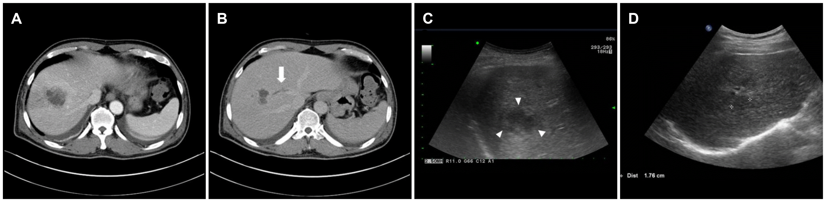 | Fig. 1(A) In case 1, the initial abdominal computed tomography showed a 3.5-cm lobulated low-attenuation lesion in segment eight in the right hepatic lobe. (B) Thrombus was shown in the middle hepatic vein (arrow). (C) Ultrasonography on aspiration revealed an ill-defined hypoechogenic lesion in the right hepatic lobe (arrowheads). (D) Follow-up ultrasonography after 6 months showed an ill-defined iso-echogenic area in the previous abscess pocket (asterisks).
|
Case 2
A previously healthy 58-year-old woman was admitted with complaints of fever, chills, and left subcostal pleural pain for 2 days. She had no history of diabetes mellitus or cancer. She had undergone a colonoscopic polypectomy 6 days before at an outside clinic (no available data). Her blood pressure was 120/80 mmHg, and her body temperature was 38.3℃. Tenderness was detected in the epigastric area. Her white blood cell count was 18,380/mm3 (neutrophil 82%) and CRP level was 4.69 mg/dL. Abdominal CT revealed a 2.5-cm lobulated low-attenuated mass abutting the hepatic capsule in the left lateral segment of the liver. Internal septum-like enhancement and hyperemic changes in the adjacent parenchyma were noted (Fig. 2A). She was diagnosed with a PLAs and treated with intravenous cefotaxime and metronidazole. Sono-guided aspiration was performed, and 15 mL pus was drained (Fig. 2B). No pathogen was identified in her blood or pus culture. The antibody to Entamoeba histolytica was negative. She was switched to oral cefixime and discharged. Follow-up CT was performed 2 weeks later, which showed improvement in the abscess (Fig. 2C).
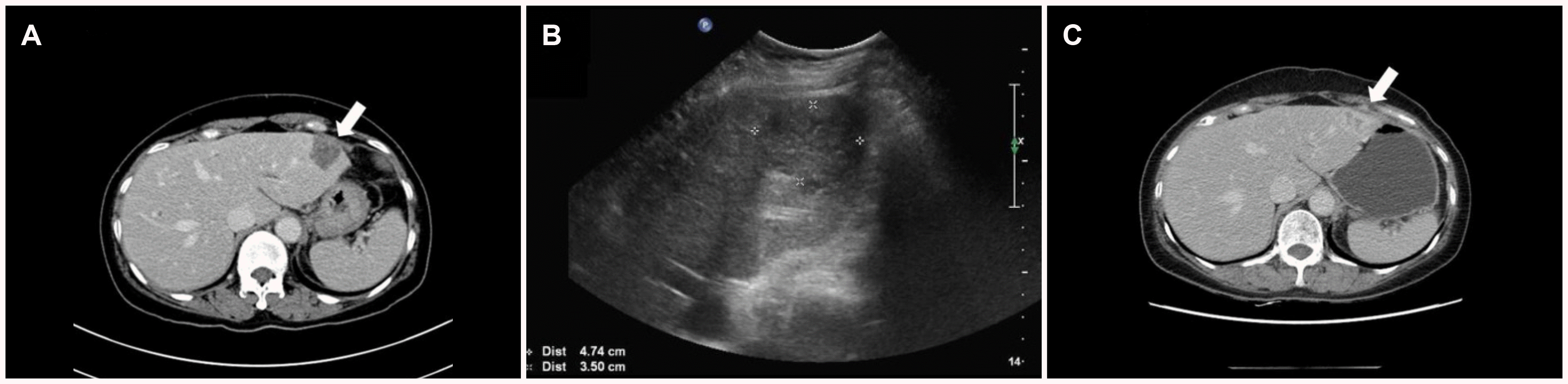 | Fig. 2(A) In case 2, the initial abdominal computed tomography (CT) showed a 2.5-cm lobulated low-attenuated mass abutting the hepatic capsule in the left lateral segment of the liver (arrow). (B) On sono-guided aspiration, the lesion was observed as a heterogeneous echogenic mass with a low echogenic soap-bubble-like mass (asterisks). (C) Follow-up CT was performed 2 weeks later. The abscess showed improvement (arrow).
|
Case 3
An 84-year-old man was referred to the ER for intermittent fever (up to 39℃) and general weakness in a geriatric hospital. He had no specific medical history, including diabetes mellitus or cancer, except that he had undergone a colonoscopy for constipation in this hospital 2 weeks before admission. His symptoms developed a few days after the colonoscopic procedure. He was treated in a geriatric hospital, but his symptoms did not improve. Therefore, he was transferred to this hospital. The colonoscopy performed 2 weeks before showed two polyps in the transverse colon. One was a 7-mm Is type polyp, which was removed by an endoscopic mucosal resection. The other was a 3-mm Is type polyp that was removed by cold biopsy forceps. Both polyps were reported as tubular adenoma (Fig. 3). The quality of his bowel preparation was excellent (Boston bowel preparation scale: 9). Six months before, he had undergone an abdominal CT scan for constipation. Multiple simple hepatic cysts were noted; the largest was 5 cm (Fig. 4A). Follow-up abdominal CT upon admission revealed the largest cyst to have peripheral wall thickening and hyperemic changes in the adjacent liver parenchyma (Fig. 4B). On admission, his blood pressure was 137/70 mmHg, and his body temperature was 37.0℃. Mild tenderness was noted in the right upper abdomen. His white blood cell count was 5,490/mm3 and the CRP level was 2.58 mg/dL. He was diagnosed with an infected hepatic cyst. Intravenous ceftriaxone was started. Two days later, a percutaneous drainage tube was inserted. One week later, CT revealed a decrease in the size of the lobulated hepatic cystic lesion (Fig. 4C). He was discharged with oral cefpodoxime after catheter removal. No pathogen was identified in his blood or drained fluid culture. The antibody to Entamoeba histolytica was negative.
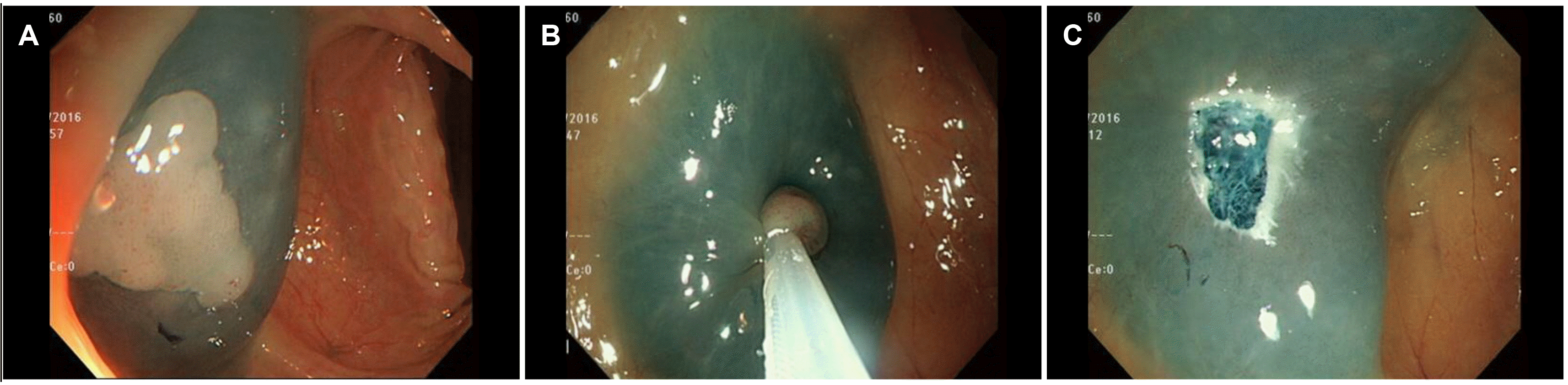 | Fig. 3(A-C) In case 3, colonoscopy 2 weeks before showed a transverse colon polyp, which was removed by snare polypectomy.
|
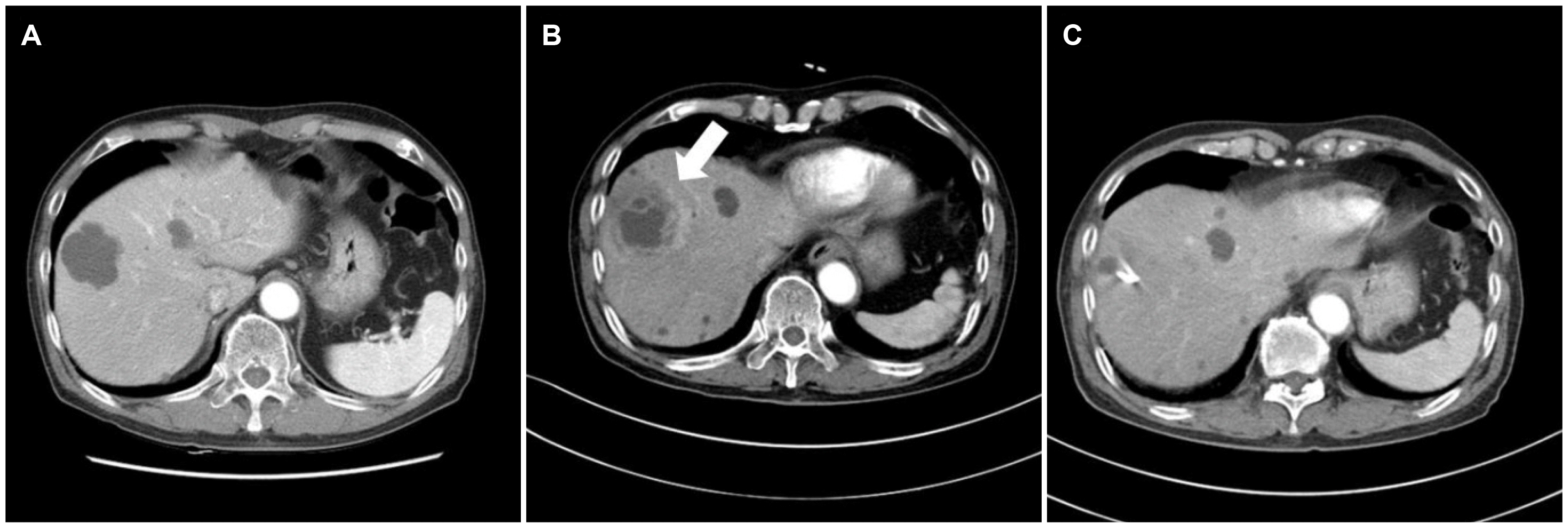 | Fig. 4(A) In case 3, abdominal computed tomography (CT) performed 6 months prior to admission showed multiple simple hepatic cysts. (B) Upon admission, CT showed that the wall of the largest cyst was enhanced with adjacent hyperemic change (arrow). (C) One week later, the size of the complicated cyst with the catheter decreased.
|
Go to :
DISCUSSION
The incidence of PLA ranges from 10 to 20 cases per 100,000 hospital admissions.4 The prognosis of PLA has improved with the advances in diagnostic and therapeutic techniques. Nevertheless, early diagnosis of PLA is essential because its mortality rate remains at approximately 10%.5 On the other hand, its diagnosis can be delayed because of its largely nonspecific presentation unless the clinician maintains a high level of suspicion. A retrospective study reported that only 59%, 39%, and 14% of PLA patients present with fever, right upper quadrant pain, and peritoneal signs, respectively. Therefore, its diagnosis is often delayed for an average of 1 week from symptom onset.6
The routes of invasion include the biliary tree, portal and systemic circulation, direct intraperitoneal extension, and penetrating trauma. In the past, the most common cause of PLA was appendicitis complicated by pylephlebitis. With the introduction of effective antibiotic therapy, this has become a rare cause of PLA. The most common cause of PLA is biliary tract disease, such as cholangitis.7 Nevertheless, approximately 35% of PLA cases remain cryptogenic.6 Some cryptogenic PLA cases are probably associated with colonic mucosal lesions caused by a range of conditions, such as colon neoplasm,7-9 inflammatory bowel disease,10 and intestinal tuberbulosis.11 In addition, there are some reports of PLAs related to colonic procedures,2,3,12-14 in which most cases have underlying disease disrupting colon mucosa, such as ulcerative colitis,12 tubulovillous adenoma,13 and adenocarcinoma.2,3 One case occurred 1 week after a screening colonoscopy without intervention. This case, however, had sigmoid diverticulosis, which might be a route for bacteremia after a colonoscopic examination.14 For cases of PLA following colonoscopic polypectomy, advanced adenoma (tubulovillous adenoma)13 and adenocarcinoma2,3 can cause a mucosal defect that provides a route for the bacteria to invade the portal system or spread intraperitoneally to the liver. Therefore, PLA in these cases might have originated from an underlying colon disease rather than from a colonoscopy. In contrast, in the present cases, there was no evidence of colon neoplasm or inflammation that could disrupt the colon mucosa. Two patients, cases 1 and 3, had only small (<1 cm) tubular adenoma. The other case (case 2) denied a colon malignancy for her endoscopic pathology (no available data). Therefore, their PLA or liver cyst infection might have been due to mucosal disruption occurring after colonoscopic polypectomy.
A liver cyst infection is rare, occurring mostly in patients with autosomal dominant polycystic kidney disease.15 Only one report of a liver cyst infection after colon endoscopic mucosal resection in a patient with autosomal dominant polycystic kidney disease could be found.16 The present patient had no predisposing factors except for old age. To the best of the authors’ knowledge, this is the first case of a simple liver cyst infection after a colonoscopic polypectomy.
Klebsiella pneumoniae is the most common pathogen isolated from PLA. Several studies have suggested that Klebsiella pneumoniae is associated with PLA of a colonic origin.7,17 In case 1, Klebsiella pneumoniae was identified in the blood culture, suggesting the possible hematogenous spread of bacteria via the mucosal defect caused by the colonoscopic polypectomy. In cases 2 and 3, the negative results in blood and pus culture could be due to the following two reasons: 1) the pathogen might be an anaerobe that was a colonizer in the colon; 2) the patient had been partially treated at an outside hospital.
In the present cases, the risk factors of PLAs, such as diabetes mellitus, previous biliary operation, or immune suppression, could not be found. In the literature, the rate of bacteremia related to colonoscopy was 0-25%. This was not associated with infectious complications.18 One hypothesis was that inadequate bowel preparation might influence bacteremia from the colon. However, no study has dealt with this topic yet. In this context, more studies will be needed to determine the risk factors for PLA after a colonoscopy.
In conclusion, the present cases suggest that colonic mucosal lesions caused by a colonoscopic polypectomy should be considered possible causes of cryptogenic PLA. Clinicians should be aware of the possibility of PLA if a patient complains of fever and abdominal pain after a colonoscopic polypectomy.
Go to :
 XML Download