

 PDF
PDF Citation
 Print
Print


INTRODUCTION
Peptic ulcer disease can be complicated by inflammation, ulceration, bleeding, or perforation. The condition can be diagnosed easily if there is a history of ulcer or acute characteristic pain in the abdomen. On the other hand, it is difficult to diagnose patients without the characteristic symptoms of peptic ulcer or elderly patients who are insensitive to pain. Penetration into the liver is a rare peptic ulcer disease complication that can lead to severe complications, such as upper gastrointestinal bleeding or abscess formation.1 This paper reports a 72-year-old female who developed a silent gastric ulcer perforation to the liver. The diagnosis was made by a histological examination of endoscopic biopsies, and surgical treatment was performed.
Go to : 
CASE REPORT
A 72-year-old female visited the hospital because of a two-week history of weakness, dizziness, poor oral intake, and vomiting. She appeared pale and had anemic conjunctiva. Her abdomen was soft without organomegaly or mass, and she did not suffer from abdominal pain or distension. On the other hand, she complained of tenderness on the pressure of the epigastric area. The bowel sounds were normo-active. The rectal examination did not reveal any palpable mass. Five months before her visit to the hospital, she purchased painkillers at the pharmacy and used them because of sustained pain on the left shoulder. She had anemia in the blood test at a local hospital 3 months ago and was taking hypertension medication.
Upon arrival to the emergency room, her initial vital signs were a regular heart rate at 102 beats/min, a blood pressure of 100/60 mmHg, and 36.5ºC of body temperature. The following are the results of her complete blood and biochemistry tests at the time of her visit: complete blood count, white blood cells count 18.75×10⁹ L (95% neutrophils, 4% lymphocytes); hemoglobin 2.5 g/dL; hematocrit 9.1%, platelet 882 k/mm3; liver function tests; PT 11.7 sec; total bilirubin 0.3 mg/dL; AST 17 U/L; ALT 6 U/L; ALP 199 U/L; BUN 53 mg/dL; creatinine 1.15 mg/dL; glucose 174 mg/dL; amylase 26 U/L; lipase 14 U/L; and CRP 8.11 mg/dL.
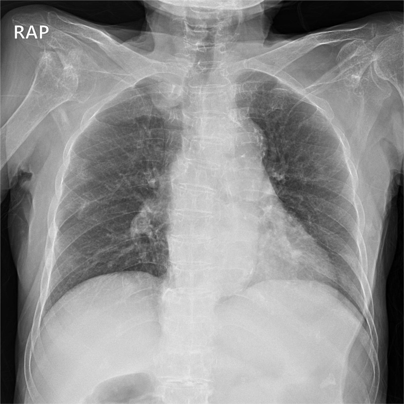
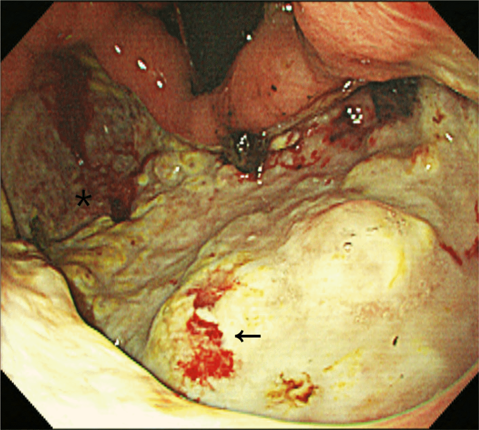
There was no free air in the chest X-ray (Fig. 1). CT revealed diffuse wall thickening in the stomach with a suspected ulcerative lesion in the lesser curvature side of the gastric antrum, possible sealed-off perforation, and perigastric infiltration (Fig. 2). Upper gastrointestinal endoscopy revealed a large ulcer (A1 stage), which was identified by CT, approximately 10 cm in size at the lesser curvature side of the body (Fig. 3). The pylorus had a deformity because of an ulcer scar and had stenosis that impeded the endoscope. The duodenal bulb and second portion were normal. Biopsies from the center and margin of the gastric ulcer were taken. The rapid urease test for Helicobacter pylori was positive. A histology examination of the biopsies revealed ulcer debris and hepatic parenchyma (Fig. 4). Considering the size of the ulcer and the risk of developing a liver abscess, it was decided that surgery should be performed. Hemigastrectomy was performed, and the pathology of the surgical specimen was a chronic ulcer with perforation and fibrous adhesion to the liver. The patient started sips of water and diet on the 5th day of surgery and was discharged on the 13th day of surgery without postoperative complications.
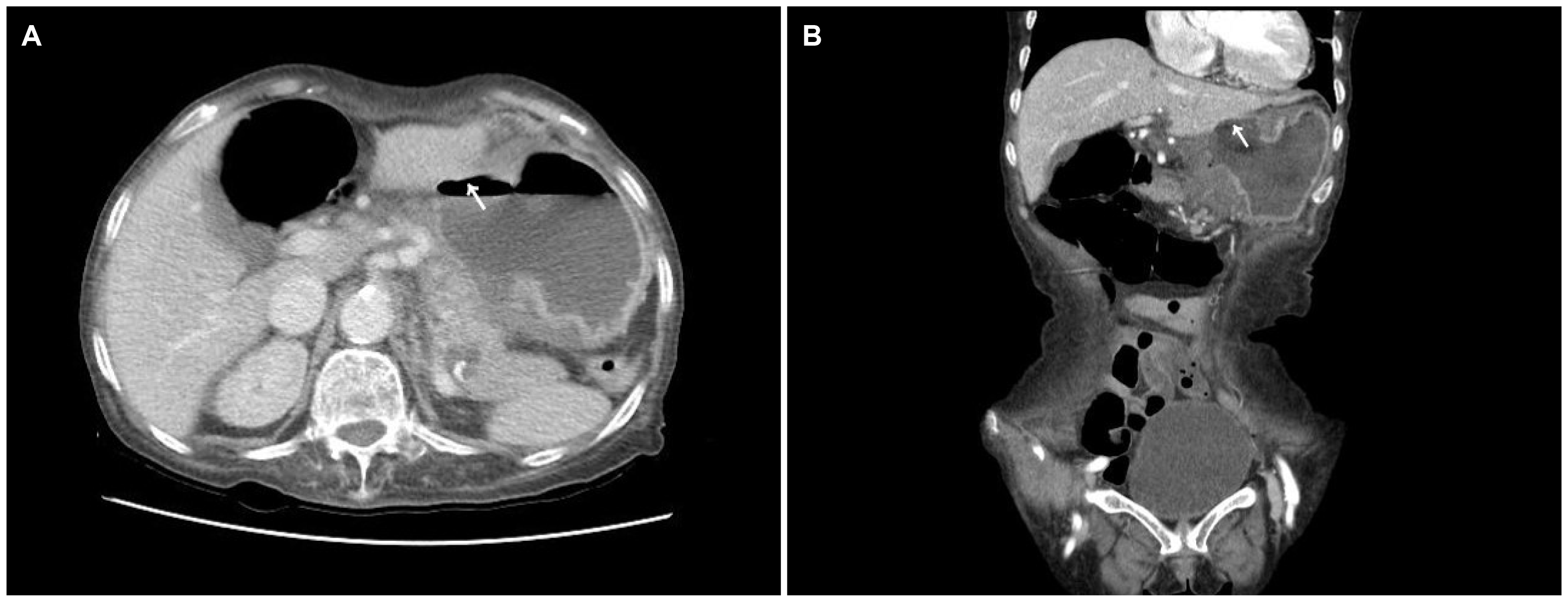 | Fig. 2(A, B) Abdominal computed tomography shows diffuse wall thickening in the stomach with a suspected ulcerative lesion (arrows) in the lesser curvature side of the gastric antrum and perigastric infiltration.
|

Go to :
DISCUSSION
In a systematic review, the reported annual incidence of peptic ulcer perforation ranged from 3.8 to 14 per 100,000 individuals.1 Thirty-day and 90-day mortality rates of up to 20% and 30%, respectively, have been reported.2 In developing countries, patients tend to be young male smokers. In developed countries, however, patients tend to be elderly with multiple co-morbidities and the associated use of NSAIDs or steroids. NSAIDs, Helicobacter pylori, physiological stress, smoking, corticosteroids, and previous history of peptic ulcer disease are risk factors for perforated peptic ulcer.2 The clinical features of peptic ulcer perforation include the sudden onset of abdominal pain, tachycardia, and abdominal rigidity in the early stages, followed by abdominal distension, pyrexia, hypotension, fever, and vomiting.3 The prognosis for a diagnosis within the first 6 hours is good, but adverse effects increase markedly when the delay exceeds 12 hours. Therefore, a rapid diagnosis is essential.4 Abdominal plain film, CT, and ultrasonography may have a diagnostic role in confirming peptic ulcer perforation.5
Organ penetration to the pancreas, gastro-hepatic omentum, biliary system, and liver was reported in 8% of 417 patients who had undergone surgical treatment due to peptic ulcer disease.6,7 Liver penetration is a rare but severe complication of peptic ulcer disease. Generally, the diagnosis is made by surgery or endoscopic biopsy.7-12 Non-surgical treatment using antibiotics and intravenous proton pump inhibitors may be possible if the patient has no leakage, is clinically stable, and shows signs of improvement. On the other hand, urgent surgery is essential if the patient’s condition deteriorates, regardless of a leak.
In 2004, Kayacetin and Kayacetin8 described 14 cases of gastric ulcer penetrating to the liver. All the patients showed severe gastrointestinal bleeding, as in the present case. Abdominal pain was described in only four patients. Therefore, in the present case, abdominal pain did not appear to be a common finding. Most cases were located at the anterior wall or lesser curvature of the corpus or antrum. Nine of the 14 cases were treated with surgery operation.
Most cases had normal liver function tests. This may reflect that local hepatic injury does not cause abnormalities in the liver functions. The diagnostic value of liver function tests in cases of ulcer penetration into the liver is limited.
In this case, the patient showed severe anemia at the time of hospitalization and complained of dizziness, vomiting, inadequate oral intake, and epigastric tenderness. A physical examination did not show any signs indicating a perforation, such as rebound tenderness and peritoneal irritation sign, and there was no free air in the simple abdominal image. In the present case, the history of painkiller medication, advanced age, and Helicobacter pylori could be the cause of the gastric ulcer perforation. A small gastric ulcer perforated to the liver. As the liver tissue around the perforation became inflamed and fibrous, it caused adhesion, which concealed the patient’s perforation symptoms. As the size of the gastric ulcer gradually grew, it is believed that ulcer bleeding caused severe anemia.
In conclusion, a high index of suspicion to make a diagnosis is necessary when patients have the risk factors of a perforated peptic ulcer because they may have a silent perforated peptic ulcer that is not accompanied by sudden physical symptoms.
Go to :
 XML Download