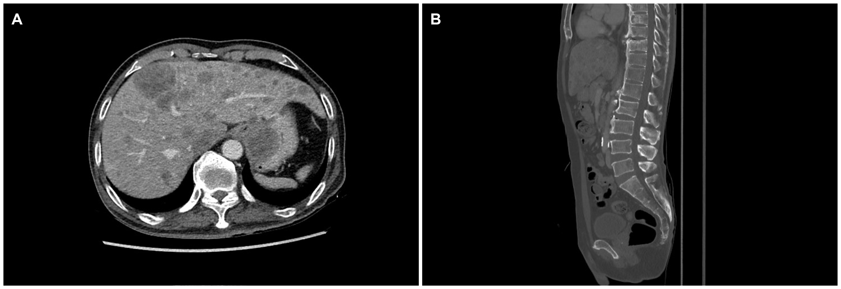
A 64-year-old man presented to Ewha Womans University Seoul Hospital with complaints of right chest, abdominal pain and dysphagia. He had experienced multiple traumatic rib fractures 6 months prior and he received conservative management at a local hospital. Recently, the pain had not subsided even when administering opioid painkillers, and he was transferred to Ewha Womans University Seoul Hospital for further evaluation. He complained of progressive dysphagia to solid foods rather than liqids. He denied having undergone previous surgeries or any past history or family history of cancer. He had an initial blood pressure of 139/97 mmHg, a pulse rate of 143 beats/min, a body temperature of 36.6℃, and a respiratory rate of 22 breaths/min. The physical examination revealed direct tenderness at the right ribs. Non-specific findings were noted on the abdominal physical examination. The laboratory test results were as follows: a white blood cell count of 9,580 cells/mm
3, a hemoglobin level of 12.6 g/dL, and a platelet count of 193×10
3 cells/mm
3. No abnormality of his electrolytes was noted. Elevated levels of AST (98 U/L), ALT (121 U/L), ALP (457 U/L), and LDH (2,636 U/L) were showed. The serum levels of tumor markers such as CEA (47 ng/mL) and CA 19-9 (65 U/mL) were increased, whereas the AFP level (3.5 ng/mL) was within the normal range. His chest X-ray showed multiple acute fractures of the right ribs. Chest CT revealed no abnormalities. A contrast-enhanced CT scan of the abdomen and pelvis showed a mass measuring 3.6 cm around the gastric fundus portion and multiple liver and lymphatic metastases in the portal hepatis and periportal areas. Dissemimated bone metastasis at whole spines, sternum, ribs, and both pelvic bones were noted on the abdomen and pelvis CT (
Fig. 1). Esophagogastroduodenoscopy was performed and a semicircular longitudinal ulcerative mass was found to be located at the distal esophagus (34-37 cm from the upper incisor). A mass measuring about 4 cm in diameter with central ulceration was noted at the cardia (
Fig. 2). An intensely hypermetabolic esophageal mass involving the gastroesophageal junction (standardized uptake value [SUV] 11.0) was noted on PET-CT. In addition, multiple hypermetabolic lesions were detected in the whole liver (SUV 14.5), bones, and lymph nodes (SUV 11.5) at the left gastric, portocaval, and porta hepatis areas (
Fig. 3). Histopathological examination of an endoscopic esophageal biopsy specimen showed small cell neuroendocrine carcinoma (NEC) and SCC in a colliding pattern. The gastric mass in the cardia was grossly suspected to be a malignant gastrointestinal stromal tumor, but the finding was inconclusive. Immunohistochemistry showed p63 (+) and p16 (+) in the squamous carcinoma cells and cluster of differentiation-56 (+), synaptophysin (+), and thyroid transcription factor-1 (+) in the NEC cells (
Fig. 4). Therefore, the final diagnosis was a collision tumor of the distal esophagus (T3N3M1, clinical stage IV). He was transferred to the oncology department for palliative chemotherapy. The patient was treated with palliative chemotherapy using a combination of etoposide (100 mg/m
2 for 3 days) and cisplatin (60 mg/m
2 for 3 days). He died 2 months later due to his suppressed immune condition after the first course of chemotherapy.
 | Fig. 1Abdominal computed tomography findings. (A) About a 3.6 cm mass around the gastric fundus portion and also liver metastasis. (B) Multiple bone metastasis in the vertebra and sacrum. 
|
 | Fig. 2Endoscopic images of the esophageal neoplasm. (A, B) A semicircular longitudinal ulcerative mass at the distal esophagus. (C) About a 4 cm sized subepithelial mass with central ulceration at the fundus. 
|
 | Fig. 3Positron emission tomography-computed tomography findings. (A-C) A hypermetabolic lesion in the esophagus and cardia, and several tumors in the whole liver, pancreas, and bone. 
|
 | Fig. 4Histologic features of the esophagus specimen. (A) The positive for the component of the neuroendocrine carcinoma adjacent to a squamous carcinoma without intermigling (H&E, ×400). (B) Strong staining of p63 (+) for the squamous carcinoma cells (Immunohistochemical staining, ×400). (C) Strong staining of CD56 (+) for the neuroendocrine carcinoma cells (Immunohistochemical staining, ×400). 
|


 PDF
PDF Citation
 Print
Print


 XML Download