

 PDF
PDF Citation
 Print
Print


INTRODUCTION
Clostridioides difficile, previously known as Clostridium difficile, is a anaerobic gram-positive spore-forming bacterium that is prevalent in soil [1]. Active C. difficile infections (CDIs) are a leading cause of antibiotic-associated nosocomial diarrhea and colitis in hospitals in the industrialized world [2]. Current diagnostic methods [3, 4] for CDI recommend a combination of a toxin enzyme immunoassay (EIA), nucleic acid amplification test, and glutamate dehydrogenase assay, but these tests do not provide direct information on intestinal inflammation status [5-7]. As fecal biomarkers are more reflective of intestinal inflammation status than serum-based biomarkers, there is a need for easily measurable fecal biomarkers that reflect intestinal inflammation status [8]. Among fecal biomarkers, fecal neutrophil gelatinase-associated lipocalin (NGAL), calprotectin, and lactoferrin are the most researched biomarkers related to intestinal inflammation; although they are not yet considered gold standards for CDI diagnosis and monitoring, research is ongoing [9-11]. The absence of evidence-backed biomarkers for CDI has led to the dependence on clinical symptoms and other non-specific laboratory tests for determining disease severity and treatment progress. As CDI tends to occur in multimorbid patients, dependence on non-specific indicators is highly problematic and the results may be potentially confusing. In this study, we demonstrated that fecal NGAL and calprotectin levels were higher in patients with CDI than in healthy controls. We also evaluated the utility of these biomarkers for determining disease severity, as measured by existing international clinical guidelines for CDI, and for predicting the clinical outcomes of CDI.
Go to : 
MATERIALS AND METHODS
1. Study population and sample preparation
A total of 147 leftover fecal samples, consisting of 97 samples from patients with CDI and 50 samples from routine healthcare checkups, were obtained between November 2017 and March 2018. This study protocol was approved by the Institutional Review Board of our institution before the enrolment of the first patient (KUH1200089). Fecal NGAL and calprotectin levels were measured in residual fecal samples that would otherwise have been discarded, and no further tests were performed. Therefore, we were given an exemption for written informed consent from the enrolled patients. CDI diagnosis was established by testing symptomatic patients (diarrhea) with a C. difficile glutamate dehydrogenase assay and real-time polymerase chain reaction (PCR) assay for tcdB toxin genes using the Xpert C. difficile Assay (Cepheid, Sunnyvale, CA, USA). The demographic and clinical characteristics of the study population are shown in Table 1. The medical records of the tcdB-positive patients were reviewed and the patients were divided into the subgroups described in Table 2. A cycle threshold (Ct) value of 26.3 was used as the cut-off for high or low toxin tcdB gene load [4]. Leukopenia was defined as a white blood cell (WBC) count of less than <4×10⁹/L, while treatment failure was defined as the persistence of symptoms after treatment or any change in the medication from oral metronidazole to oral vancomycin. CDI severity was assessed as prescribed by the Infectious Diseases Society of America/Society for Healthcare Epidemiology of America (IDSA/SHEA) [3] and European Society of Clinical Microbiology and Infectious Diseases (ESCMID) [12]. The clinical criteria of CDI severity as defined by these two societies are shown in Table 3.
Table 1
Demographic and clinical characteristics of the study population
![]()
Table 2
Criteria for subgroups of patients with CDI
| Leukopenia (WBC count <4×10⁹/L) |
| Low/high tcdB gene load (Ct value ≤26.3) |
| Antibiotic usage within 3 days |
| IDSA/SHEA and ESCMID severity |
| Low/high fecal NGAL and calprotectin levels* |
*Defined as being lower or higher than median values (6.51 and 88.82 μg/g forNGAL and calprotectin, respectively).
Abbreviations: WBC, white blood cell; IDSA/SHEA, Infectious Diseases Society ofAmerica/Society for Healthcare Epidemiology of America; ESCMID, European Society of Clinical Microbiology and Infectious Diseases; NGAL, neutrophil gelatinase-associated lipocalin; CDI, Clostridioides difficile infection.
![]()
Table 3
Clinical criteria of CDI severity
| IDSA/SHEA clinical definition | ESCMID clinical definition* | |
|---|---|---|
| Non-severe | Leukocytosis (WBC count <15 ×109/L) and serum creatinine level <1.5 mg/dL | - |
| Severe | Leukocytosis (WBC count ≥15×109/L) or serum creatinine level ≥1.5 mg/dL | Age ≥65 years or Leukocytosis (WBC count ≥15×109/L) or serum creatinine level ≥1.5 mg/dL or decreased blood albumin (<3.0 g/dL) or other comorbidity |
| Fulminant Hypotension or shock, ileus, megacolon | - | |
![]()
2. Measurement of fecal calprotectin and NGAL levels
Fecal samples were prepared using a Fecal Calprotectin Sample Collection Kit (Epitope Diagnostics, San Diego, CA, USA). The collected fecal matter was added to 10 mL of sample extraction buffer and frozen at −70°C before being used in the ELISA assay. Fecal calprotectin and NGAL concentrations were measured using a Quantitative Fecal Calprotectin ELISA Kit and Quantitative Fecal NGAL ELISA Kit (Epitope Diagnostics, San Diego, CA, USA), respectively [13, 14].
3. Statistical analysis
Data are expressed as the median and interquartile range. Groups were compared using the Mann-Whitney U and Kruskal-Wallis tests. Receiver-operating characteristic (ROC) curves and area under the curve (AUC) were used to determine cut-off levels. Microsoft Excel (version 14.0.7113.5005, Microsoft, Seattle, WA, USA) and MedCalc Software (version 16.8.4, MedCalc Software bvba, Ostend, Belgium) were used for statistical analyses. A P value less than 0.05 was considered statistically significant.
Go to :
RESULTS
1. Fecal biomarker levels in patients and controls
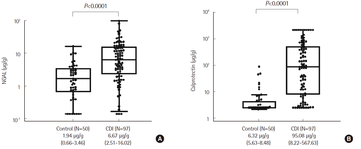
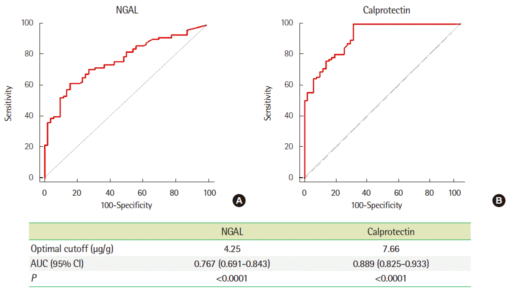
We compared the levels of both fecal biomarkers in patients with CDI and healthy controls. The concentrations of the fecal biomark-ers were higher in patients than in healthy controls (P <0.0001, Fig. 1). We then performed ROC curve analysis to determine the optimum cut-off value for differentiating between healthy controls and patients with CDI (Fig. 2). The optimal cutoff and AUC values were 4.25 μg/g and 0.767, respectively, for NGAL, and 7.66 μg/g and 0.889, respectively, for calprotectin. Pearson’s correlation analysis of the two fecal biomarkers also confirmed the high correlation between the two biomarkers (r =0.608, P <0.0001).
2. Fecal biomarker levels in patient subgroups
We compared fecal NGAL and calprotectin levels in subgroups of patients with CDI (Table 4). Fecal NGAL and calprotectin levels were significantly different in the high and low tcdB gene load groups (P =0.005 and 0.006, respectively). As both fecal biomarkers are found in neutrophils, we then compared their levels in non-leukopenia and leukopenia patients. While no significant difference was observed in NGAL levels (P =0.553), the difference in calprotectin levels was statistically significant (P =0.004). No statistically significant differences in fecal biomarker levels were associated with antibiotic usage within 3 days of diagnosis or for IDSA/SHEA clinical severity grade (P =0.137 and 0.430, respec-tively). For ESCMID clinical severity, the differences in fecal NGAL levels were not statistically significant, but the differences in fecal calprotectin levels were significantly different (P =0.16 and 0.021 respectively).
Table 4
Evaluation of fecal NGAL and calprotectin levels in subgroups of patients with CDI
| NGAL (μg/g) | P | Calprotectin (μg/g) | P | ||
|---|---|---|---|---|---|
| Leukopenia | Yes (N=18) | 3.80 (3.27-10.16)* | 0.553 | 12.43 (2.89-41.26)* | 0.004† |
| No (N=79) | 7.19 (2.12-16.03)* | 139.82 (13.95-677.76)* | |||
| Antibiotic use within 3 days | Yes (N=62) | 9.06 (3.51-18.21)* | 0.098 | 77.78 (4.24-419.67)* | 0.253 |
| No (N=35) | 5.16 (1.56-14.83)* | 141.83 (14.22-674.51)* | |||
| tcdB gene load | High (N=39) | 10.27 (4.01-21.01)* | 0.005† | 274.05 (41.17-1,019.61)* | 0.006† |
| Low (N=58) | 4.60 (1.46-11.94)* | 35.91 (6.86-304.96)* | |||
| IDSA/SHEA severity | Severe (N=23) | 10.27 (3.34-22.47)* | 0.137 | 141.83 (3.74-1,339.82)* | 0.430 |
| Non-severe (N=74) | 5.40 (2.43-14.46)* | 82.48 (8.67-419.67)* | |||
| ESCMID severity | Severe (N=77) | 7.19 (3.30-16.36)* | 0.160 | 112.05 (13.53-624.66)* | 0.021† |
| Non-severe (N=20) | 3.39 (1.25-15.38)* | 11.95 (4.30-224.64)* | |||
![]()
3. Comparison of fecal biomarkers for the prediction of clinical outcomes
We analyzed the correlation between fecal NGAL and calprotectin levels and the clinical outcomes among the patient groups (Table 5). There was no statistically significant difference in 30-day all-cause mortality associated with either fecal NGAL or calprotectin (P =0.260 and 0.159, respectively). Excluding the 11 patients that failed to show up for follow-up, a significant association with treatment failure observed for fecal calprotectin (P =0.033) but not for fecal NGAL (P =0.13).
Table 5
Fecal NGAL and calprotectin levels by clinical outcome
| NGAL (μg/g) | P | Calprotectin (μg/g) | P | ||
|---|---|---|---|---|---|
| 30-day all-cause mortality | Yes (N=10) | 7.72 (4.45-23.76)* | 0.26 | 296.95 (40.47-1,094.96)* | 0.159 |
| No (N=87) | 6.44 (2.12-15.5)* | 79.46 (7.46-442.77)* | |||
| Treatment failure‡ | Yes (N=6) | 8.53 (4.19-19.08)* | 0.13 | 382.19 (63.97-1,503.52)* | 0.033† |
| No (N=80) | 5.46 (1.98-15.38)* | 77.78 (7.08-426.49)* | |||
![]()
Go to :
DISCUSSION
This is the first study to explore the relationship between fecal NGAL and calprotectin levels in patients with CDI. As both fecal biomarkers are known to be upregulated in the intestine in inflammatory conditions, the increase in the levels of both fecal biomarkers in the intestine and the feces is reasonable [5-8, 15]. As both fecal NGAL and calprotectin are primarily found in neutrophils, we hypothesized that their levels in leukopenia patients differ [16, 17]. In comparison with non-leukopenia patients, CDI patients with leukopenia had lower fecal calprotectin levels. Thus, fecal calprotectin levels may be an unreliable CDI marker in leukopenia patients. We also calculated the best cut-off value to distinguish between patients with and without CDI for both fecal biomarkers. However, fecal calprotectin levels are also known to increase in response to Campylobacter infections and inflammatory diseases of the intestine [5, 15]. Hence, these biomarkers may be able to be used to estimate disease severity or monitor treatment progress. Towards this end, we analyzed the association between fecal biomarker levels and disease severity using existing clinical severity guidelines [3, 12], and found that fecal calprotectin level, but not NGAL level, could be used to stratify patients according to the ESCMID guidelines. Our finding indicates that fecal calprotectin may serve as a more useful biomarker than fecal NGAL. However, neither fecal biomarker could be used stratify patients according to the IDSA/SHEA guidelines. This failure to successfully stratify patients according to the IDSA/SHEA guidelines may be related to the definition of severity criteria and the characteristics of the patient population involved in this study [3]. Unlike the ESCMID guidelines, the IDSA/SHEA guidelines do not include age as a criterion and have a third category for fulminant severity. In our study group, only two patients had fulminant CDI, thereby rendering any statistical analysis futile owing to the low numbers.
Fecal NGAL and calprotectin are both non-specific markers; although their use as diagnostic markers may be limited, these molecules are still useful for predicting disease severity and outcomes [6, 8, 15, 18, 19]. The finding that C. difficile tcdB PCR Ct values were lower in patients with high biomarker levels may be indicative of a link between disease severity and fecal NGAL and calprotectin levels. However, some previous studies have shown that the EIA toxin is a more accurate predictor of patient mortality than tcdB PCR [9, 20]. A statistically significant relationship was observed between higher calprotectin, but not fecal NGAL, levels and treatment failure. When only non-leukopenia patients were included in the analysis, there was also a statistical association between higher fecal calprotectin levels and higher 30-day all-cause mortality. These results should be confirmed in future studies, as this was a single-center study on a low number of patients [20, 21]. As both fecal biomarkers are primarily secreted by neutrophils [16, 17], it is logical to theorize that their levels cannot increase in leukopenia patients. Therefore, fecal calprotectin should not be used as a biomarker for clinical outcomes in leukopenia patients. Furthermore, while hypervirulent ribotypes have been reported in Korea [22], their prevalence is lower than that in countries outside of East Asia, leading to lower mortality [23, 24].
The present study has a few limitations. It is a retrospective study and we could not monitor the changes in fecal NGAL and calprotectin levels [25], as the patients underwent CDI treatment. We also did not measure serum NGAL and calprotectin levels. We could have potentially employed the fecal toxin EIA to differentiate between actual CDI and colonization [9, 21, 26]. As we used leftover samples, we could not confirm whether the healthy controls were free of C. difficile. Future studies should enroll patients with non-CDI diarrhea as a control group to clarify the relationship between NGAL and calprotectin levels and CDI. In addition, when classifying patients based on CDI severity, the relative lack of patients with severe CDI limited the statistical power of our analysis. While the Xpert C. difficile assay presumptively identies ribotype 027, with none identified among our samples, the fact that we did not perform genotyping for C. difficile ribotypes is another limitation of this study. Peretz et al. [27] noted that fecal calprotectin levels were higher in patients with ribotype 027; future studies should aim to distinguish between ribotypes. Finally, considering the poor state of fecal samples from patients experiencing diarrhea, it is possible that the fecal NGAL and calprotectin levels detected by ELISA are highly variable. Another factor re-lated to fecal samples is that the initial sample was diluted in 10 mL of sample extraction buffer; this volume was larger than the8-mL reagent used in the nucleic acid amplification test assay [13, 14, 28].
In conclusion, this is the first study to evaluate the utility of both fecal NGAL and calprotectin levels in determining disease severity and treatment status of CDI. We demonstrate that fecal NGAL and calprotectin levels were higher in patients with CDI than in healthy controls and highlight their significant relationship with tcdB gene load. High fecal calprotectin levels were significantly associated with disease severity and treatment failure. Fecal calprotectin levels were significantly lower in leukopenia patients, suggesting the need for the cautious interpretation of fecal calprotectin levels in leukopenia patients. Further studies with a larger group of patients are warranted.
Go to :
 XML Download