

 PDF
PDF Citation
 Print
Print


Go to : 
INTRODUCTION
Hepatocyte transplantation is a promising alternative to liver transplantation in patients with acute liver failure (ALF) and metabolic liver disease [1]. Hepatocyte transplantation is a safer and less invasive procedure for patients when compared to whole organ transplantation. Animal studies have clearly shown the efficacy of hepatocyte transplantation; however, this has not translated into clinical practice where there is a limited benefit [2]. Successful translation demands high-quality cell preparation, adequate cell numbers, viability, and efficient delivery [3]. In the case of ALF, the cell dose needed to rescue/reverse the outcome is far more than that for metabolic liver diseases [4]. Our results demonstrate that adequate rates of cellular infusion can increase engraftment of transplanted hepatocytes in a D-galactosamine-induced acute liver injury rat model [5]. However, the engraftment efficiency was only about 2.5% 1 week after hepatocyte transplantation. In addition, it is difficult for hepatocytes to proliferate in the hostile microenvironment associated with ALF [6]. Considering the urgent need for large amounts of hepatocytes to treat ALF, there still much room for improvement in the cell based therapeutics.
In a retrorsine-treated liver injury rat model, immediate transplantation with either hepatocyte or bone marrow derived cells can ameliorate liver injury via a number of different mechanisms including hepatocyte proliferation in the former, and paracrine effects in the later [7]. Mesenchymal stem cells (MSCs), a major component of bone marrow derived cell mixtures, have been shown to exhibit multiple beneficial effects in vitro relevant to the liver injury therapeutic context, including (1) hepatocellular functional support (improved albumin secretion, ureagenesis, hepato-specific gene expression, cytochrome P450 activity) [8], (2) secretion of molecules that inhibit hepatocyte apoptosis (such as stromal-cell-derived factor-1 and vascular endothelial growth factor) [9-12] and stimulation of hepatocyte proliferation (via secretion of hepatocyte growth factor [HGF], epidermal growth factor, interleukin 6 [IL-6], and tumor necrosis factor-α [TNF-α]), (3) modulation of the acute phase response and suppression of the inflammatory responses including IL-1 receptor antagonists and upregulation of anti-inflammatory cytokines like IL-10 [11], and finally (4) secrete several extracellular matrix molecules, including collagen, fibronectin and laminin necessary for liver reconstruction [13,14]. These observations suggest that MSC-derived cytokines could potentially protect the liver during injury.
In vivo, MSC or MSC-conditioned media can attenuate inflammation and augment cytokine and growth factor concentrations improving cell proliferation and providing an avenue for preventing fulminant hepatocyte failure [9,15-19]. MSC transplantation following solid organ transplantation, both clinically and experimentally, can also reduce the rate of acute rejection [19,20]. MSC transplantation alone, however, is not expected to exert any effect on AFL because the hostile microenvironment of ALF is not a good niche for MSCs and long-term engraftment rates are low [15]. Transplanted hepatocytes were unable to function, or even survive well, without stromal cell support. Thus, the addition of bone marrow-derived mesenchymal stromal cells (MSCs) during transplantation could support the proliferation and functionality of the transplanted hepatocytes [13].
There are over 280 registered clinical trials examining the application of MSC, 28 of which focus on the treatment of liver disease [15]. While, no severe side-effects have been reported so far, the long-term benefits of these treatments remain uncertain [15]. Li et al. [18] evaluated the transplantation of human bone-marrow-derived MSCs in a porcine model of acute liver failure (ALF induced with D-galactosamine) without the use of immunosuppressants. Most (13/15) achieved long-term survival (>6 months) while all of the animals that did not have MSC treatment died [18]. Up to 30% of the hepatocytes, in this study, were shown to be derived from the bone marrow MSCs [18]. The underlying mechanisms for this response remain unknown, and are the subject of further investigation [16,21].
We have previously described the optimal rate for hepatocyte transplantation in an acute liver injury rat model to ensure optimal engraftment and repopulation [5]. If transplanted hepatocytes can proliferate properly, cell populations will double overtime improving the likelihood that there will be enough functional cells to compensate for the rapid loss of native hepatocytes and thus rescue the host’s liver function. It is, therefore, reasonable to assume that cotransplantation of hepatocytes and MSCs could provide enough support to facilitate improved survival and proliferation of the transplanted hepatocytes, enhancing repopulation. The ability to properly repopulate the deteriorating liver is crucial in effective clinical intervention and thus the purpose of this study was to investigate the effects of MSC and hepatocyte cotransplantation in rats with acute liver injury.
Go to :
METHODS
Ethics Statement
All animal experiments were approved by the Institutional Laboratory Animal Care and Use Committee of the National University of Taiwan. All animals received humane care in accordance with the guidelines set out by the National Science Council of Taiwan (1997) and the Guide for the Care and Use of Laboratory Animals (National Institutes of Health publication 86–23, 1985 revision). All procedures were also performed in accordance with these guidelines.
Animals
Male Sprague-Dawley (SD) rats were used as recipient animals. Fluorescent SD rats (aged 8–10 weeks, 200–250 g) were purchased from the National Laboratory Animal Center in Taiwan and used as donor animals. These animals were bred in-house and maintained on standard laboratory chow and daily 12-hour light/dark cycles. All animal experiments were approved by the Institutional Laboratory Animal Care and Use Committee of the National Taiwan University (No. 20130523 and 20150405).
Isolation of Hepatocytes and MSCs for Transplantation
In situ liver perfusion, collagenase digestion, and differential centrifugation were used to purify hepatocytes from GFP transgenic SD rats as previously described [22,23]. The viability and purity of each preparation were assessed using trypan blue exclusion on a hemocytometer. Isolated hepatocytes were resuspended 1×10⁷ cells/mL in phosphate buffered saline (PBS) without serum. Marrow cells, from both femurs and tibias were flushed from the bones of the DsRed transgenic SD rats using a syringe with a 26-G needle. Bone marrow mononuclear cells were isolated using Percoll gradient density centrifugation. MSCs were collected by depleting the cell suspensions of hematopoietic cells (CD45+) using anti-CD45 coated magnetic beads. Isolated MSCs were then resuspended to 1×10⁶ cells/mL in PBS without serum. MSCs were plated at a concentration of 106 cells/mL in murine MesenCult medium (Stemcell Technologies, Vancouver, Canada) and incubated at 37°C in a 5% humidified CO2 atmosphere for 3 hours and the unattached cells were then removed. Cells were put through a second round of purification if necessary [24].
Hepatic Tissue Histology and the Determination of Liver Repopulation
Fresh liver sections were fixed in formalin, embedded in paraffin, sectioned, and stained using hematoxylin and eosin to evaluate the histology of these samples. To identify transplanted hepatocytes in the recipient liver, DsRed expression was determined using fluorescence or enzyme histochemistry in liver cryosections. To analyze the liver repopulation, three to four sections from multiple liver lobes per rat were stained for DsRed activity. Microphotographs were obtained from consecutively adjacent areas to represent the whole section under ×100 magnification using a digital camera. The relative occupation of these sections by the transplanted hepatocytes was evaluated and quantified using J-Image software (National Cancer Institute, Bethesda, MD, USA).
Experimental Design
The D-galactosamine (Sigma, St. Louis, MO, USA) working solution was prepared as previously described [22] and used immediately after preparation. Acute hepatic injury was induced by D-galactosamine treatment (0.9 g/kg, intraperitoneal injection [IP]) in male SD rats 24 hours before transplantation. Isolated hepatocytes (1×10⁷/mL) from GFP+ SD rats were transplanted intraportally 24 hours after treatment with D-galactosamine at an infusion rate of 70 seconds. DsRed+ MSCs (0.5, 1, or 2×10⁶/0.5 mL) were transplanted intraportally 1 hour after hepatocyte transplantation. The surviving rats were sacrificed and their livers harvested at 1, 2, and 4 weeks after transplantation (Fig. 1A).
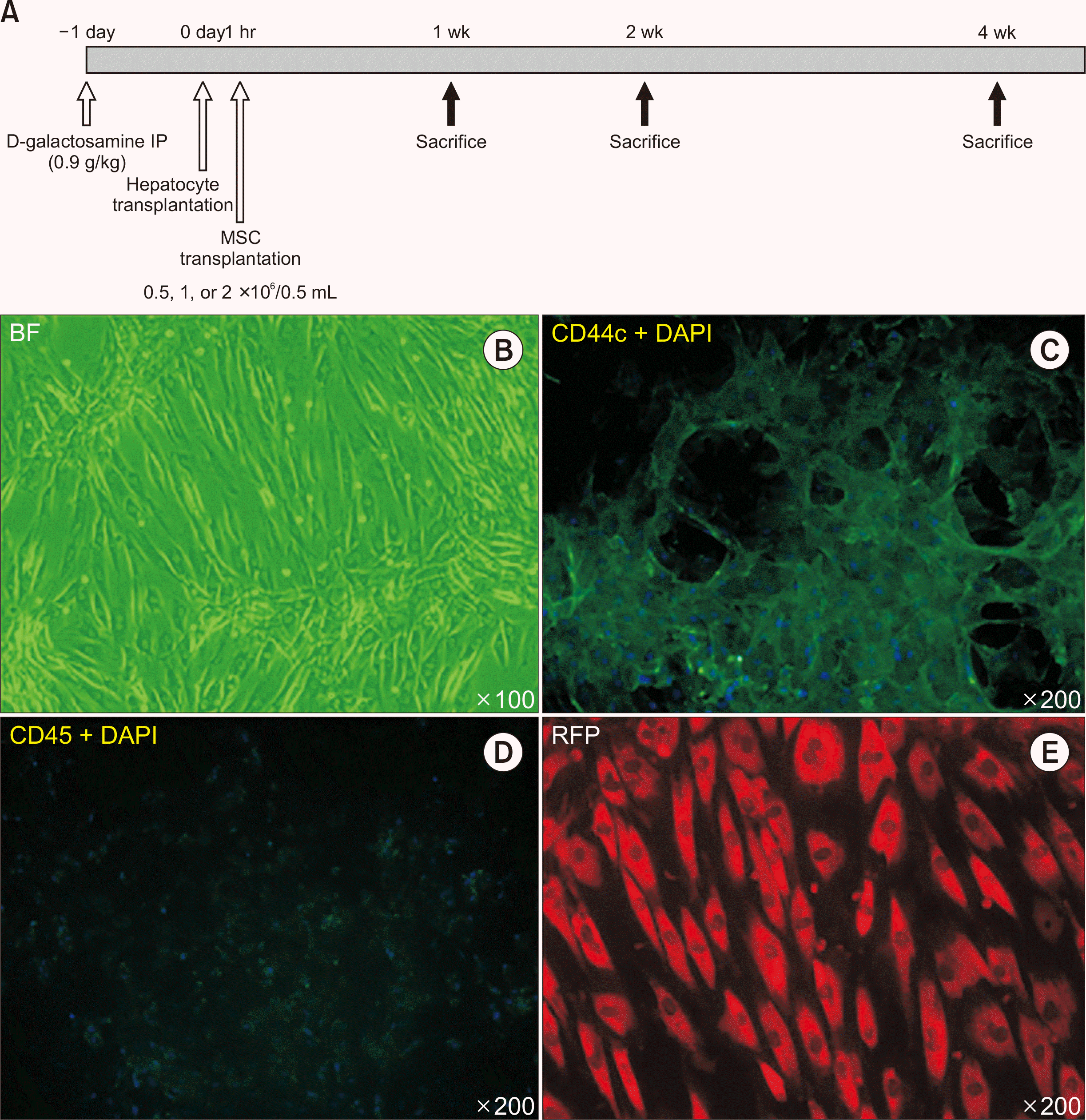 | Fig. 1Model of acute liver failure (ALF) used in the cotransplantation study. (A) Experimental design for the cotransplantation of rat hepatocytes and mesenchymal stem cells (MSCs) in a rat model of ALF. (B-E) Characterization of MSC. The MSCs were spindle shaped (B) and expressed CD44c (C) but not CD45 (D). They were fluoro-red (+) (E) since they were derived from bone marrow aspirates of DsRed transgenic Sprague-Dawley rats. IP, intraperitoneal injection; BF, blank field; DAPI, 4', 6-diamidino-2-phenylindole; RFP, red fluorescent protein.
|
Liver Tissue Evaluations
All immunofluorescent/immunohistochemical staining was performed according to previously described protocols [25]. Quantitative Reverse Transcriptase polymerase chain reaction was used to evaluate tissue specific expression of HGF, EGF, SCF, IL-6, IL-10, TNFα, TGFβ1, and collagen I and was performed as previously [25].
Serological Assay
Hepatic venous blood was sampled after the recipient rats were sacrificed. Biochemical analyses (aspartate aminotransferase [AST], alanine aminotransferase [ALT], alkaline phosphatase [ALP], lactate dehydrogenase [LDH], ammonia, albumin, and blood urea nitrogen [BUN]) were performed in an animal laboratory using the standard automated assays, as previously described [5].
Statistical Analysis
At least four animals per treatment were evaluated. Data are shown in a qualitative manner or presented as the mean, as appropriate. No animal data were excluded.
Go to :
RESULTS
Characterization of Donor MSCs
MSCs in culture before transplantation are shown in Fig. 1B-E. They maintained the classic spindle-shape morphologically (Fig. 1B), expressed CD44 (Fig. 1C) and did not express hematopoietic cell marker CD45 (Fig. 1D). They were derived from the bone marrow aspirations from the DsRed SD rats and were therefore fluoro-red positive.
Histopathological Changes after Hepatocyte and MSC Co-transplantation
One day after D-gal administration, livers of SD rats showed extensive hepatocyte necrosis, periportal focal expression of γ-glutamyltransferase, oval cell marker (OV-6), and pan-cytokeratin. No fibrosis was evident at this stage. Multiple CD163+ macrophages were present in the parenchyma sparing the portal area (Fig. 2A). After cotransplantation, the liver parenchyma was seen to undergo progressive restoration with reduced inflammatory cell infiltration (Fig. 2B). However, biliary ductular proliferation can still be observed 4 weeks after cotransplantation.
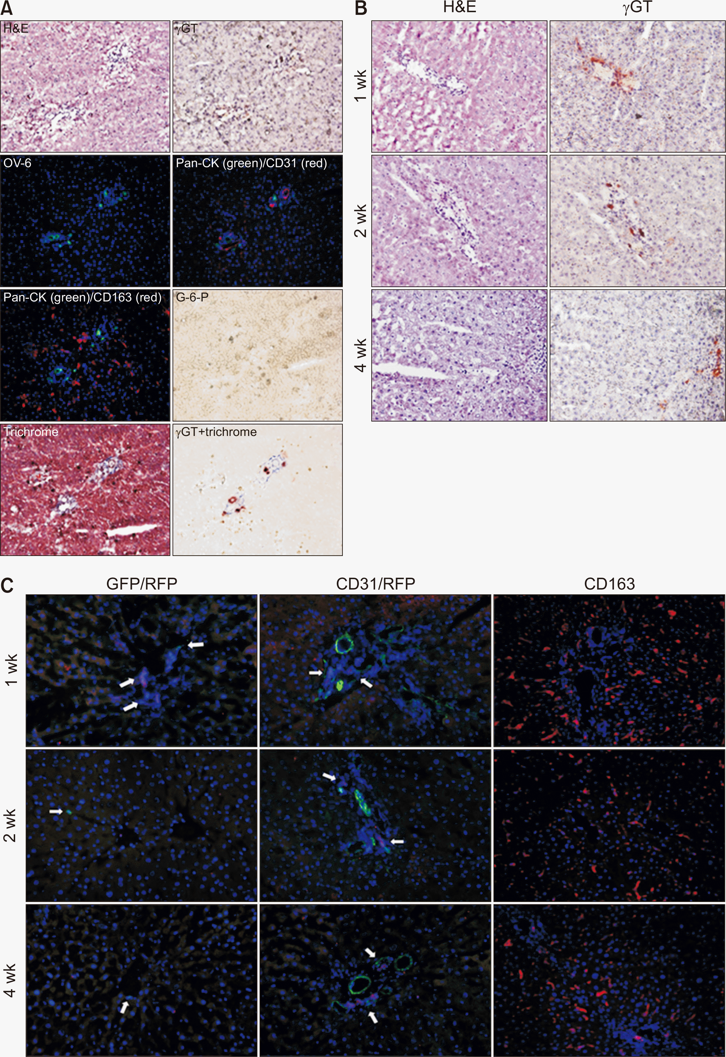 | Fig. 2Histopathological changes after hepatocyte and mesenchymal stem cell (MSC) cotransplantation in acute liver injury. (A) Control liver. Acute liver injury developed, 1 day after D-galactosamine treatment. Ductular reactions were noted by oval cell marker (OV-6), expression near the portal vein. Prominent CD163+ macrophage and low G-6-P expression were also observed. Fibrosis was limited to the periportal region. (B) One, 2 and 4 weeks after cotransplantation, γ-glutamyltransferase (γGT), was expressed in the ductular cells along with obvious signs of liver recovery. (C) Tracing of transplanted donor cells after cotransplantation. Donor hepatocytes were labelled green and MSCs were labelled red. CD163 marker expression was used to determine macrophage infiltration after cotransplantation and is shown on the right. Arrows indicate the donor cells. CD31 marks the endothelial inner lining of the portal vein. GFP, green fluorescent protein; RFP, red fluorescent protein. Magnification, all ×200.
|
Tracing the Origins of the Transplanted Hepatocytes and MSCs
Transplanted MSCs (fluoro-red) were located within the periportal area, 1, 2, and 4 weeks after cotransplantation (Fig. 2C). Transplanted hepatocytes (fluoro-green) were found scattered throughout the parenchyma but did not show any signs of proliferation after engraftment (1, 2, and 4 weeks) (Fig. 2C). Transplanted MSCs did not appear to differentiate into hepatocytes or proliferate during this study period.
Serological Changes and Synthetic Functional Recovery Following Hepatocyte and MSC Transplantation: Comparing Different Doses of MSCs
After cotransplantation, we evaluated changes in the serological markers for liver injury (AST, ALT, ALP, LDH; indicating liver injury) and synthetic markers (albumin, BUN) with the results shown in Fig. 3A and B, respectively. When we compared the serological results after 1 and 2 weeks of cotransplantation with MSCs, at doses of 0.5 or 2×10⁶/0.5 mL, we were able to determine that 1×10⁶ MSCs was the correct dose for future cotransplantation studies.
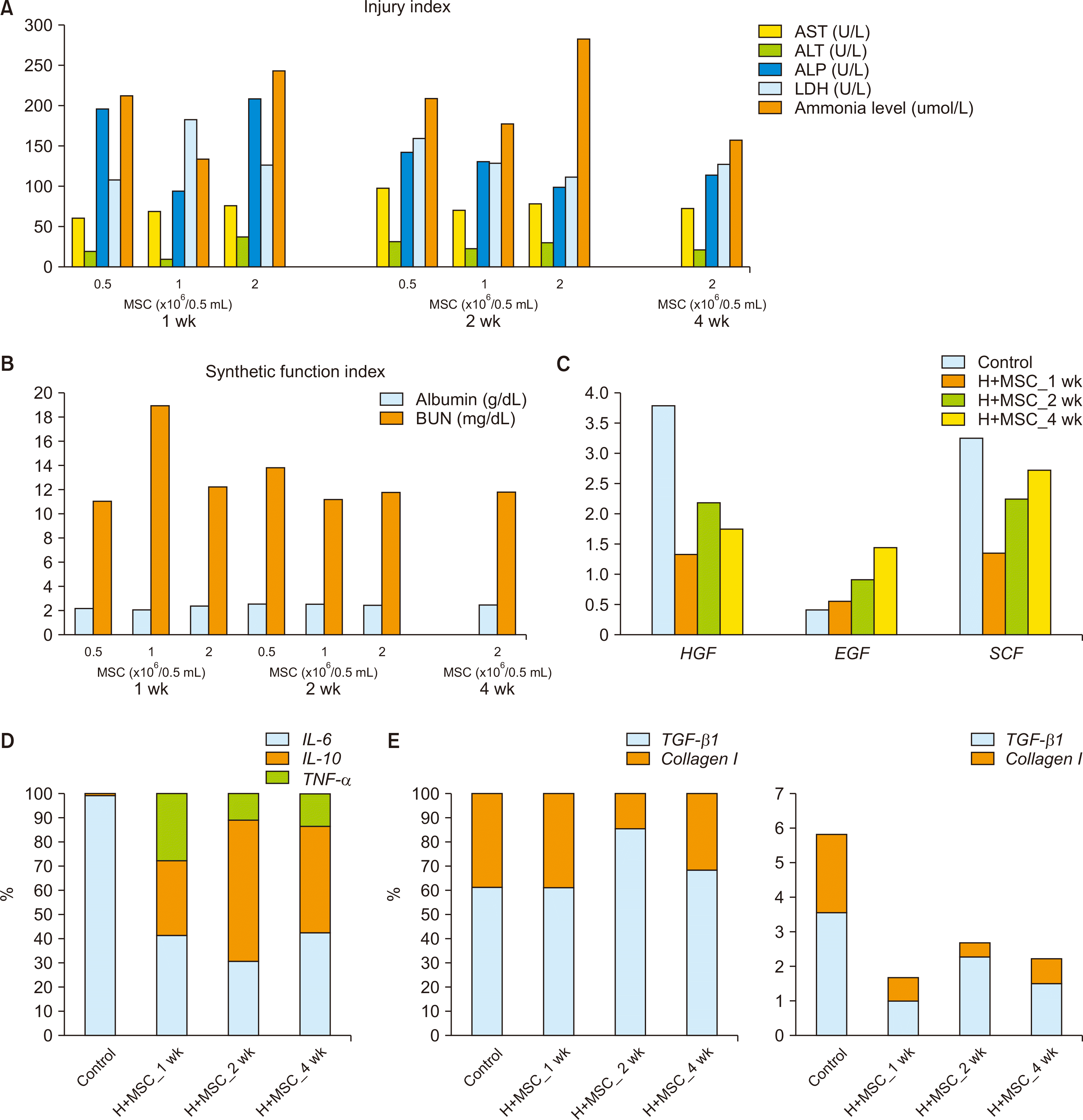 | Fig. 3Serological marker and gene expression changes. Serological changes after cotransplantation of hepatocyte and mesenchymal stem cells (MSCs) in rats with acute liver injury. Markers of injury index (A) and synthetic function index (B). Gene expression of growth factors (C), inflammation (D) and fibrosis (E) after cotransplantation. Control: acute liver injury 1 day after D-galactosamine treatment; Baseline expression: glyceraldehyde 3-phosphate dehydrogenase; AST, aspartate aminotransferase; ALT, alanine aminotransferase; ALP, alkaline phosphatase; LDH, lactate dehydrogenase; BUN, blood urea nitrogen; H, hepatocyte; HGF, hepatocyte growth factor; EGF, epidermal growth factor; SCF, stem cell factor; IL, interleukin; TNF-α, tumor necrosis factor-α; TGF-β1, transforming growth factor-β1.
|
Gene Expression in Liver Tissues Following Cotransplantation
Gene expression profiles of genes associated with growth factors, inflammation-related cytokines, and fibrosis were evaluated at 1, 2, and 4 weeks after cotransplantation (Fig. 3C-E). HGF and SCF were highly expressed in the D-galactosamine induced acute liver injury (control) samples. After cotransplantation, EGF and SCF progressively increased over the 4-week follow-up period (Fig. 3C). In addition, IL-10 expression increased, and expression of both IL-6 and TNF-α, progressively decreased, within the first 2 weeks following cotransplantation (Fig. 3D). The gene expression profiles for TGF-β1 and Collagen I, associated with fibrosis, remained similar in the first week after cotransplantation, after which they decreased and then fluctuated for the rest of the study period (Fig. 3E).
CD163+ Macrophages after Cotransplantation in D-Gal-Induced Acute Liver Injury Rats
When we compared CD163 expression at 1 week after cotransplantation, we observed a reduction in the proportion of cytoplasm and CD163+ macrophages at both 2 and 4 weeks after cotransplantation (Fig. 2C).
Go to :
DISCUSSION
This study made four key observations. MSC cotransplantation did not directly benefit donor hepatocyte proliferation. Transplanted MSCs, mostly resided in the periportal niche, and did not differentiate into liver cells. IL-10 gene expression, the anti-inflammatory cytokine, increased proportionally 1 week after cotransplantation. While, CD163+ macrophages were prominent with large amounts of cytoplasm.
Transplanted MSCs did not differentiate into liver cells in our study. Although other studies have demonstrated that MSCs can differentiate into cells from the terminal organs in vivo, there is growing scepticism about their functional differentiation beyond their differentiation to adipocytes, osteocytes and chondrocytes [26,27]. Current rationales have shifted toward an emphasis on the paracrine effects and immunomodulation of MSC cell therapies within the tissue microenvironment [26,28]. Wang et al. [27] showed that IL-10 secreted by MSCs attenuated ALF by inhibiting pyroptosis in mice. Our study showed significant changes in the cytokine profile of liver tissues 1 and 2 weeks after cotransplantation with a definite decrease in the expression of the inflammatory cytokines. IL-10 gene expression increased in direct proportion to IL-6 expression after cotransplantation. Whether these induced mRNAs are derived from the transplanted MSCs needs further investigation.
ALF is characterized by sequential and overlapping episodes of hepatocyte death-related inflammation, followed by the induction of the anti-inflammatory response with or without resultant immune paralysis and sepsis, followed by liver repair and recovery, if the host survives [29]. In our ALF model, the gene expression of IL-6 was very high, one day after D-galactosamine treatment (data not shown). Meanwhile, IL-10 was elevated and CD163+ macrophage appeared in the liver microenvironment shortly thereafter. At 2 weeks after cotransplantation, although the proportion of IL-10 increased, this expression gradually decreased compared to the initial values. This observation suggests that the microenvironment settled down soon after cotransplantation. Consistently, gene expression associated with inflammation-related cytokines was reduced 1 week after cotransplantation when we compared MSC cotransplantation and hepatocyte-only transplantation (data not shown).
Clinical challenges of MSC application include variabilities among the large number of disease categories, different routes of delivery, range of doses, and types of MSCs being used [30]. The low success rate for meeting primary outcomes in clinical trials actually underscores the need for new designs [30]. Our study provides a reasonable expectation for potential human effect of MSC cotransplantation (reducing micro-environmental inflammation of ALF) that could aid in setting reasonable outcomes for future trial designs (such as inflammation score).
This study has one major limitation, the data generated in this study was insufficient to generate an interactive mechanism for the MSCs and the microenvironment, which is an inherent issue with rat studies of this nature. In summary, hepatocyte and MSC cotransplantation is feasible and safe in an acute liver injury rat model. Transplanted MSCs were located within the periportal niches up to 4 weeks after transplantation without any signs of enhancing transplanted hepatocyte proliferation or their differentiation into hepatocytes themselves. Inflammation was ameliorated by cotransplantation.
Go to :
 XML Download