

 PDF
PDF Citation
 Print
Print


INTRODUCTION
In living donor liver transplantation (LDLT), every anatomical structure of the recipient liver should be carefully dissected because it can be used for graft reconstruction. If bleeding is observed during the procedure, then anatomical dissection becomes difficult primarily due to excessive bleeding from the dissected surfaces. Recipient hepatectomy also becomes difficult to perform if portal hypertension exists or liver surgery was performed previously [1,2].
To manage excessive intraoperative bleeding, prolonged occlusion of the hepatoduodenal ligament can be performed [3]. This approach is the same as the Pringle maneuver. In the Pringle maneuver, temporary occlusion is repeated to prevent ischemic damage in the liver [4]. By contrast, the native liver would be sacrificed in LDLT; thus, ischemic damage is not a matter of concern, and inflow control can be prolonged over several hours. This retrospective case-control study aimed to analyze the effect of prolonged hepatic inflow occlusion (PHIO), considering blood loss volume during bleeding, when applied during recipient hepatectomy in LDLT.
Go to : 
METHODS
This study protocol was approved by the Institutional Review Board of Asan Medical Center (IRB No. 2019-0599).
Study Groups
This study was designed to be a retrospective case-control study to compare the amount of bleeding during recipient hepatectomy of LDLT. The amount of blood loss during bleeding was defined as the sum of the amount of the transfused blood components (packed red blood cells and fresh frozen plasma). The study group comprised patients who underwent PHIO due to anticipated difficult dissection with the Model for End-Stage Liver Disease (MELD) scores ranging from 26 to 35. The two control groups included patients who did not undergo PHIO. These patients were selected according to their MELD scores considering that the MELD score is closely associated with extensive bleeding [5]. Patients with MELD scores of 15–20 were assigned in the low-MELD score group, while patients with MELD scores of 26–35 were assigned in the high-MELD score group. Patients who required cell saver were excluded because the assessment of bleeding is difficult in these patients. We also excluded patients who underwent PHIO aimed at preventing intraoperative metastasis caused by hepatocellular carcinoma (HCC) because most of these patients had a low MELD score or had no portal hypertension.
Patient Selection
The clinical application of PIHO in LDLT was performed since 2014, and technical refinement was completed at the end of 2016. Thus, we set the study period for 30 months, from January 2017 to June 2019. We used our institutional LDLT database to select patients meeting the inclusion criteria. Twenty patients were included in the study group. Considering a 1 to 2 matching, 40 patients were distributed into the two control groups depending on their scores. Patients’ medical records were retrospectively reviewed. The input and output records at the anesthesia record sheet and operation nursing chart were comprehensively collected and integrated to calculate the amount of blood transfused.
Techniques for PHIO
For PHIO, we attached a single curved intestinal clamp to the hepatoduodenal ligament without dissecting the hepatoduodenal ligament. The clamping power of the curved intestinal clamp was set at 2 or 3 jaw steps according to the thickness of the hepatoduodenal ligament (Fig. 1). PHIO was performed according to two steps. First, we dissected the retrohepatic inferior vena cava to detach the liver under PHIO. Second, if brisk bleeding was anticipated during the dissection of the hepatoduodenal ligament, then PHIO was intermittently performed at the distal part of the hepatoduodenal ligament to reduce bleeding during dissection. PHIO could be temporarily released to palpate the pulsation of the right hepatic artery.
 | Fig. 1Concept of prolonged hepatic inflow occlusion. (A) Interruption of the main portal flow and hepatic arterial flow in a patient with liver cirrhosis, and portal hypertension does not induce significant splanchnic congestion because of portal bypass through venous collaterals. Adapted from Choi et al. Ann Hepatobiliary Pancreat Surg 2019;23:61-4 [3]. (B) A curved intestinal clamp is attached to the hepatoduodenal ligament for right liver mobilization.
|
Statistical Analysis
Dissection duration was simply calculated as the time from skin incision to completion of dissection of the native liver. Our recipients usually underwent greater saphenous vein harvest after liver dissection; thus, the time for this extra-abdominal procedure was excluded when calculating the total dissection duration. The amount of bleeding was calculated as the total amount of the transfused blood components (sum of packed red blood cells and fresh frozen plasma).
All numerical data were presented as mean values with standard deviations. Incidence variables were compared with chi-square test or Fisher’s exact test. Continuous variables were compared with Student t-test. Statistical analyses were performed using IBM SPSS ver. 22.0 (IBM Corp., Armonk, NY, USA).
Go to :
RESULTS
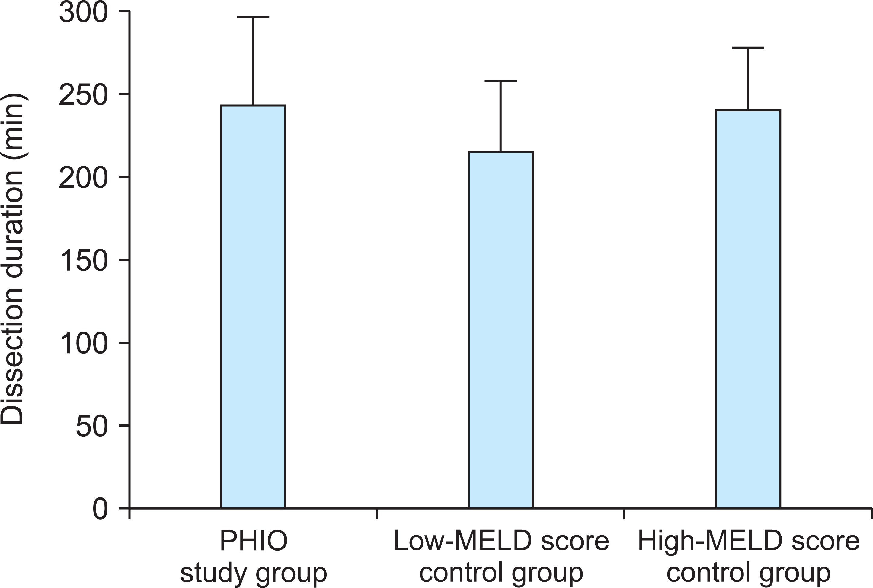
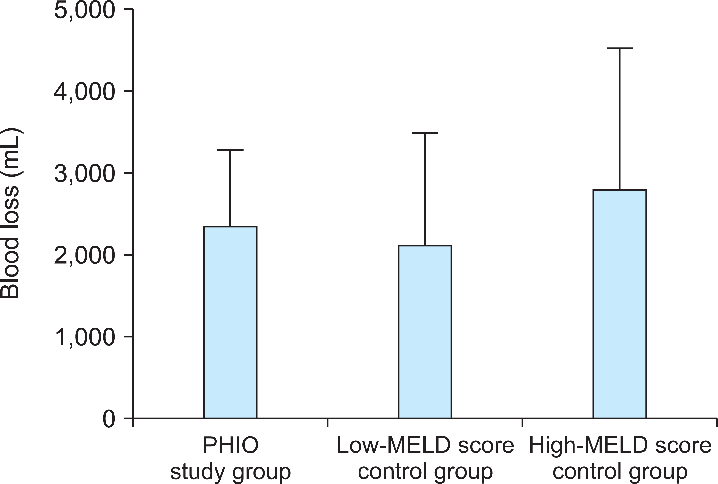
The clinical profiles of patients in the study group and the two control groups are summarized in Table 1. These profiles were relatively similar in all groups except for the MELD score and its three components. In the PHIO study group, mean total dissection duration and mean PHIO duration were 226.3±59.4 and 68.2±19.1 minutes, respectively. Twelve patients (60%) underwent PHIO twice: one for liver mobilization and detachment from the retrohepatic inferior vena cava and the other for the dissection of the hepatoduodenal ligament. Their mean total dissection duration and mean total PHIO time were 243.5±55.3 and 78.7±18.5 minutes, respectively. The other eight patients underwent PHIO once for liver mobilization and detachment from the retrohepatic vena cava, and their mean total dissection duration and mean total PHIO duration were 201.5±65.3 and 51.9±24.3 minutes, respectively. The mean amount of blood loss in all 20 patients was 2,350.0±951.9 mL.
Table 1
Clinical profiles of the PHIO study group and two control groups
![]()
In the low-MELD score control group, mean total dissection duration and mean amount of blood loss were 216.0±43.9 minutes and 2,112.5±1,614.9 mL, respectively. In the high-MELD score control group, mean total dissection duration and mean amount of blood loss were 241.0±41.9 minutes and 2,815±1,813.9 mL, respectively. The PHIO study group and the low-MELD score control group showed similar total dissection duration (226.3±59.4 vs. 216.0±43.9 minutes, P=0.82) and similar blood loss during dissection (2,350.0±951.9 vs. 2,112.5±1,614.9 mL, P=0.17). The PHIO study group and the high-MELD score control group showed similar total dissection duration (226.3±59.4 vs. 241.0±41.9 minutes, P=0.71), but the PHIO group showed a significantly lower blood loss during dissection than the high-MELD score group (2,350.0±951.9 vs. 2,815.0±1,813.9 mL, P=0.002) (Figs. 2 and 3).
During LDLT operation using PHIO, major serosal peritoneal tearing-associated bleeding and hepatic artery dissection did not develop in all patients. Six of the 20 patients (30%) showed noticeable edematous change after PHIO for more than 1 hour, but this was immediately resolved after releasing the intestinal clamp. The other 14 patients (70%) did not show noticeable signs of splanchnic congestion such as bowel edema or mesenteric discoloration. None of the patients experienced posttransplant acute pancreatitis.
Go to :
DISCUSSION
Excessive bleeding is considered a serious complication of LDLT operation because of difficult dissection, resulting in bleeding tendency. Thus far, we have performed more than 5,000 LDLT operations, and a non-negligible number of patients required massive transfusion due to excessive bleeding during LDLT operation. Intraoperative bleeding is common during LDLT when compared to that during deceased donor liver transplantation because the whole retrohepatic inferior vena cava should be preserved and all perihilar structures should be meticulously dissected to preserve the small hepatic artery branches and hilar bile duct openings [1,5,6]. Excessive bleeding and massive transfusion can cause several adverse effects on intraoperative management and posttransplant recovery [7]. Thus, intraoperative blood loss should be reduced as much as possible [8,9]. There are a few reports on transfusion-free liver transplantation [5,10]. The MELD score is considered to be one of the important risk factors for massive transfusion despite the controversy regarding this assumption [5,11]. Intraoperative cell salvage with autologous transfusion using a cell saver machine is effective in managing massive bleeding [12]. Preventing bleeding is more important than blood replacement. Various surgical techniques including Pinch-Burn-Cut techniques, high hilar dissection, and dissection with energy devices have been developed to reduce intraoperative bleeding during LDLT operations [13-15]. The Pringle maneuver is one of the essential approaches used in liver surgery. Pringle [16] reported the arrest of hepatic hemorrhage due to trauma in 1908. Thereafter, the Pringle maneuver has been recognized as one of the standard procedures in liver surgery. Thus far, its application was confined to liver surgery requiring hepatic transection. Our previous report suggested that prolonging the Pringle maneuver was effective in reducing bleeding during LDLT operation [3]. This study proved that PHIO is effective in reducing bleeding during hepatic mobilization and dissection, although it did not reduce the total dissection duration. In clinical practice, PHIO provides only a better operative field; thus, every step in the surgical procedure should be meticulously and comprehensively performed.
In PHIO, simple application of a curved intestinal clamp to the hepatoduodenal ligament is the only procedure we can perform. The reason why we use such a curved intestinal clamp is that it is atraumatic even at its maximal grasping power. It is feasible to use an umbilical tape with a vascular tourniquet set, but we do not recommend this method because it can induce excessive squeezing at the hanging point if the hepatoduodenal ligament is edematous. Some surgeons used high hilar dissection techniques for LDLT, in which hepatic artery dissection can be developed because of clamping of the hepatoduodenal ligament with a vascular clamp [14]. At this point, we emphasize that a vascular clamp with or without protective rubber shoes should not be used for PHIO because such clamps have greater squeezing power than that of intestinal clamps. To the best of our knowledge, the intestinal clamp is the most appropriate instrument that should be used when performing PHIO in LDLT operation because it is atraumatic.
A simulative hemodynamic analysis revealed that the occlusion of the hepatoduodenal ligament in liver transplantation recipients is considered a temporary measure to weaken the bleeding-prone effect from portal hypertension [3]. If brisk bleeding is observed after damage of venous collaterals around the liver, promptly initiating a local bleeding control is usually difficult. If venous collaterals exist proximal to the main portal vein, occlusion of the hepatoduodenal ligament will prevent bleeding. In patients with portal hypertension, there may be collaterals to compensate portal hypertension. Such a situation may prevent potential PHIO-induced splanchnic congestion because portal blood flow will bypass through the preexisting collaterals.
One of the potential indications for PHIO is prevention of intraoperative tumor spread [3]. If surgeons manipulate the HCC-containing liver excessively, it increases the risk of hematogenous tumor cell spread into the bloodstream. We hypothesize that PHIO can be performed during right liver mobilization to minimize the hematogenous spread of HCC cells. We also hypothesize that the primary indication for PHIO is the presence of an HCC greater than 5 cm because this tumor size is one of the most significant prognostic factors in LDLT.
Acute pancreatitis rarely develops after liver transplantation [17-19]. Prolonged prehepatic portal venous congestion or sinistral portal hypertension can be a potential risk factor of acute pancreatitis [20]. Therefore, routine or irrelevant application of PHIO is not recommended, particularly in patients without portal vein collaterals.
This study has some limitations. First, this study has a small sample size, and difficult-to-dissect cases were intentionally selected for objective comparison, possibly leading to non-negligible selection bias. Second, the total dissection duration and total blood loss volume were retrospectively estimated using only the patients’ medical records. In conclusion, our findings suggest that PHIO is a simple effective method to reduce intraoperative bleeding during hepatic mobilization and dissection during LDLT operation.
Go to :
 XML Download