Journal List > Korean J Transplant > v.34(1) > 1145092

Yoon, Kwon, Jeong, Lee, Oh, Huh, Jang, Joh, Kim, Choi, Kim, Park, Lee, Sinn, Choi, and Kang: Comparative evaluation of QuantiFERON-TB Gold Plus for diagnosis of latent tuberculosis infection during solid organ transplantation

Abstract

Background

The diagnosis of latent tuberculosis infection (LTBI) in solid organ transplantation (SOT) patients under lifelong immunosuppression has profound effects on preoperative and postoperative management. Interferon-gamma release assay (IGRA) is widely used to screen LTBI before or after transplantation.

Methods

We evaluated the effect of posttransplantation immunosuppression on IGRA and influencing factors by measuring interval change of QuantiFERON-TB Gold Plus (QFT-Plus) between pretransplantation (pre-QFT-Plus) and posttransplantation (post-QFT-Plus) state in 20 patients who previously had reactive IGRA but not taken LTBI treatment.

Results

Eleven (55%) out of 20 pre-QFT-Plus reactive patients became nonreactive state in repeated QFT-Plus (post-QFT-Plus) at 194–413 days (median, 257 days) after transplantation (discordant group). Even in persistently reactive group (concordant group), interferon-gamma (IFN-γ) levels after transplantation were decreased about 34% and 36% of their pretransplantation levels for TB1 and TB2, respectively. The only significant factors that affect interval change of QFT-Plus between pre- and post-SOT status were the concentrations of IFN-γ in pre-QFT-Plus (6.93 vs. 0.44 IU/mL in TB1 and 7.33 vs. 0.71 IU/mL in TB2).

Conclusions

The reactivity of QFT-Plus is significantly compromised by immunosuppressive therapy, which increase the risk of false negative, particularly in patients with low level of IGRA reactivity. Therefore, interpretation of IGRA under immunosuppressive treatment require a caution and eventually, more sensitive tuberculosis-specific cytokine markers might be needed.

HIGHLIGHTS
  • Interferon-gamma release assays (IGRAs) is widely used to screen latent tuberculosis infection before or after transplantation.

  • The reactivity of QuantiFERON-TB Gold Plus can be significantly compromised by immunosuppressive therapy (IST).

  • Interpretation of IGRA under IST requires a caution and more sensitive tuberculosis-specific cytokine markers.

INTRODUCTION

Latent tuberculosis infection (LTBI) is an infection of Mycobacterium tuberculosis which is clinically asymptomatic with negative finding on M. tuberculosis culture and smear, but with positive results on tuberculin skin test (TST) and/or interferon-gamma release assays (IGRAs) [1]. Incidence rate and mortality of M. tuberculosis infection in solid organ transplantation (SOT) group are 20 to 70 times and 10 times higher than those in the immunocompetent group, respectively [2,3]. Therefore, accurate diagnosis of LTBI in time is crucial to manage complication and survival rates of SOT group [2-5]. Although concentration of interferon-gamma (IFN-γ) is expected to be low when immunosuppressive therapy (IST) is introduced [6-8], a comparative study of IGRA levels between pre-SOT status and post-SOT has not been reported yet. Thus, the objective of this study was to determine quantitative interval change of QuantiFERON-TB Gold Plus (QFT-Plus) in SOT patients who previously had reactive IGRA but not taken LTBI treatment.

METHODS

The study protocol was approved by Samsung Medical Center Ethics Review Committee (No. 2016-04-076). Written informed consent was obtained from each patient.
We prospectively included 169 patients with primary organ failure and awaiting SOT from February to August 2017 at Samsung Medical Center (Seoul, Korea). Diagnosis of LTBI was made primarily by IGRA using QFT-Plus. Among 169 patients, 20 patients diagnosed with LTBI were included consecutively in this study for evaluating quantitative interval change of QFT-Plus. Based on clinician's discretion, these 20 patients did not receive LTBI treatment before transplantation. Patients' medical records including previous tuberculosis history, microbiological studies, and concomitant immunosuppression were reviewed. QFT-Plus assays (Qiagen, Hilden, Germany) were performed according to the manufacturer's instructions. Within 4 hours of whole blood collection in lithium heparin tubes, each milliliters of whole blood was transferred to M. tuberculosis antigen tubes (TB1 and TB2) Nil, and Mitogen tubes. These tubes were incubated immediately at 37°C for 20 hours. Enzyme-linked immunosorbent assay (ELISA) for quantitation of IFN-γ was performed using a DS-2 automated ELISA processor (Dynex, Chantilly, VA, USA). Results were interpreted as positive when the antigen tube (either TB1 or TB2 for QFT-Plus) minus Nil IFN-γ concentration was ≥0.35 IU/mL and ≥25% of the Nil value. Test results with Nil IFN-γ concentration over 8.0 IU/mL or mitogen IFN-γ concentration less than 0.5 IU/mL were considered as indeterminate. Data are expressed as a number of positive results (percentage) or median, range, and/or interquartile range of quantitative IFN-γ results. All data were analyzed using IBM SPSS ver. 23.0 (IBM Corp., Armonk, NY, USA). All reported P-values were two-tailed and calculated with statistical significance set at a P-value of less than 0.05.

RESULTS

Patient Characteristics

Among a total of 169 patients who were awaiting SOT, 63 patients (37.3%) were diagnosed LTBI based on reactivity in QFT-Plus performed before transplantation (pre-QFT-Plus). Initial characteristics of SOT candidates are described in Table 1. Median concentrations of IFN-γ in reactive group were 1.49 UI/mL (range, 0.01–10.0 UI/mL) and 2.22 UI/mL (range, 0.05–10.0 UI/mL) for TB1 tube and TB2 tube, respectively. Of 63 patients who had diagnosed with LTBI before transplantation, 20 patients had not received TB prophylaxis were subjected to second QFT-Plus after transplantation (post-QFT-Plus) at a median of 257 days (range, 194–413 days). Of 20 patients, 14 patients had kidney transplantation and six patients had liver transplantation (Table 2).

Distribution of IFN-γ Values of Pre- and Posttransplantation QFT-Plus

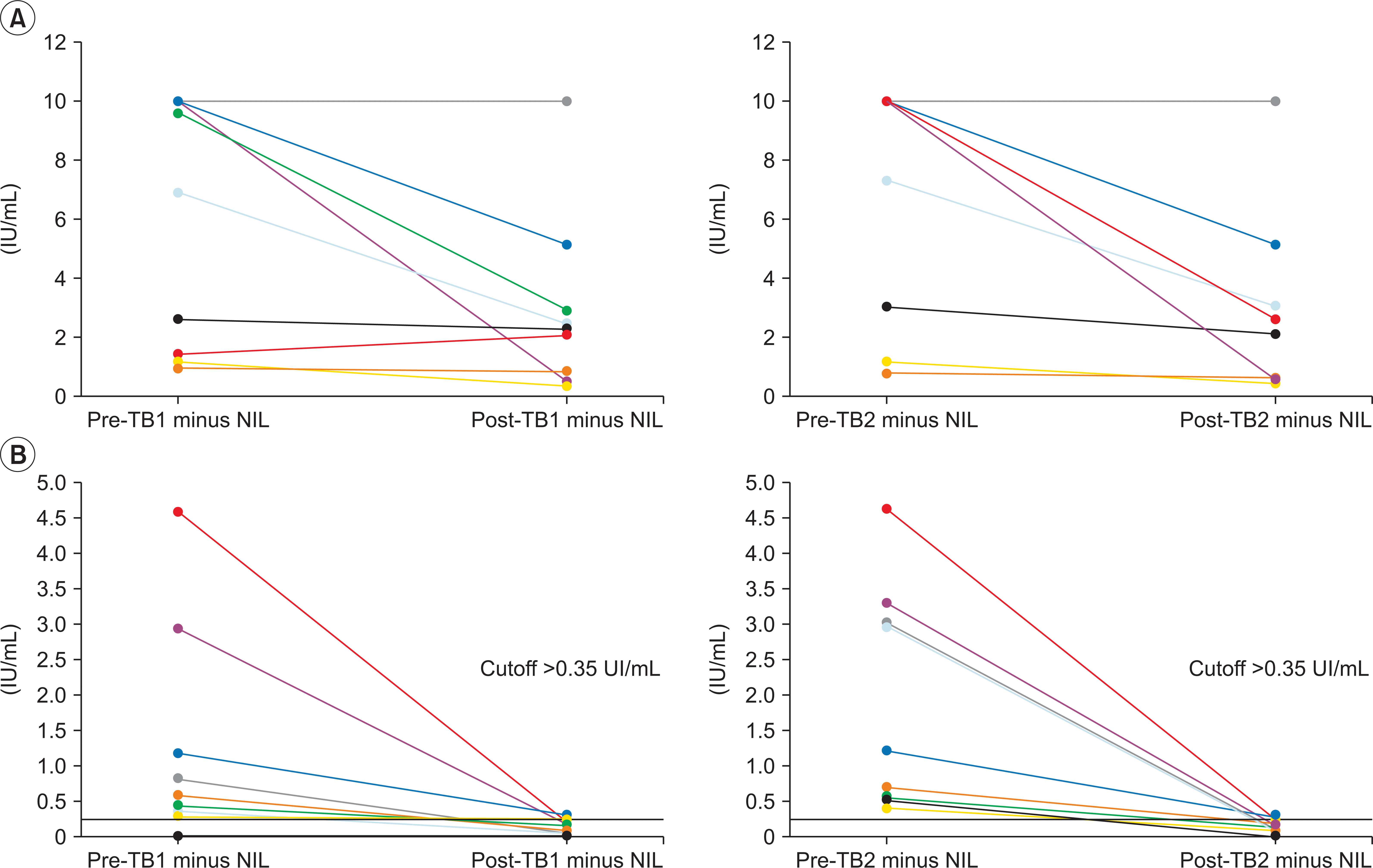
Among 20 recipients, 11 cases (55%) became nonreactive in post-QFT-Plus, which is discordant reactivity with pre-QFT-Plus (discordant group). Although the qualitative results had not changed in nine patients (concordant group), reduction rate of IFN-γ concentrations after transplantation were 34% and 36% for TB1 and TB2, respectively. Pretransplantation IFN-γ concentrations of TB1 and TB2 tubes were 0.44 IU/mL (median, 0.01–4.58 IU/mL) and 0.71 IU/mL (median, 0.4–4.62 IU/mL), respectively, in the discordant group. On the other hand, IFN-γ concentrations of TB1 and TB2 tubes were 6.93 IU/mL (median, 1.03–10.0 IU/mL) and 7.33 IU/mL (median, 0.84–10.0 IU/mL), respectively, in the concordant group (Table 2, Fig. 1).

Factors Affecting the Posttransplantation QFT-Plus Results

Factors that might affect reversion of QFT-Plus reactivity were analyzed (Table 3). Patient demography, type of transplantation organ and time to perform post-QFT-Plus were not different between concordant and discordant groups. Well known factors affecting IGRA results such as lymphocyte count, CD4 count, albumin level and immunosuppressive treatment were not related to the reversion of QFT-Plus reactivity. The only significant factors that affect interval change of QFT-Plus between pre- and post-SOT status in concordant and discordant groups were the concentrations of IFN-γ in pre-QFT-Plus (6.93 vs. 0.44 IU/mL in TB1 and 7.33 vs. 0.71 IU/mL in TB2, respectively).

DISCUSSION

Introduction of immunosuppressant in SOT made it possible to prevent acute graft rejection and to help achieving high survival rate without devastating immune reaction [9,10]. On the other hand, instead of permitting immune tolerance, immunosuppressant also arouses many kinds of complication such as neurotoxicity, nephrotoxicity, and other metabolic side effects [11]. Most of all, immunosuppression can stir up the immune system and make a patient susceptible to infection [10,11]. Therefore, SOT patients taking IST are still at risk of active or latent TB infection by exposure [2-4]. IGRA becomes useful tool to monitor and to diagnose LTBI not only in immunocompetent individuals but also in SOT patients. Diagnosis and treatment guidelines for latent tuberculosis have been revised to insist the role of IGRA in many countries including Korea [4,12]. Because negative TST or IGRA cannot exclude the potential LTBI, it is recommended to use both diagnostic tools complementarily and to combine the radiological findings and previous treatment history, particularly in immunocompromised subjects. LTBI treatment is indicated in SOT patients who are taking immunosuppressant or being planned to take immunosuppressant. Previous reports have shown the reduced reactivity in immunosuppressed patients including SOT recipients [13], however, sequential studies of pre-SOT and post-SOT IGRA in same individuals are rarely reported. For this, we've studied 20 patients who did not receive LTBI treatment before transplantation base on clinician's decision. While TB prophylaxis is recommended to patients who are diagnosed LTBI and awaiting organ transplantation according to 2017 revised Korean guideline which published at the beginning of 2018 “Diagnosis and Treatment of Latent Tuberculosis Infection: The Updated 2017 Korean Guidelines [12],” the study designed and prospectively recruit the patients with primary organ failure from February to August 2017 and TB prophylaxis was determined according to past history, imaging of the chest with computerized tomography scan or X-ray.
Herein, we compared results between pre-SOT status and post-SOT status with QFT-Plus. In this sequential test, eleven (55%) out of 20 patients had nonreactivity after transplantation. Discrepant result between pre-SOT status and post-SOT status with QFT-Plus and about 34% of reduction rate between pre- and posttransplantation status in the same reactivity group may indicate that the risk of false-negative could expressively increase after IST and this study would be a good example of IST effect on IGRA. Although effects on QFT-Plus induced by types of donor, combination of IST regimen, leukopenia and hypoalbuminemia were uncertain in this report, significant differences between concordant and discordant groups were observed in distributed levels of IFN-γ. Patients who had low IFN-γ levels in pretransplantation work-up (less than 1.07 IU/mL for TB1 and less than 3.02 IU/mL for TB2) became nonreactive during posttransplantation period (which were considered false negative results). Thus, clinicians need to interpret results of posttransplantation IGRA carefully by combining clinical findings with other laboratory results such as TST and chest radiology.
By presenting significant interval changes of IFN-γ between pre- and posttransplantation work-up in this report, we emphasize not only the importance of LTBI work-up before transplantation, but also the necessity of more sensitive test to detect MTB-specific immune response. QFT-Plus has two M. tuberculosis-specific antigen coated tubes called TB1 and TB2. TB1 contains long peptides of ESAT-6 and CFP-10 while TB2 contains six short peptides in addition to the same components in TB1, thus inducing both CD4+ and CD8+ T-cell immune responses [13]. Although QFT-Plus is a new generation of QuantiFERON-TB Gold in Tube which is expected to improve the sensitivity [14,15], QFT-Plus seems to be insufficient for detecting MTB-specific cytokine responses in SOT patients undertaking IST. Several studies have suggested that diagnostic efficacy can be improved by measuring more than two cytokines or by combining specific cytokines such as interleukin-2, monocyte chemotactic protein 2, interferon gamma inducible protein-10 (IP-10), granzyme B, and tumor necrosis factor superfamily member 14 [16,17]. Among them, IFN-γ IP-10 has been shown to be more sensitive than IFN-γ, particularly in immunosuppressed patients [17-19]. Thus, in the near future, we need TB-specific immune test to measure new cytokine alone or in combination with IFN-γ that is optimized for immune compromised patient population. This study included a relatively small number of patients with a short follow up period. Thus, larger longitudinal study with longer follow-up may be needed to elucidate the clinical consequence of altered sensitivity of current IGRA. To the best of our knowledge, this is the first comparative study of IGRA levels between pre-SOT status and post-SOT in the same individuals.

ACKNOWLEDGMENTS

Conflict of Interest
No potential conflict of interest relevant to this article was reported.
Author Contributions
Conceptualization: HYO, WH, HRJ, JWJ, JMK, GSC, SJK, JBP, KWL, DHS, JOC. Data curation: MJJ. Formal analysis: SJY, WKK. Methodology: JL. Project administration: ESK. Visualization: SJY. Writing-oritianl draft: SJY. Writing- review & editing: ESK.

REFERENCES

Dheda K., Barry CE 3rd., Maartens G. 2016. Tuberculosis. Lancet. 387:1211–26. DOI: 10.1016/S0140-6736(15)00151-8. PMID: 26377143.
crossref
Aguado JM., Herrero JA., Gavaldá J., Torre-Cisneros J., Blanes M., Rufí G, et al. 1997. Clinical presentation and outcome of tuberculosis in kidney, liver, and heart transplant recipients in Spain. Spanish Transplantation Infection Study Group, GESITRA. Transplantation. 63:1278–86. DOI: 10.1097/00007890-199705150-00015. PMID: 9158022.
Singh N., Paterson DL. 1998. Mycobacterium tuberculosis infection in solid-organ transplant recipients: impact and implications for management. Clin Infect Dis. 27:1266–77. DOI: 10.1086/514993. PMID: 9827281.
Bumbacea D., Arend SM., Eyuboglu F., Fishman JA., Goletti D., Ison MG, et al. 2012. The risk of tuberculosis in transplant candidates and recipients: a TBNET consensus statement. Eur Respir J. 40:990–1013. DOI: 10.1183/09031936.00000712. PMID: 22496318.
crossref
Pai M., Behr MA., Dowdy D., Dheda K., Divangahi M., Boehme CC, et al. 2016. Tuberculosis. Nat Rev Dis Primers. 2:16076. DOI: 10.1038/nrdp.2016.76. PMID: 27784885.
crossref
Auguste P., Tsertsvadze A., Pink J., Court R., McCarthy N., Sutcliffe P, et al. 2017. Comparing interferon-gamma release assays with tuberculin skin test for identifying latent tuberculosis infection that progresses to active tuberculosis: systematic review and meta-analysis. BMC Infect Dis. 17:200. DOI: 10.1186/s12879-017-2301-4. PMID: 28274215. PMCID: PMC5343308.
crossref
Kim EY., Lim JE., Jung JY., Son JY., Lee KJ., Yoon YW, et al. 2009. Performance of the tuberculin skin test and interferon-gamma release assay for detection of tuberculosis infection in immunocompromised patients in a BCG-vaccinated population. BMC Infect Dis. 9:207. DOI: 10.1186/1471-2334-9-207. PMID: 20003535. PMCID: PMC2801508.
crossref
Sester M., van Leth F., Bruchfeld J., Bumbacea D., Cirillo DM., Dilektasli AG, et al. 2014. Risk assessment of tuberculosis in immunocompromised patients. A TBNET study. Am J Respir Crit Care Med. 190:1168–76. DOI: 10.1164/rccm.201405-0967OC. PMID: 25303140.
crossref
Ekberg H., Tedesco-Silva H., Demirbas A., Vítko S., Nashan B., Gürkan A, et al. 2007. Reduced exposure to calcineurin inhibitors in renal transplantation. N Engl J Med. 357:2562–75. DOI: 10.1056/NEJMoa067411. PMID: 18094377.
crossref
Watson CJ., Dark JH. 2012. Organ transplantation: historical perspective and current practice. Br J Anaesth. 108(Suppl 1):i29–42. DOI: 10.1093/bja/aer384. PMID: 22194428.
crossref
Collett D., Mumford L., Banner NR., Neuberger J., Watson C. 2010. Comparison of the incidence of malignancy in recipients of different types of organ: a UK Registry audit. Am J Transplant. 10:1889–96. DOI: 10.1111/j.1600-6143.2010.03181.x. PMID: 20659094.
crossref
Lee SH. 2018. Diagnosis and treatment of latent tuberculosis infection: the updated 2017 Korean guidelines. Korean J Med. 93:509–17. DOI: 10.3904/kjm.2018.93.6.509.
crossref
Ryu MR., Park MS., Cho EH., Jung CW., Kim K., Kim SJ, et al. 2018. Comparative evaluation of QuantiFERON-TB Gold In-Tube and QuantiFERON-TB Gold Plus in diagnosis of latent tuberculosis infection in immunocompromised patients. J Clin Microbiol. 56:e00438–18. DOI: 10.1128/JCM.00438-18. PMID: 30135226. PMCID: PMC6204680.
crossref
Theel ES., Hilgart H., Breen-Lyles M., McCoy K., Flury R., Breeher LE, et al. 2018. Comparison of the QuantiFERON-TB Gold Plus and QuantiFERON-TB Gold In-Tube interferon gamma release assays in patients at risk for tuberculosis and in health care workers. J Clin Microbiol. 56:e00614–18. DOI: 10.1128/JCM.00614-18. PMID: 29743310. PMCID: PMC6018330.
crossref
Yi L., Sasaki Y., Nagai H., Ishikawa S., Takamori M., Sakashita K, et al. 2016. Evaluation of QuantiFERON-TB Gold Plus for detection of Mycobacterium tuberculosis infection in Japan. Sci Rep. 6:30617. DOI: 10.1038/srep30617. PMID: 27470684. PMCID: PMC4965764.
crossref
De Groote MA., Higgins M., Hraha T., Wall K., Wilson ML., Sterling DG, et al. 2017. Highly multiplexed proteomic analysis of quantiferon supernatants to identify biomarkers of latent tuberculosis infection. J Clin Microbiol. 55:391–402. DOI: 10.1128/JCM.01646-16. PMID: 27852671. PMCID: PMC5277508.
crossref
Gunluoglu G., Seyhan EC., Kazancioglu R., Gunluoglu Z., Veske NS., Yazar EE, et al. 2015. Diagnosing latent tuberculosis in immunocompromised patients measuring blood IP-10 production capacity: an analysis of chronic renal failure patients. Intern Med. 54:465–72. DOI: 10.2169/internalmedicine.54.3245. PMID: 25758071.
crossref
Jenum S., Dhanasekaran S., Ritz C., Macaden R., Doherty TM., Grewal HM, et al. 2016. Added value of IP-10 as a read-out of mycobacterium tuberculosis: specific immunity in young children. Pediatr Infect Dis J. 35:1336–8. DOI: 10.1097/INF.0000000000001328. PMID: 27642776. PMCID: PMC5108305.
Wergeland I., Assmus J., Dyrhol-Riise AM. 2016. Cytokine patterns in tuberculosis infection; IL-1ra, IL-2 and IP-10 differentiate borderline QuantiFERON-TB samples from uninfected controls. PLoS One. 11:e0163848. DOI: 10.1371/journal.pone.0163848. PMID: 27685462. PMCID: PMC5042373.
crossref

Fig. 1
Distribution of interferon-gamma (IFN-γ) values of pre- and posttransplantation QuantiFERON-TB Gold Plus (QFT-Plus) in concordant (A) and discordant (B) patient groups. Each line presents the changes of IFN-γ between pre-QFT-Plus and post-QFT-Plus in each patient. TB, tuberculosis; Pre-TB1 minus NIL, TB1 antigen tube minus Nil IFN-γ concentration before SOT; Post-TB1 minus NIL, TB1 antigen tube minus Nil IFN-γ concentration after SOT; Pre-TB2 minus NIL, TB2 antigen tube minus Nil IFN-γ concentration before SOT; Post-TB2 minus NIL, TB2 antigen tube minus Nil IFN-γ concentration before SOT.
KJT-34-008-f1.tif
Table 1
Initial characteristics of solid organ transplantation candidates who performed pretransplantation QFT-Plus
Characteristic Total (n=169) Kidney (n=116) Liver (n=41) Heart (n=9) Lung (n=3)
Age (yr) 54 (22–76) 55 (24–78) 56 (25–75) 63 (51–69) 58
Male sex 124 (73.4) 83 (71.6) 33 (80.5) 5 (55.6) 3 (100)
Previous tuberculosis medication 11 (6.5) 8 (6.9) 1 (2.4) 1 (1.1) 1 (33.3)
Concomitant steroid and/or immunosuppressant 17 (10.1) 13 (11.2) 2 (4.9) 0 2 (66.7)
Reactivity in pretransplantation QFT-Plus 63 (37.3) 44 (37.9) 13 (31.7) 5 (55.6) 1 (33.3)
Concentration of IFN-¥ã in reactive results (IU/mL)
TB1-NIL 1.49 (0.01–10.0) 3.78 (0.1–10.0) 1.22 (0.13–10.0) 0.49 (0.28–6.39) 1.1
TB2-NIL 2.22 (0.05–10.0) 4.09 (0.05–10.0) 1.75 (0.35–10.0) 0.77 (0.39–6.0) 1.02

Values are presented as median (range) or number (%).

QFT-Plus, QuantiFERON-TB Gold Plus

Table 2
Analysis of factors affecting the posttransplantation QFT-Plus results in patients having initially reactive pre-QFT-Plus but not taking TB prophylaxis
Characteristic Patient group (pre-QFT-Plus/post-QFT-Plus) P-value

Concordant group (R/R, n=9) Discordant group (R/NR, n=11)
Age (yr) 59 (40–67) 55 (41–68) 0.531
Male sex 9 (100) 10 (91) 0.910
Living donor 7 (77.8) 9 (81.8) 0.734
Kidney transplantation 6 (66.7) 8 (72.7) 0.842
Liver transplantation 3 (33.3) 3 (27.3) 0.842
Time of post-QFT-Plus (day) 331 (204–410) 257 (194–413) 0.228
Double immunosuppression regimena) 5 (55.6) 6 (54.6) 0.684
Triple immunosuppression regimenb) 4 (44.4) 5 (43.4) 0.683
Leukopenia (<3,000/uL) 1 (11.1) 4 (36.4) 0.435
Lymphopenia (<500/uL) 2 (22.2) 5 (45.5) 0.538
CD4 count (/uL) 660 (271–1,011) 638 (192–1,949) 0.920
Hypoproteinemia (<6 g/dL) 0 0 NA
Hypoalbuminemia (<3.5 g/dL) 0 0 NA
CRP (>0.5 mg/dL) 1 (11.1) 1 (9.1) 0.548
IFN-γ value (IU/mL)
Pre-QFT-Plus TB1-NIL 6.93 (1.03–10.0) 0.44 (0.01–4.58) 0.002
Pre QFT-Plus TB2-NIL 7.33 (0.84–10.0) 0.71 (0.4–4.62) 0.005

Values are presented as median (range) or number (%).

QFT-Plus, QuantiFERON-TB Gold Plus; Pre-QFT-Plus, QFT-Plus performed before transplantation; TB, tuberculosis; Post-QFT-Plus, QFT-Plus performed after transplantation; R/R, reactive/reactive; R/NR, reactive/nonreactive; NA, not applicable; CRP, C-reactive protein; IFN-γ, interferon-gamma; TB1-NIL, TB1 antigen tube minus Nil IFN-γ concentration; TB2-NIL, TB2 antigen tube minus Nil IFN-γ concentration.

a) Includes tacrolimus or sirolimus/mycophenolic acid or steroid; b)Includes tacrolimus or sirolimus/mycophenolic acid/steroid.

Table 3
Distribution of IFN-γ concentrations between concordant and discordant results in solid organ transplantation groups
Pre-/post-QFT-Plus result No. of patients Measurement point Concentration of IFN-γ (IU/mL)

TB1 minus NIL TB2 minus NIL


Median Range IQR Median Range IQR
R/R 9 Pretransplantation 6.93 1.03 to 10.0 1.43 to 10.0 7.33 0.84 to 10.0 1.74 to 10.0
Posttransplantation 2.32 0.41 to 10.0 0.8 to 3.54 2.63 0.47 to 10.0 0.6 to 3.67
R/NR 11 Pretransplantation 0.44 0.01 to 4.58 0.30 to 1.07 0.71 0.40 to 4.62 0.53 to 3.02
Posttransplantation 0.11 0.00 to 0.28 0.04 to 0.18 0.11 –0.03 to 0.29 0.07 to 0.17

IFN-γ, interferon-gamma; Pre-QFT-Plus, QuantiFERON-TB Gold Plus performed before transplantation; Post-QFT-Plus, QuantiFERON-TB Gold Plus performed after transplantation; IQR, interquartile range; R/R, reactive/reactive; R/NR, reactive/nonreactive.

TOOLS
Similar articles