

 PDF
PDF Citation
 Print
Print


INTRODUCTION
Middle hepatic vein (MHV) reconstruction with vascular graft interposition is regarded as one of the standard procedures for living-donor liver transplantation (LDLT) using a modified right lobe graft. Various interposition materials have been used so far, including homologous and autologous vessels and prosthetic vascular grafts [1-4]. Theoretically, homologous vein allografts are the best material for MHV reconstruction. However, their supply is very limited due to an extreme shortage of tissue donors in Korea. To overcome the discrepancy between the demand and supply of homologous vessel allografts, prosthetic vascular grafts have been used instead. We previously presented that the short-term patency rate of ringed polytetrafluoroethylene (PTFE) grafts was acceptably high in comparison with that of cryopreserved iliac vein and aorta allografts [5]. However, the long-term patency rate of PTFE grafts remains unclear because there are only a few studies regarding this topic. In addition, accidental unwanted migration of a PTFE graft into the adjacent hollow viscus has been reported from several high-volume transplantation centers that frequently perform LDLT [5-11]. In this study, we presented the real-world long-term patency rates and complications of PTFE grafts used for MHV reconstruction of LDLT in a single high-volume liver transplantation (LT) center.
Go to : 
METHODS
This study protocol was approved by the Institutional Review Board of Asan Medical Center (IRB No. 2019-1347).
Study Design
This was a retrospective single-arm study on the outcomes of PTFE graft interposition for MHV reconstruction. The primary aim of this study was to present the real-world 5-year patency rates of PTFE grafts. The secondary aim was to present the actual incidence of unwanted PTFE migration up to 5-year posttransplant.
Therefore, we designed our study to recruit and assess 100 consecutive LDLT recipients who survived for more than 5 years after primary LDLT with PTFE graft interposition. Mortality cases were intentionally excluded, and the recipients showing hepatocellular carcinoma recurrence were also excluded to avoid unnecessary bias.
During a study period of 36 months from January 2011 to December 2013, we selected 100 cases LDLT in adults using PTFE grafts for the study group. All patients were alive at the end of 2018 and underwent regular follow-up at the outpatient clinic of our institution.
Selecting PTFE Grafts for Reconstruction of MHV
After we established the techniques for MHV reconstruction in 1997, we have tried to reconstruct most sizable MHV branches that are ≥5 mm in diameter. Our indication for MHV reconstruction has been described elsewhere [1-5]. When any adequately large-sized vessel allograft was not available at our institutional tissue bank, we had to finally select a PTFE graft. Vessel graft material was selected primarily by the graft-recipient weight ratio (GRWR) and model for end-stage liver disease (MELD) score.
Surgical Techniques
We used ringed PTFE grafts (GORE-TEX; W. L. Gore and Associates, Newark, DE, USA) of an internal diameter of 10 mm. After making a small niche to enlarge the orifices of V5 and V8 (segments 5 and 8 of the hepatic vein branches), an intervening allograft patch was attached for the end-to-side anastomosis of MHV branches (Fig. 1). This PTFE graft was anastomosed to the middle-left hepatic vein trunk stump of the recipient. Nonabsorbable monofilaments made of expanded PTFE that reduced needle-hole bleeding from the PTFE graft were used to make redundant composite patch venoplasty for end-to-side branch anastomosis, especially for V8. Details of these procedures have been presented elsewhere [5].
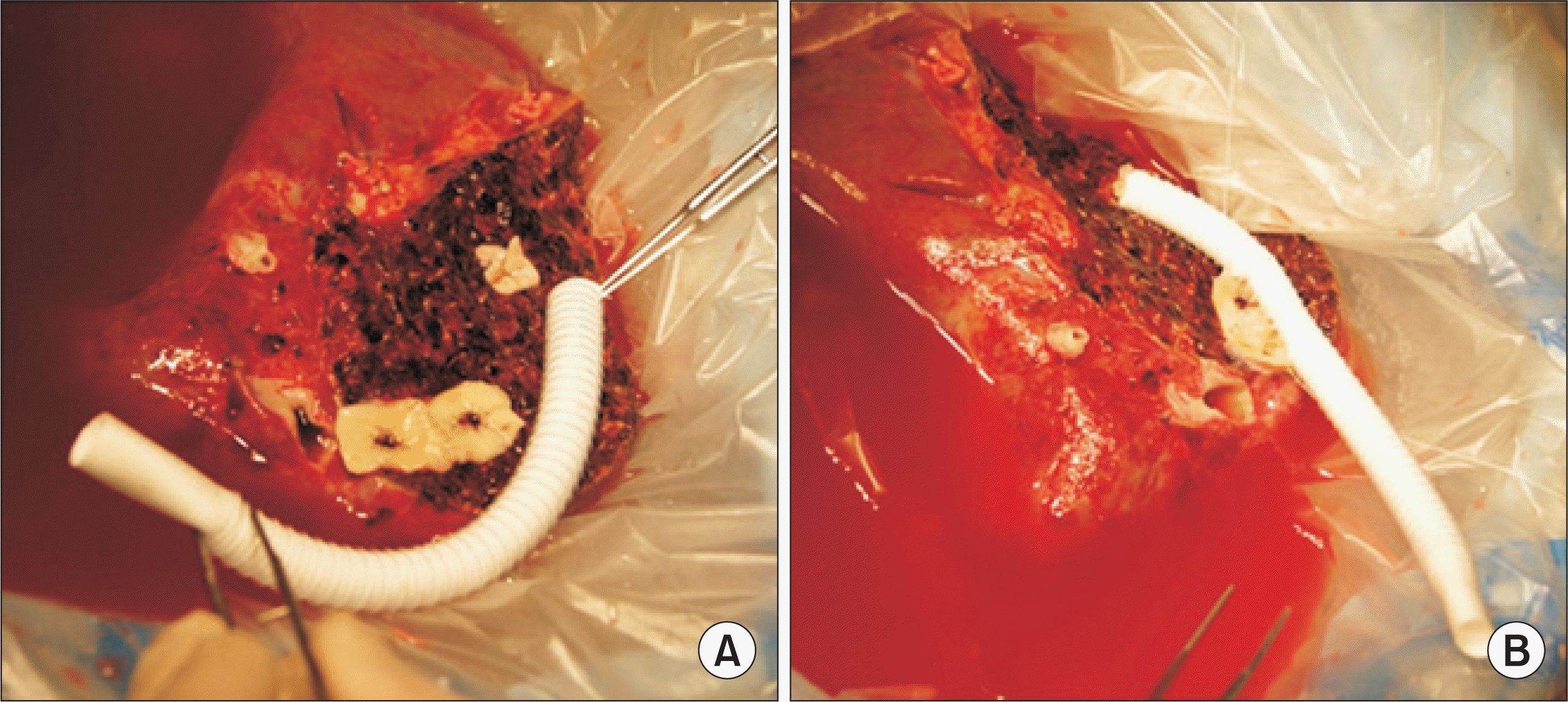 | Fig. 1Intraoperative photographs showing the standardized techniques of middle hepatic vein reconstruction using a composite graft of ringed polytetrafluoroethylene (PTFE) and cryopreserved iliac artery patch. (A) The hepatic vein branch orifices at the liver cut surface were widened by a ventral cut, and then an arterial patch was sutured to each orifice. (B) A 10-mm-sized ringed PTFE graft was prepared and end-to-side anastomosis was done between the PTFE graft and arterial patch, making a funnel-shaped intervening arterial patch.
|
Evaluation of PTFE-Interposed MHV Patency and Indications for Stenting
According to our LDLT management protocol, posttransplant dynamic computed tomography (CT) scans were routinely performed every week while patients were in the hospital, and at 1, 3, 6, and 12 months after LDLT. Thereafter, follow-up abdomen CT scans were repeated annually for 5 years and biannually after 5 years. We define MHV occlusion as a nonvisualization of blood flow in the PTFE graft conduit between V8 (or V5 when only V5 was reconstructed) and the inferior vena cava on dynamic liver CT. When V5 was thrombosed but V8 was patent, we considered it as patent. When CT scan was not taken due to impaired renal function, information from Doppler ultrasonography was used instead. Interventional stenting of the thrombosed PTFE graft was indicated when significant MHV occlusion-related perfusion abnormality developed [12,13]. We classified MHV stenting as MHV occlusion in this study, although MHV patency was regained after stenting.
Statistical Analysis
All numerical data were presented as a mean±standard deviation. Patency rates were determined using the Kaplan-Meier method and compared using a log-rank test. Statistical analyses were performed using SPSS IBM ver. 22.0 (IBM Corp., Armonk, NY, USA).
Go to :
RESULTS
Patient Profiles
The clinical profiles of 100 patients that underwent LDLT using a modified right lobe graft, with MHV reconstruction using PTFE grafts, were as follows: the mean age was 53.5±5.4 years; male to female sex ratio was 73:27; primary diagnoses were hepatitis B virus infection (n=71) and other (n=28); MELD score was 16.2±8.3; ABO blood-incompatible LDLT was 15 (15%); and the GRWR was 1.12±0.3.
Configurations of MHV Reconstruction Using a PTFE Graft
The sources of vessel patches attached at the V5/V8 orifices were patched cryopreserved iliac arteries and veins, and autologous saphenous and portal veins. Patch unification of two or three small V5/V8 branches was preferentially used because it enabled us to make a single anastomosis to the interposition PTFE graft. V5 reconstruction was done in a single anastomosis (n=85; 85%, including unification venoplasty), and double anastomoses (n=14; 14%), and no reconstruction (n=1; 1%). V8 reconstruction was done in a single anastomosis (n=75; 75%, including unification venoplasty), and double anastomoses (n=0; 0%), and no reconstruction (n=25; 25%).
Patterns of MHV Graft Occlusion
Serial follow-up CT scans showed that luminal thrombosis occurred within PTFE grafts around the V5 anastomosis. V5 outflow was gradually reduced, which resulted in concentric thickening of luminal thrombus. At this phase, a PTFE graft with a 10-mm inner diameter was transformed to a narrow conduit with an inner diameter of 3–5 mm. At last, the graft lumen between the V5 and V8 orifices was occluded. Meanwhile, the V8 outflow was maintained for a longer period (Fig. 2).
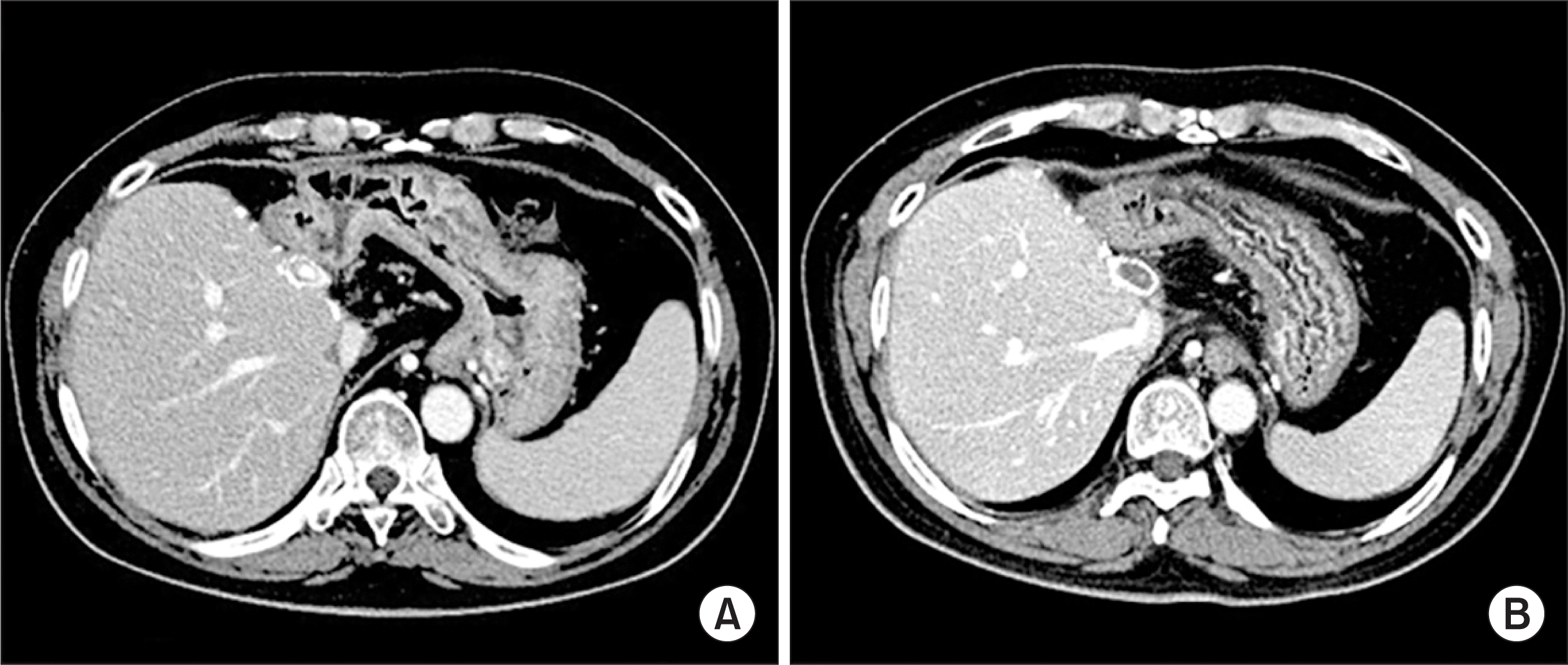 | Fig. 2Computed tomography images showing progressive occlusion of the lumen within the interposed polytetrafluoroethylene graft, taken after 3 months (A) and 6 months (B). Despite the deprivation of middle hepatic vein outflow, noticeable hepatic venous congestion was not developed due to intrahepatic venous collateral formation.
|
PTFE-Interposition MHV Patency
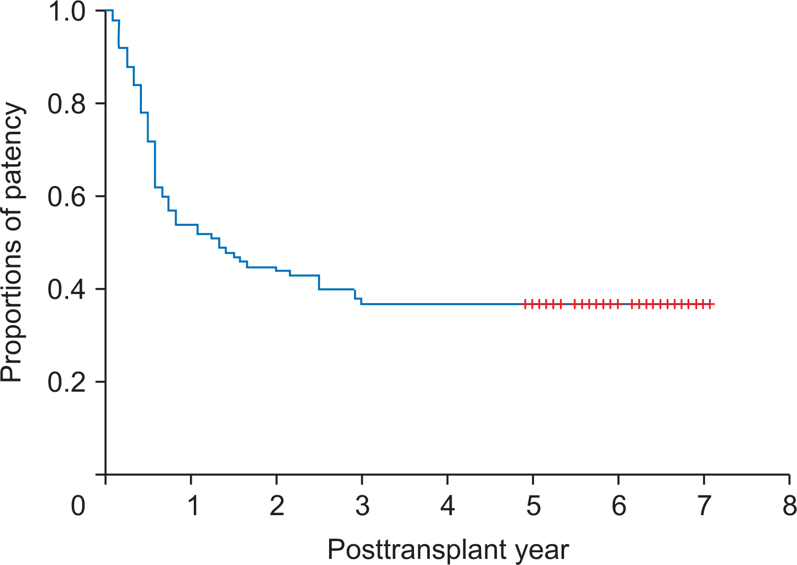
During a mean follow-up of 6 years, three patients (3%) required MHV stenting. All of them underwent early stenting within 2 weeks after LT and MHV flow was restored. After 3 months, there were no episodes of congestion-associated hepatic infarct, regardless of MHV patency. The actual patency rates of PTFE-interposed MHV were 54.0%, 44.0%, 37.0%, and 37.0% at 1, 2, 3, and 5 years, respectively (Fig. 3).
Unwanted Migration of PTFE Graft into the Adjacent Hollow Viscus
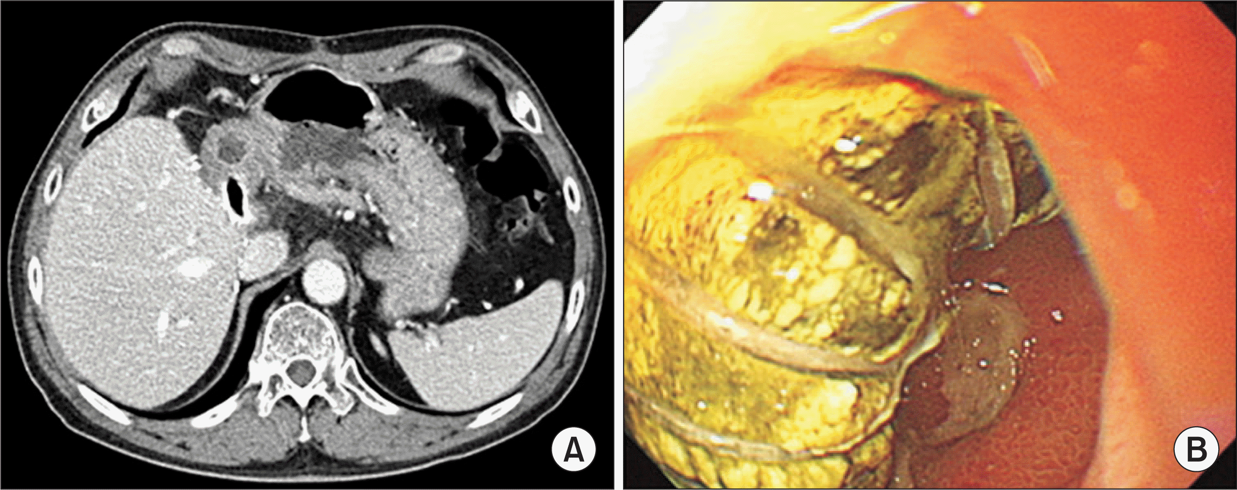
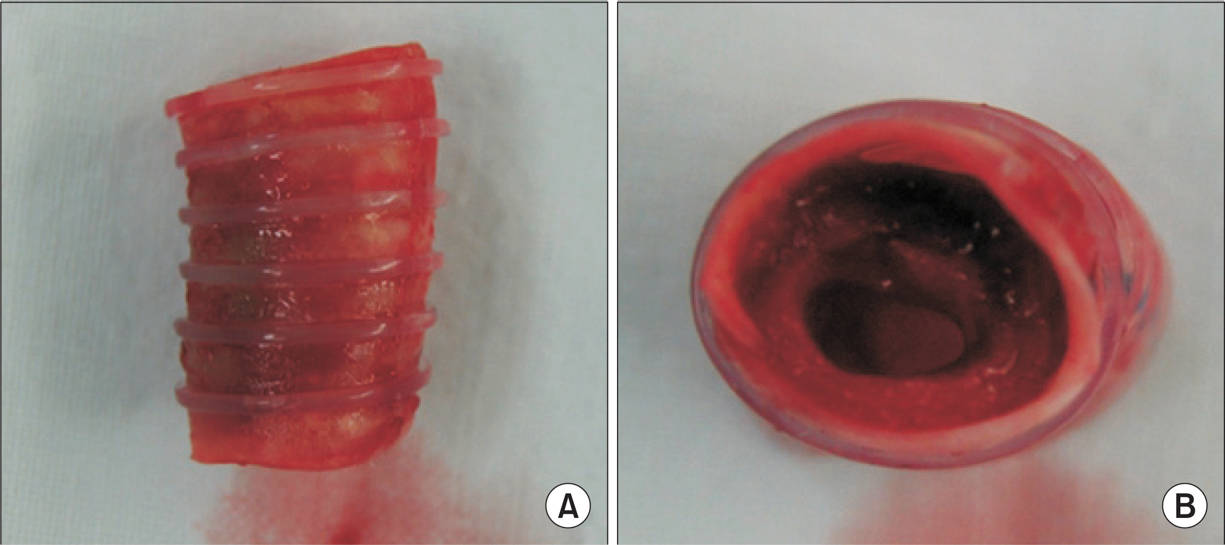
Ringed PTFE grafts in two patients accidentally penetrated the gastric wall and each had to be removed by exploratory laparotomy at 6 months (Fig. 4) and 3 years (Fig. 5) posttransplant [6]. These two patients did not show any symptoms or signs indicating vascular complications at the time of detection. PTFE migration was detected on routine follow-up CT scans and gastric luminal penetration was confirmed by endoscopic examination. In this study, the actual incidence of unwanted migration of the PTFE graft at 5 years was 2%. A majority of the occluded PTFE grafts remained silent as foreign bodies even after thrombotic luminal obliteration.
Go to :
DISCUSSION
MHV reconstruction resulted in a new demand for vascular allografts in the field of LDLT. Moreover, the increase in LDLT volume lead to relative shortages in the supply of vessel allografts. We have used every available vessel material for MHV reconstruction. Cryopreserved iliac vein allografts have been traditionally regarded as the most suitable interposition material for MHV reconstruction. However, the most serious problem is its limited availability.
In regard to availability, prosthetic vessel grafts have a definite merit of unlimited supply. The short- and long-term patency rates of ringed PTFE grafts were acceptably high, as shown in this study. We previously reported that the 6-month patency rates of MHV interposition grafts were 75.3% with cryopreserved iliac veins, 35.2% with iliac arteries, 92.3% with aortas, and 76.6% with ringed PTFE grafts [5]. We think that such high patency rates are primarily due to the surgical techniques that use ringed PTFE grafts for the following reasons: the protective effect of the outer rings against extrinsic compression; offset against the stenosis-inducing effects from tissue reactions after placing a composite artery patch between the V5/V8 orifice and PTFE grafts [14]; and the construction of a streamline-shaped, endothelial cell-lined internal tunnel within the luminal thrombus of the PTFE graft [5]. Such an internal pathway acts like a narrow neointima-lined conduit with an internal diameter that is adjusted by the passing blood flow volume [15].
PTFE grafts have the definite advantage of unlimited supply and appear promising due to improved luminal patency. Although PTFE grafts have some nonnegligible disadvantages such as longer bench work time and requirement of a small vessel patch for composite grafts, most of these demerits are acceptable or manageable.
However, unwanted migration of a PTFE graft into the hollow viscus is an unexpected serious complication [6,7]. Migration of such a foreign body into the stomach or duodenum can induce life-threatening complications that require surgical removal. We presume that the underlying mechanism of unwanted PTFE graft migration is based on inflammation against a foreign body. Inflammation-induced adhesion might facilitate the migration of a PTFE graft into the adjacent hollow viscus. Its real-world 5-year incidence was estimated to be 2% in this study. A majority of the occluded PTFE grafts remained silent as foreign bodies after thrombotic luminal obliteration, which can be a potential source of other rare complications later.
Hsu et al. [7] reported that PTFE-related complications developed in 1.5% (4/262) of patients. One patient developed complete thrombosis with sepsis at 24 months and died due to multi-organ failure. Three patients developed graft migration into the second portion of the duodenum, without overt peritonitis. Surgical exploration and PTFE graft removal was done in all three patients. One patient died due to overwhelming sepsis.
Considering the benefits and complications of PTFE grafts, the grafts are currently regarded as vascular substitutes of “necessary evil.” Considering that massive hepatic venous congestion from exclusion of MHV deprivation is one of the leading causes of graft failure, the abovementioned demerits from PTFE-associated complications are not great enough to abandon the use of PTFE grafts. In conclusion, ringed PTFE grafts combined with small-artery patches demonstrated acceptably high short- and long-term patency rates. Since the risk of unwanted PTFE graft migration is not negligibly low, lifelong surveillance is necessary to detect unexpected rare complications.
Go to :
 XML Download