

 PDF
PDF Citation
 Print
Print


INTRODUCTION
Syndesmotic injuries commonly occur when ankle joint is involved in rotational force or direct injury. The syndesmotic ligaments can be also disrupted even without fracture, which is mainly high ankle sprain.1,2) And its prevalence is reported up to 20%.3,4) This injury could lead to chronic ankle pain or arthritis, which cause the need of syndesmosis reduction and fixation surgery when injury.5)
However, there remain still some debates over syndesmosis fixation.6-8) Many believe weight bearing should be permitted only after removal of syndesmotic screw because the screw could limit normal physiologic motions of talus as well as distal tibiofibular joint. Even more, it may lead the breakage of syndesmotic screw. Once it occurs, there may lead some problems including additional surgical time caused by broken screw removal, additional incision, or medical sue problem.
So, there are several concerns regarding screw breakage such as when weight bearing should be permitted, whether syndesmotic screw should be removed after syndesmotic healing, and if so, when the screw should be removed. Even whether tri- or quadricortical fixation, and 3.5 mm or 4.5 mm cortical screw should be purchased are also related to this concern. Tricortical technique is known not to require routine removal of screw before full weight bearing. And 4.5 mm cortical screw was reported to have greater resistance to shear stress at the syndesmosis. Suture button technique is recently popularized, however, this also has own limitation; no multidirectional stability, complications such as osteomyelitis, and so on.9-11)
Therefore, the purpose of this study was to know whether a screw with novel design would have more fatigue strength compared to conventional cortical screw in the fixed state of syndesmosis. Our hypothesis was that this screw with novel design would have less tendency of fatigue breakage than conventional cortical screw has in syndesmosis fixation.
Go to : 
MATERIALS AND METHODS
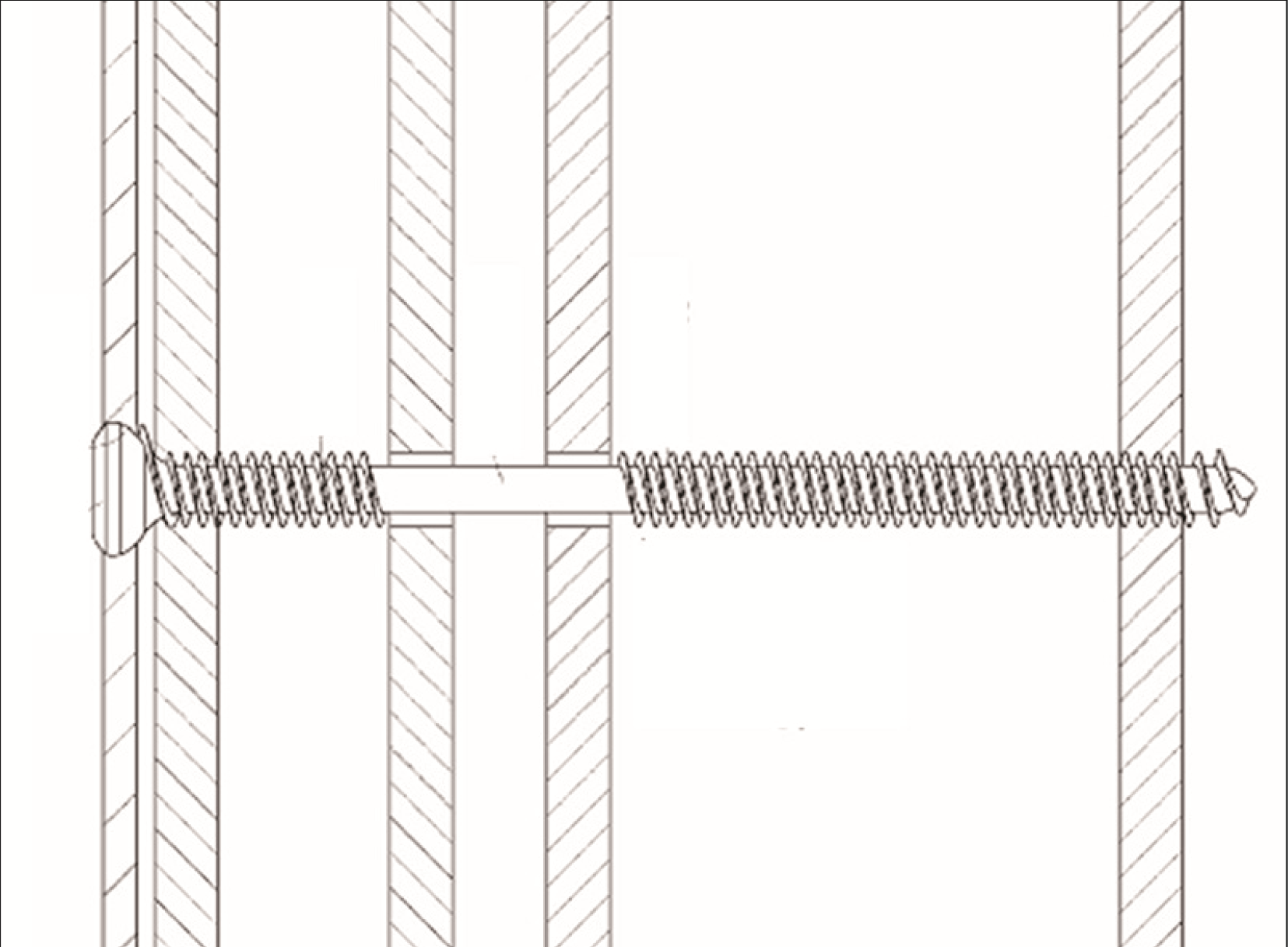
This study was approved by Institutional Review Board of Myongji Hospital. Three kinds of screws made of titanium were tested which had four samples of each kind of screw with the technique of four-cortex fixation. As the first group, conventional cortical screw of 3.5 mm diameter, of which core diameter was 2.4 mm, was set as control group. Here, we made a screw of novel design. A thinner thread-free part, mid-portion of the screw in the central cortex area which consists of far cortex of fibula and near cortex of tibia, that creates room for movement as the surrounding bone is drilled in with the wider diameter of the external parts of the screw (Fig. 1). Second group was fixed using this newly-designed screw with core diameter of mid portion 2.4 mm. Third group was the same kind of the newly-designed screw but its core diameter of mid portion was thinner than second group; 2.0 mm.
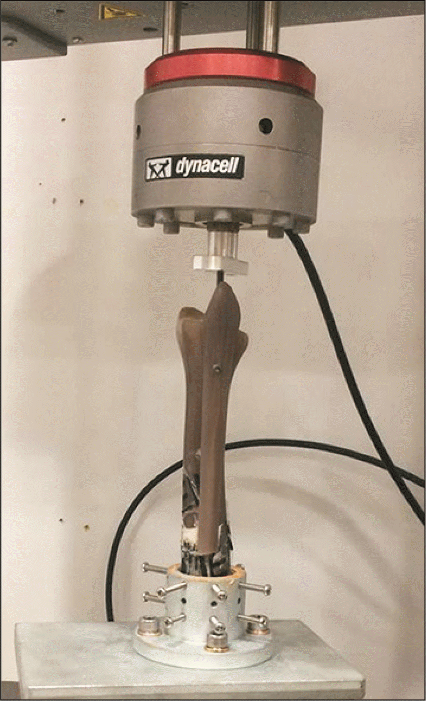
Preliminary test was performed to determine an amount of cyclic load using stress machine (Instron Corp., Norwood, MA, USA). Tibia of Sawbone model (Sawbones, Vashon, WA, USA), which had hollow canal and cortical bone structure, was fixed upside down at the basement of machine with the manner of previous literature (Fig. 2).12) And fibula of the same model was transfixed by 3.5 mm cortical screw with the usual manner of syndesmosis fixation. The distal fibula was loaded at a displacement rate of 5 mm/minute in compressive direction. As a result, we set repetitive loading as 400 N.
In fatigue test, 400 N was applied to a fibula model and at each test we record cycles which lead to screw failure; bending or breakage of the screw. Each group had 4 samples, so we did a total of 12 fatigue tests.
After performing all tests, statistical analysis was done. For comparison of fatigue endurance of all screws, Kruskal-Wallis H test was performed. And Tukey’s method for post hoc analysis after ANOVA analysis using ranking variable were used. And p-value less than 0.05 was considered as significant. All analysis was performed using SPSS software (ver. 20; IBM Corp., Armonk, NY, USA).
Go to :
RESULTS
Each number of cycles to metal failure was obtained on all 12 specimens after all fatigue tests (Table 1). Significant difference was noticed among three groups (p=0.018). Screws of second group showed the most fatigue strength (Table 1). First group (3.5 cortical screws) could not showed significant difference of fatigue strength compared to screws of third group (p=0.401; Table 1).
Table 1
Numbers of Cycle Until Metal Failure in Fatigue Tests of Three Group Screws
![]()
Go to :
DISCUSSION
In this biomechanical study, screw with thread-free mid-shank of 2.4 mm diameter, which is the same core diameter with that of 3.5 mm cortical screw, has the most fatigue strength among three groups. Cortical screw followed that, and screw with thread-free mid-shank of 2.0 mm diameter was the least fatigue strength although these two screws did not show significant difference.
Syndesmosis screw breakage is one of complications which surgeons want to avoid after retention of syndesmosis screw although there are still debates over removal or retention of screw. Therefore, there have been a number of studies regarding the breakage of syndesmosis screw using cadavers or sawbone models.12-14) And retention of syndesmosis screw may limit the motion of ankle joint.15) So, screw removal or broken state was known to show better clinical outcome than the retention of unbroken screw.16) Another study with 30 patients recommended the removal of syndesmotic screw because two of screw retention group showed screw breakage although both groups showed similar clinical outcomes.4) In contrast, some studies showed no difference of clinical outcome between removed or retention state of syndesmotic screw.6,17) However, their population was relatively small as series with 20 to 50 patients.
We had hypothesized that thread-free mid-shank with 2.0 mm diameter might have more endurance to fatigue stress than that with 2.4 mm diameter because it had more flexibility to have room for screw to bend in middle space; far cortex of fibula and near cortex of tibia. However, screw with large diameter (2.4 mm) of thread-free mid-portion had the most fatigue strength in this study. Previous study comparing 3.5- and 4.5-mm cortical screw fixation already showed that larger diameter screw provides greater resistance to an applied the distal syndesmosis. Meanwhile, screw with 2.4 mm diameter of thread-free mid-portion had more fatigue strength than cortical screw with the same core diameter. Considering these outcomes, both the strength and flexibility of screw are all important to avoid the fatigue breakage of screw.
Some surgeons prefer to use partial-threaded cannulated screw available in clinical practice when fixing syndesmosis. Partial-threaded cannulated screw, which has no thread in proximal portion which is placed in fibula, has similar flexibility to our novel design. So, the outcome of this study could support their preference to use partial-threaded cannulated screw in some extent. However, partial-threaded cannulated screw could bring the effect of compression to fibula toward tibia when fixing the syndesmosis because that has no thread in proximal portion of the screw. Moreover, pull-out strength of partial-threaded screw is theoretically less than the screw of novel design, which would necessitate require future study comparing them.
Before initial preliminary test, we axially loaded 800 N to 3.5 mm cortical screw fixed in syndesmosis of sawbone model for 225,000 cycles, which applied had been calculated by multiplying the daily values retrieved from step counting studies over 63 days, in a materials testing machine.13,18) However, this condition was not enough to cause metal failure, so we modified test condition as above; preliminary test and fatigue test.
There were some limitations in this study. First, we did not use cadaver in biomechanical test and did not perform clinical study, which might show different outcome from our results. Future study may be warranted when the screw with this novel design is available by manufacturers. Second, we did not apply external rotational force or anteroposterior translation force to specimen due to the limitation of machine ability. Third, this study was performed with specimen of small volume, which might affect statistical significance of the outcome. So, more mechanical study with large volume should be needed to know if this newly-designed screw would have the similar syndesmosis-holding ability compared to the conventional cortical screw because it had thread-free portion in its mid-shank.
Go to :
 XML Download