

 PDF
PDF Citation
 Print
Print


I. Introduction
Angiosarcoma is an extremely rare malignant mesenchymal tumor of vascular or lymphatic origin
1
. This type of tumor represent less than 1% of all malignant tumors, and about 50% of angiosarcomas are found in the head and neck region, with only approximately 1% reported to occur in the oral cavity or salivary glands
1,2
. Angiosarcoma most commonly affects the elderly and is predominant in males, with a male to female ratio of 2:1
3
. Angiosarcoma is an extremely aggressive malignancy, with a predicted 5-year survival rate for cases in the head and neck region of only 10% to 35%
4
. Optimal treatment includes extensive surgery and wide-field postoperative radiation therapy (RT). In this paper, we report a case of angiosarcoma that transformed from mucoepidermoid carcinoma as a result of RT, in a female patient originally diagnosed with an odontogenic keratocyst (OKC).
Go to : 
II. Case Report
A 58-year-old female visited Dankook University Dental Hospital (Cheonan, Korea) in January 2004, with complaints of facial swelling and gingival soreness. Swelling and pain of the right palatal area were found upon initial clinical examination. Computed tomography (CT) showed an extensive cystic lesion in the right maxilla and maxillary sinus. A clinical diagnosis of an OKC was made based on the patient’s history and clinical observations. Enucleation and iliac bone graft were performed under general anesthesia subsequent to endodontic treatment of teeth #13, #14, #15, #16, and #17. Biopsy results revealed respiratory mucosa with chronic inflammation, fibrosis, and old hemorrhage.
In September 2006, a similar lesion was observed on follow-up CT radiographs, and a clinical diagnosis of recurrent OKC was made, but a biopsy of the mass revealed mucoepidermoid carcinoma. Partial maxillectomy under general anesthesia was performed, and adjuvant postoperative RT of 6,660 cGy in 37 fractions was subsequently initiated in November 2006.
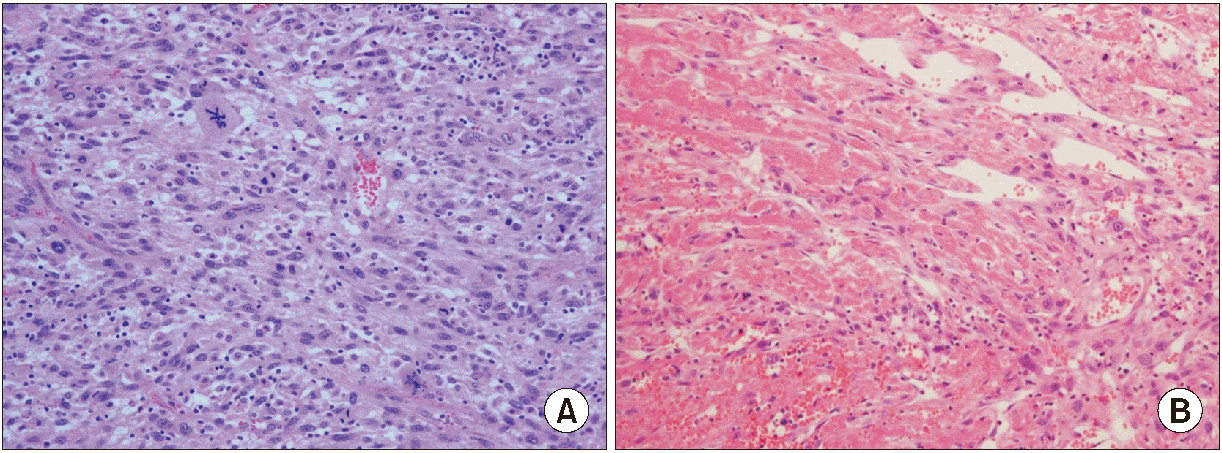
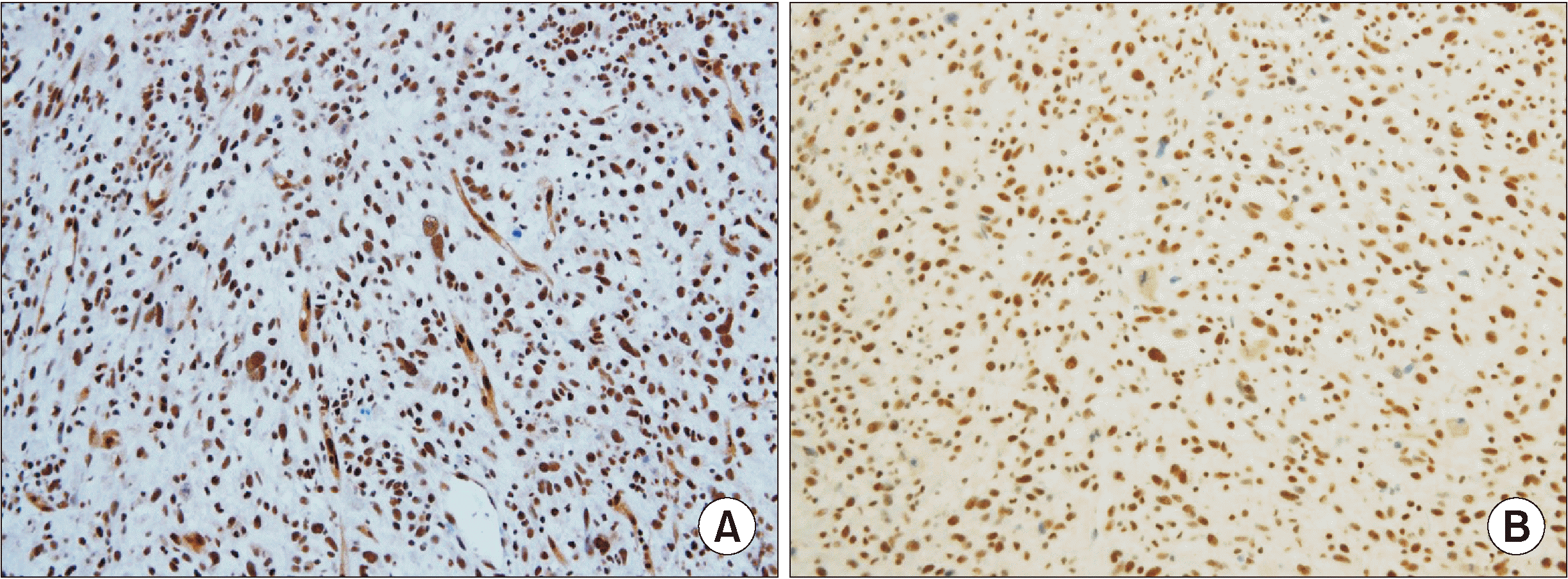
However, in September 2018, the patient returned to our hospital with a brown tumor-like lesion in the right partial maxillectomy area. Incisional biopsy revealed malignancy that was suspicious for a malignant vascular tumor. Proton emission tomography-computed tomography (PET/CT) showed a lobulating hypermetabolic lesion (SUL max 20.9) approximately 5 cm in diameter and positioned within and around the right maxillary and nasal cavity area.(Fig. 1) Hemi-maxillectomy via the Weber Ferguson approach under general anesthesia was performed (Fig. 2) with no conjunctive neck dissection, and the excised tumor was approximately 6 cm in diameter. Adjuvant RT of 6,000 cGy in 30 fractions was performed subsequent to surgery. To date, no specific symptoms or signs of recurrence have been found. Histologically, the tumor was composed of atypical spindle cells with hyperchromatic nuclei. The tumor also showed vascular formation that contained red blood cells.(Fig. 3) Immunohistochemical study revealed tumor cells that were positive for FLI-1 and INI-1.(Fig. 4) A pathological diagnosis of poorly differentiated, high-grade angiosarcoma of the maxilla was made. Based on the clinical, radiological, and pathological findings, the patient was finally diagnosed with radiation-induced angiosarcoma.
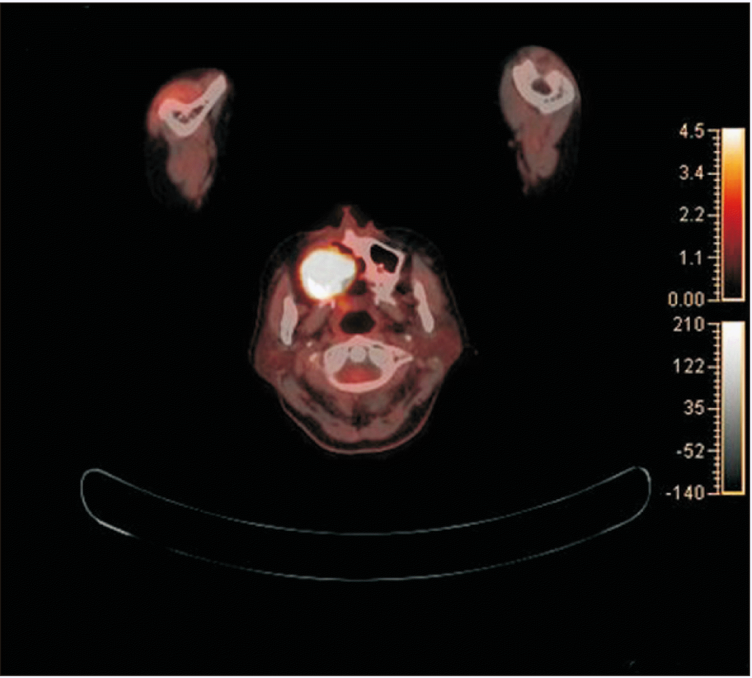 | Fig. 1Proton emission tomography-computed tomography showing hypermetabolic lesion around right maxilla and nasal cavity.
|
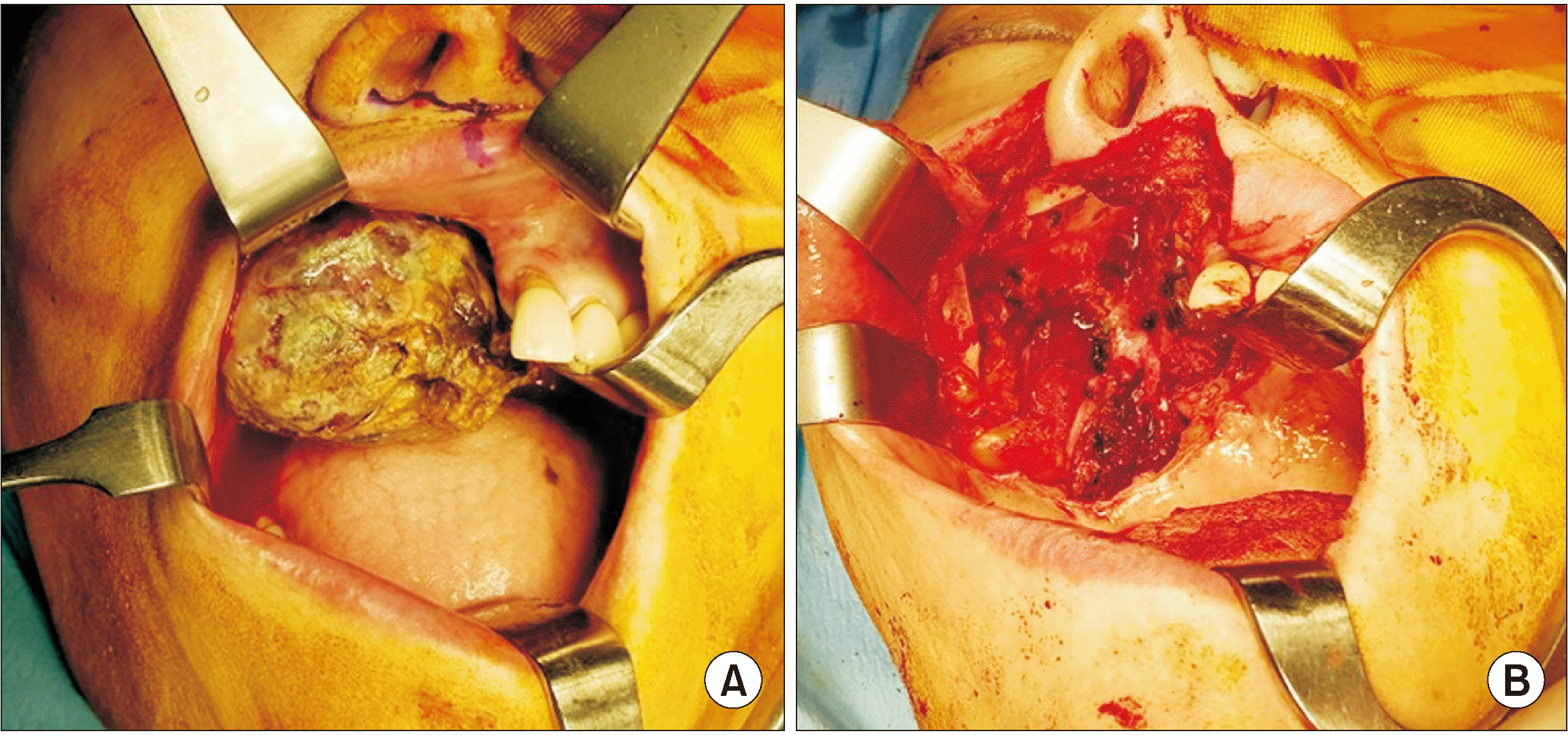 | Fig. 2A. Preoperative photograph of angiosarcoma in palate. B. Postoperative photograph of angiosarcoma in palate after resection.
|
Go to :
III. Discussion
OKC is an aggressive benign cyst that has a marked tendency for recurrence. Recurrence rates after excision are reported to range from 12% to 62.5%. Histologic examination of recurrent cysts is seldom suggestive of premalignant or malignant changes, and frank malignant degeneration is rare
5
. However, the lining of an odontogenic cyst, including an OKC, may transform into squamous cell carcinoma (SCCa) or mucoepidermoid carcinoma
6
.
Mucoepidermoid carcinoma is a malignant epithelial tumor commonly found in the salivary gland. The primary treatment modality for mucoepidermoid carcinoma is wide local resection in conjunction with neck dissection. RT is typically recommended for high-grade mucoepidermoid carcinomas. Although mucoepidermoid carcinoma diagnoses tend to show good overall prognosis, it is recommended that the postoperative follow-up period be as long as 10 years, due to the potential for late recurrence and regional metastasis
7
.
The typical histologic features of angiosarcomas include the presence of atypical endothelial cells in a number greater than required to line a vessel, as well as formation of vascular tubes with lamina that has a distinct tendency to anastomose with the delicate framework of reticulin fibers. Angiosarcomas also show rapid cell growth, local invasion, and early hematogenous spreading, which lead to poor prognosis.
Although treatment guidelines for angiosarcomas are currently minimal due to the limited number of literature reports, most authors advocate radical surgery. Because of the high risk of local recurrence, adjuvant RT in doses greater than 50 Gy is also recommended
3
. Other approaches support the use of primary RT, with surgery as an alternative or additional option8-10. Despite the risk of metastasis, no clear evidence exists for use of chemotherapy as primary treatment or as a conjunctive treatment to surgery and primary RT. Although there is currently a lack of data advocating for chemotherapy as a treatment modality for angiosarcomas, chemotherapy can be considered in unresectable cases. Overall, there are also conflicting opinions on elective treatment of the neck
10,11
.
Radiation carcinogenesis is an unfortunate toxic consequence of cancer therapy, and sarcomas can be induced as a result of the ionizing radiation
12
. The exact pathogenesis of radiation-induced angiosarcoma is unknown. The p53 protein is known to play an important role in repairing radiation-induced DNA damage
10,13
. A previous study of spontaneous angiosarcoma in p53-null mice suggested that the p53 protein acts as a tumor suppressor gene and may thus be involved in angiosarcoma carcinogenesis
10,14
.
Our case represents a rare transformation of mucoepidermoid carcinoma into angiosarcoma following RT. RT, whether primary or adjuvant, enhances treatment outcomes and is preferable in many cases. However, rare complications such as malignant transformation are known to occur long after RT. Regular long-term follow-up is therefore crucial for patients with history of RT to prevent these complications.
Go to :
 XML Download