

 PDF
PDF Citation
 Print
Print


I. Introduction
Successful management of maxillofacial fractures depends on correct reduction and precise fixation of broken segments in order to achieve a normal occlusion, resume function, and properly align the broken pieces next to each other
1
. Despite advances in maxillofacial surgery, impaired bone healing remains a concern for surgical teams
2
. Mandibular fractures are among the most common of the maxillofacial region, accounting for 23% to 97% of all facial fractures
3,4
.
Bone healing is a complex process that includes three stages: inflammation, repair, and delayed remodeling. This biological process is controlled by complex cellular and molecular mechanisms. Systemic and local factors, as well as several cell types and growth factors delivered via the adjacent tissues and blood stream, all play a role in bone healing5-7. A number of studies have evaluated growth factors, injection of medications and electrical stimulation to accelerate and enhance bone healing8-10. Other studies have indicated that some medications, such as antibiotics and bisphosphonates, delay or impair the process of bone healing. By having a comprehensive understanding of these factors and not prescribing these medications in cases of fracture, complete bone healing can be expected
11,12
. Bleeding at the site of injury is the most important factor for successful bone healing
13
. Decreased angiogenesis at the site of trauma and limited blood supply to the site are known to delay or impair the process of bone healing
14,15
. Nitric oxide and vasodilation are imperative for angiogenesis, and the positive effects of nitric oxide on wound healing are probably due its functional effects on angiogenesis and inflammation16-18.
Sildenafil, a selective phosphodiesterase-5 inhibitor, prevents the degradation of nitric oxide and is a potent stimulator of angiogenesis. Phosphodiesterase-5 results in degradation of cyclic guanosine monophosphate, which relaxes smooth muscle
19
. Sildenafil is a vasodilator of the peripheral arteries and veins and prevents the formation of thrombosis
20,21
. It is also the most commonly prescribed medication for males with erectile dysfunction
22
.
Pentoxifylline is a non-selective phosphodiesterase inhibitor that decreases inflammation and increases the blood flow and oxygenation of tissues
23,24
. Pentoxifylline also decreases platelet accumulation and formation of thrombosis
25
. Compared with other medications in this class, pentoxifylline has fewer gastrointestinal side effects and a lower cost
26,27
.
Recent studies have shown that sildenafil affects growth factors such as vascular endothelial growth factor and cysteine rich-61 and thereby enhances bone healing
14,28,29
. Histing et al.
19
reported that sildenafil enhances bone healing by increasing bone formation. Kinoshita et al.
30
showed that daily injections of pentoxifylline stimulate bone formation and increase bone mass in rats. Labib and Farid
31
indicated that pentoxifylline administration can be considered a reliable approach to manage osseointegration. Moreover, several studies have confirmed the positive efficacy of pentoxifylline for healing of osteoradionecrosis of the mandible
32,33
. Furthermore, pentoxifylline is extensively used in orthopedics to maintain viability of grafts and other vascular tissues used for regeneration treatments
34
.
Many studies have assessed the effects of sildenafil and pentoxifylline on wound healing; however, their efficacy for bone healing has not been well investigated. Moreover, no previous study has evaluated the efficacy of sildenafil and pentoxifylline to enhance healing of maxillofacial fractures. Considering this gap of information, our study aimed to assess the effect of sildenafil and pentoxifylline phosphodiesterase inhibitors on healing of mandibular fractures in rats.
Go to : 
II. Materials and Methods
The study protocol was approved by the Ethics Committee of Isfahan Islamic Azad University (approval No. IR.IAU.KHUISF.REC.1398.188). All rats received standard laboratory nutrition and were kept in a calm environment with controlled temperature and moisture (22°C±2°C and 40%-60% humidity) and 12:12 h light/dark cycle as instructed by Animal Welfare Information Center
35
. A total of 60 male 12- to 14-week-old Albino Wistar rats weighing 300-360 g were evaluated. They did not have any infection or pathological condition affecting the experiment. The rats were randomly divided into six groups of 10.
Rats in group C1 received saline orally on a daily basis after the surgery and were sacrificed after 1 week.
Rats in group S1 received 10 mg/kg sildenafil (Sildenafil 50 mg; Marham Daru, Tehran, Iran) orally via gavage on a daily basis and were sacrificed after 1 week. The rats voluntarily consumed nutritional supplement from a syringe.
Rats in group P1 received 50 mg/kg pentoxifylline (pentoxifylline 400 mg, Extended Release; Amin Pharmaceutical, Isfahan, Iran) orally via gavage on a daily basis and were sacrificed after 1 week.
Groups C4, S4, and P4 received medications as in groups C1, S1, and P1 but were sacrificed after 4 weeks.
The dosage of administered medications was determined according to previous similar studies34,36-38. Use of higher doses may cause greater vasodilation but may be associated with side effects such as hypotension, decreased tissue perfusion and severe anti-inflammatory responses. Use of lower doses may have no effect at all
39
.
1. Surgical phase
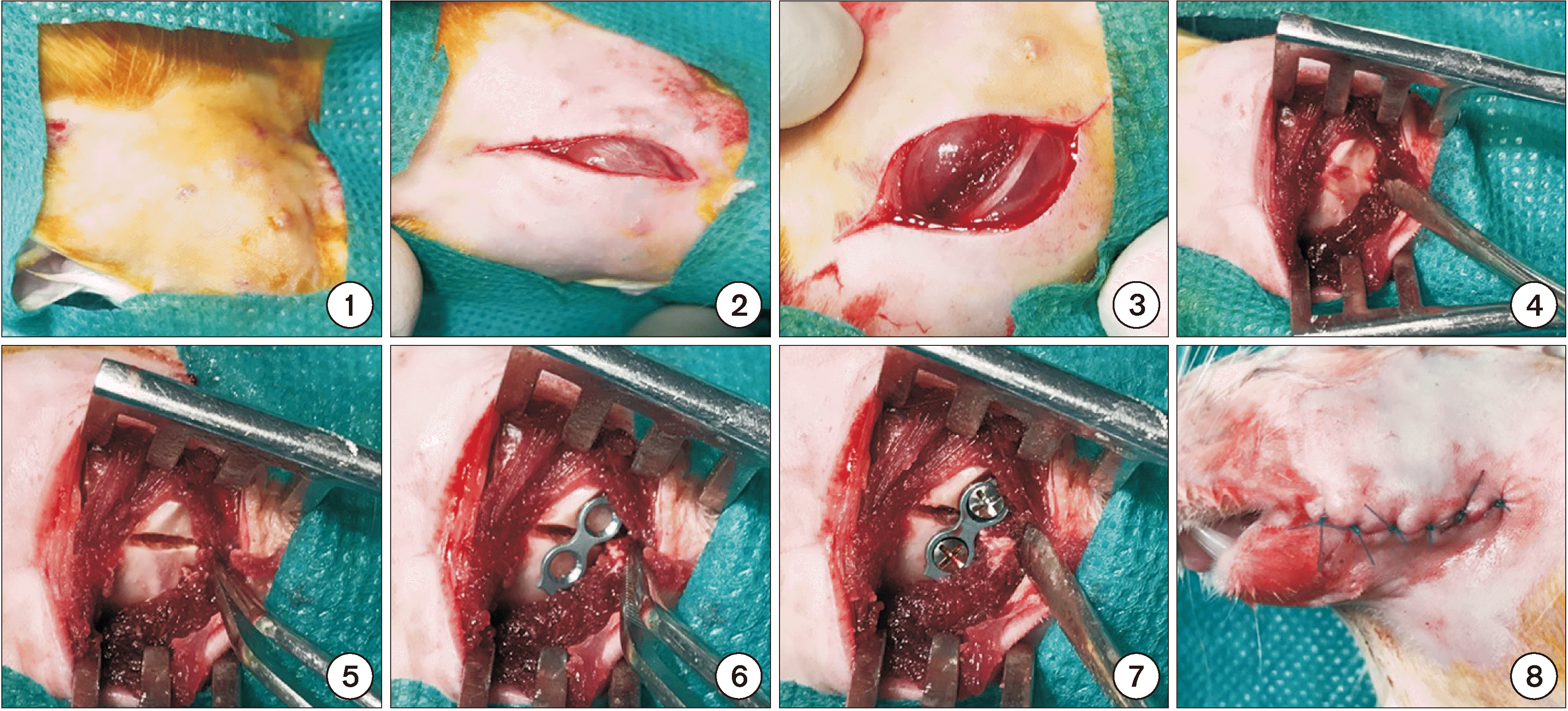
All rats were generally anesthetized by intramuscular injection of 5 mg/kg of ketamine (Ketamine 10%; Alfasan, Woerden, The Netherlands) and 0.02 mL/kg of acepromazine maleate (Neurotranq; Alfasan). Next, 0.3 mL of 2% lidocaine with 1:80,000 epinephrine (Persocain-E; Darou Pakhsh, Tehran, Iran) was injected at the surgical site for local anesthesia and hemostasis. The surgical site was shaved and disinfected (povidone iodine; Behvazan, Rasht, Iran). The rats were placed in the supine position and a 1 cm unilateral submandibular incision was made at the inferior border of the mandible under sterile conditions. After dissecting the masseter muscle, the body of the mandible was exposed. Bicortical osteotomy at the angle of the mandible was performed using a 0.5 mm dental fissure bur (Teeskavan, Tehran, Iran) under copious irrigation with sterile saline. The distance between bone segments was 0.5 mm, which was equal to the bur diameter. The fracture line was fixed using a two-hole microplate (MatrixNEURO adaption plate, thickness: 0.4 mm, pure titanium; Synthes, Oberdorf, Switzerland) and two screws of 1.5 mm diameter (MatrixNEURO screw, self-drilling, 1.5 mm diameter, 3 mm length; Synthes). Subcutaneous and cutaneous tissues were sutured using 5-0 vicryl sutures (polyglycolate coated; Supa, Tehran, Iran) and 6-0 nylon sutures (monofilament polyamide; Supa). All surgical procedures were performed by the same surgeon. All rats received 1 mg/kg tramadol (Tramadic, 50 mg/mL; Caspian Tamin, Rasht, Iran) intramuscularly for pain control and 25 mg/kg cefazolin (Ancef, Kefzol, 1 g; Razi, Tehran, Iran) intramuscularly for infection control twice a day for 5 days. All rats received soft diet for 1 week. Fig. 1 shows the surgical steps. The rats were sacrificed by administration of 200 mg/kg sodium pentobarbital (Pental; IE Ulagay, Istanbul, Turkey) after 1 week in groups C1, S1, and P1 and after 4 weeks in groups C4, S4, and P4. The respective hemimandible was then resected, and the attached soft tissue was removed. The resected hemi-mandibles were sent for histological analysis.
2. Histological analysis
Histological analysis was carried out by a pathologist who was blinded to the group allocation of samples. All specimens were fixed in 10% formalin. After fixing, the specimens were decalcified using ethylene diamine tetra-acetic acid. The specimens were then embedded in paraffin blocks and sagittally sectioned into 4 µm thick slices. They were then stained with H&E. The slides were inspected under a light microscope (Nikon Eclipse E600; Nikon, Tokyo, Japan). Each specimen was then scored based on the degree of bone healing according to the scoring system suggested by Perry et al.11 as follows:
• 1 point, only fibrous tissue
• 2 points, mainly fibrous tissue and small amount of cartilage tissue
• 3 points, equal amount of fibrous and cartilage tissue
• 4 points, completely cartilage tissue
• 5 points, mainly cartilage tissue and small amount of immature (woven) bone
• 6 points, equal amount of cartilage tissue and immature bone
• 7 points, significantly immature (woven) bone and small amount of cartilage
• 8 points, completely immature (woven) bone
• 9 points, immature bone and small amount of mature bone
• 10 points, mature (lamellar) bone
Each specimen was scored by a pathologist who was blinded to the group allocation of samples based on the degree of bone healing at the previous fracture line under a light microscope at ×100 magnification.
3. Statistical analysis
Data were analyzed using IBM SPSS Statistics (ver. 22; IBM, Armonk, NY, USA) via one-way ANOVA and Tukey’s post-hoc test to find significant differences between groups. The mean and standard deviation of the findings of histological analysis were reported. The level of statistical significance was set at P<0.05.
Go to :
III. Results
This study evaluated the effects of two phosphodiesterase inhibitors on bone healing in mandibular fractures in rats. Sixty rats were evaluated in six groups of 10. None of the rats expired during the study and no unwanted complications occurred. All rats tolerated the surgical procedure well. The mean rate of bone healing was 1.9, 3.9, and 4.6 in groups C1, S1, and P1, respectively. One-way ANOVA showed that the mean rate of bone healing in mandibular fractures was significantly different among these three groups at 1 week (P<0.001).(Table 1, Fig. 2) Tukey’s post-hoc test revealed that the mean rate of bone healing in groups S1 and P1 was significantly higher than in group C1 at 1 week (P<0.001). The mean rate of bone healing in group P1 was significantly higher than in group S1 at 1 week (P=0.04).
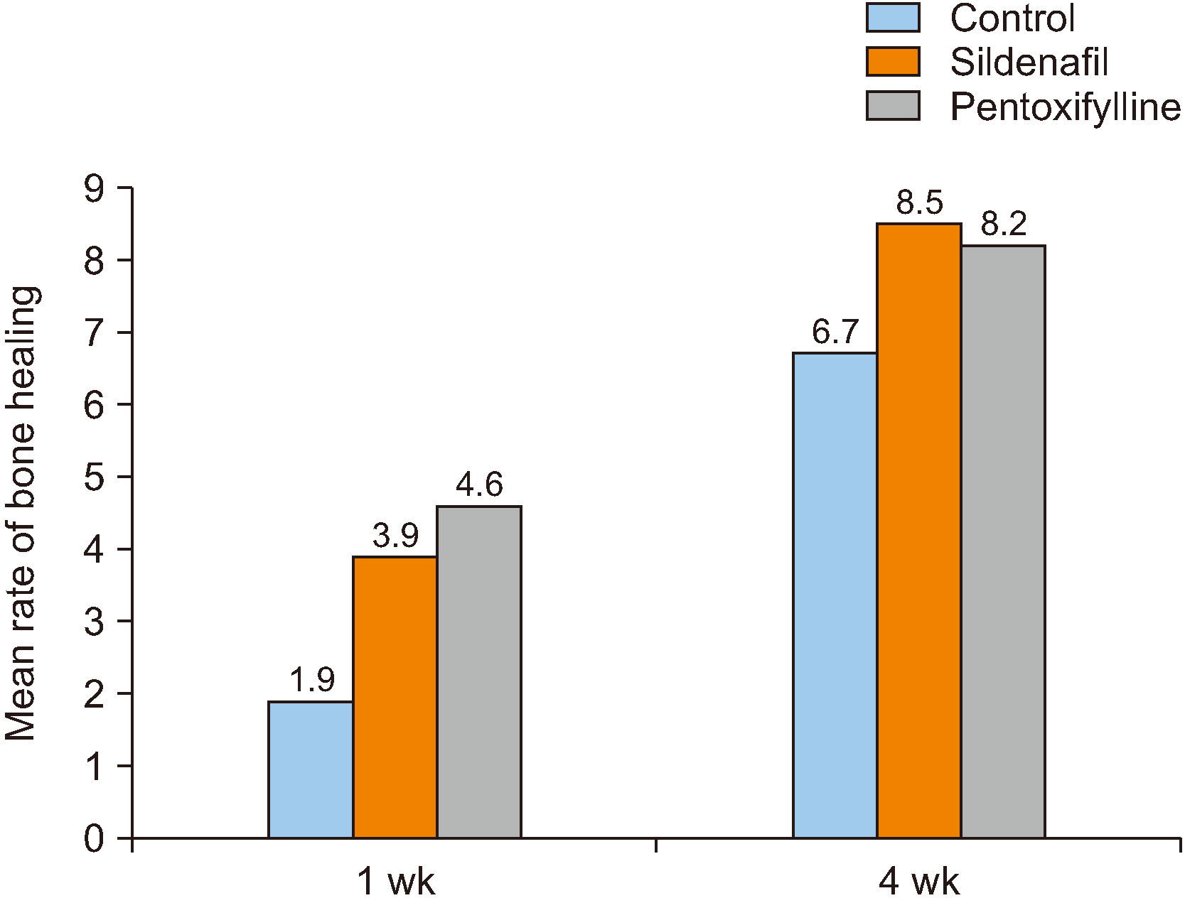
The mean rate of bone healing was 6.7, 8.5, and 8.2 in groups C4, S4, and P4, respectively. One-way ANOVA revealed that the mean rate of bone healing was significantly different among these three groups at 4 weeks (P=0.002).(Table 1, Fig. 2) Tukey’s test revealed that the mean rate of bone healing in groups S4 (P=0.001) and P4 (P=0.004) was significantly higher than in group C4 at 4 weeks but no significant difference was noted between groups P4 and S4 in this respect (P=0.53). Thus, in the present study, the lowest rate of bone healing was seen in group C1 (control group/sacrificed after 1 week) and the highest rate of bone healing was seen in group S4 (sildenafil group/sacrificed after 4 weeks).
Fig. 3 shows histological images of study groups at ×100 magnification.
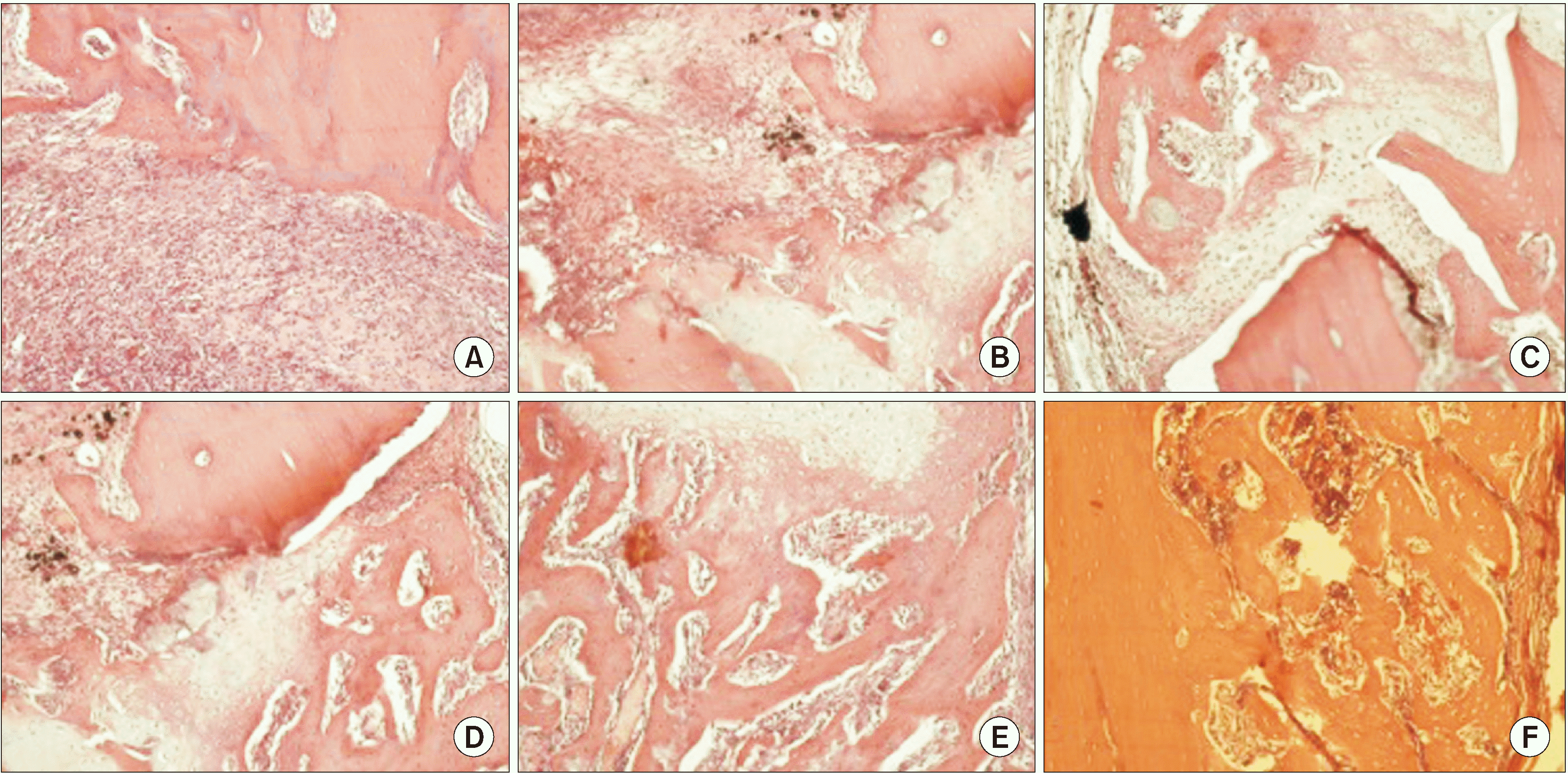 | Fig. 3Histological images of study groups (H&E staining, ×100). A. A specimen from group C1 with bone healing score 1 (healing with fibrous tissue). B. A specimen from group S1 with bone healing score 3 (healing with equal amounts of fibrous and cartilage tissue). C. A specimen from group P1 with bone healing score 5 (healing with mainly cartilage tissue and small amount of immature [woven] bone). D. A specimen from group C4 with bone healing score 6 (healing with equal amounts of cartilage tissue and immature bone). E. A specimen from group S4 with bone healing score 7 (healing with mainly immature bone and small amount of cartilage). F. A specimen from group P4 with bone healing score 9 (healing with immature bone and small amount of mature bone).
|
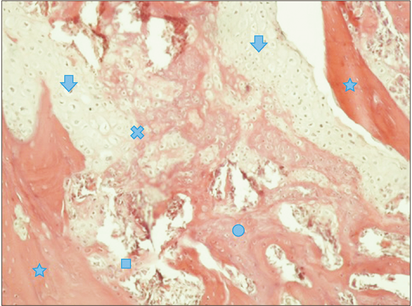
As shown in Fig. 4, bone tissue can be divided into lamellar and woven bone according to its level of maturity. An eosinophilic area containing lacunae was noted in lamellar bone tissue under a microscope (marked by asterisks) and lines confirming periodic calcification of bone could be seen. In fact, these parts were the old host bone, which was mature and seen at both sides of the fracture line. The shorter the time duration since fracture, the less mature the tissue around the mature bone margins. The size and number of lacunae are also important. In areas marked with asterisks (old bone), the number of lacunae and their size were smaller. The area marked with a circle indicates the newly forming, immature bone. This bone has a cancellous appearance and has differences with mature bone in terms of the number of lacunae (where the osteocytes are present) and their size. The area marked with a square indicates the interface of the newly formed bone and older bone. The area marked with an arrow shows the cartilage tissue. The area marked with an x indicates cartilage calcification and its conversion to bone.
Go to :
IV. Discussion
This study assessed the effects of sildenafil and pentoxifylline on bone healing in mandibular fractures in rats. Healing of fractures is an important topic in maxillofacial surgery, as the patient’s routine functions should be reinstated as soon as possible
40
. Many studies have evaluated this topic
40,41
and evidence shows that the outcome of surgical procedures for treatment of fractures is influenced by a number of factors such as patient-related factors, type of bone defect and type of surgical procedure
40
. Adequate blood supply plays a critical role in bone healing
42,43
, and impaired angiogenesis at the site leads to poor bone healing. Oxygen and nutrients are delivered to the site of the forming bone callus by the bloodstream. Moreover, the bloodstream delivers progenitor and inflammatory cells to the site of injury
20,21,28
. Nitric oxide and vasodilation are also imperative for angiogenesis
44
. The positive effects of nitric oxide on wound healing may be related to its functional effects on angiogenesis and inflammation. Sildenafil prevents the breakdown and degradation of nitric oxide, which leads to vasodilation and increased blood supply to the tissue16-18. Several studies have shown that sildenafil is effective for different pathological conditions via the nitric oxide pathway. Other studies have focused on the effects of sildenafil on tissue healing. Many clinical and animal studies have shown positive effects of sildenafil in cases of decreased blood supply to the skin and impaired vascularization as in ischemic wounds
18,21,29
. Moreover, evidence shows that enhanced bone healing by sildenafil is due to the function of cysteine-rich angiogenic inducer 61 protein, which stimulates endothelial cell migration and induces proliferation and differentiation of osteoblasts and cell adhesion
14,45
. Pentoxifylline is a phosphodiesterase inhibitor derived from xanthine, which has a vasodilatory effect. In contrast to most peripheral vasodilators, pentoxifylline has rheological effects on blood and decreases its viscosity
24
. The therapeutic effects of pentoxifylline are mainly attributed to its potential for increasing the blood flow and oxygenation of tissues due to its hemorheological property
25
. It is not clear whether pentoxifylline increases the number of osteoblasts and osteoclasts or not. Takami et al. showed that phosphodiesterase inhibitors increase the number of osteoclasts and their differentiation to osteoblasts
46
. Horiuchi et al.
47,48
demonstrated that pentoxifylline enhances new bone formation by upregulating the bone morphogenetic protein-2. Tsutsumimoto et al.
49
indicated that pentoxifylline can be used to enhance bone formation.
A number of studies have evaluated the effects of phosphodiesterase inhibitors on bone healing with results comparable to ours. However, no previous study evaluated the effects of sildenafil and pentoxifylline on maxillofacial fractures
42
. The current results revealed that pentoxifylline and sildenafil have positive effects on bone healing. Our findings regarding enhanced bone healing by sildenafil are in agreement with those of Yaman et al.
42
and Histing et al.
19
. Aydin et al.
34
used pentoxifylline at a dosage similar to ours and showed that it enhanced the formation of hematoma, which is the first phase of bone healing. This result agrees with our finding. However, in contrast to our results, they showed that the anti-inflammatory effects of pentoxifylline may delay bone healing after 3 weeks
34
. Our study showed positive effects of pentoxifylline on bone healing in the late stage. Since they evaluated femoral bone fractures, this discrepancy may be related to different rates and modes of metabolism of femoral bone and mandibular bone in the final stages of fracture healing.
Our results regarding enhanced bone healing by sildenafil were in line with those of Histing et al.
19
, with the difference that they used 5 mg/kg of sildenafil while we used 10 mg/kg according to the previously published studies. The reason behind the use of 5 mg/kg dosage of sildenafil in their study was that the speed of sildenafil metabolism is higher in rats and sildenafil has a half-life of one hour in rats and four hours in humans. The 5 mg/kg dosage for rats is 5 times the standard dosage for humans (0.7 to 1.5 mg/kg). Despite the different dosages used in the two studies, the outcomes were the same. Atalay et al.
40
used the same dosage of pentoxifylline as ours. The mean histological score of bone healing in their study was 7.8 after 4 weeks while it was 8.2 in our study. This difference is probably due to the difference in defect size in the two studies. The bone gap created in their study after osteotomy was 1 mm while in our study it was 0.5 mm. In the present study, bone healing in the pentoxifylline group was significantly higher than in sildenafil group after 1 week but the score of bone healing in the sildenafil group was higher than in the pentoxifylline group after 4 weeks. It seems that the rheological effects of pentoxifylline and reduction of blood viscosity by this medication in the first week enhanced the primary phase of healing and formation of hematoma at the site of fracture. However, the authors believe that at the end of week 4, the anti-inflammatory effects of pentoxifylline, similar to those of other non-steroidal anti-inflammatory drugs, delayed the process of bone healing. For this reason, at the end of week 4, bone healing in the sildenafil group was higher than in the pentoxifylline group. Infection also plays a role in bone healing. Aydin et al.
34
showed lower bone healing at similar time points and use of equal dosage of pentoxifylline compared to our study (7.1 vs 8.2), which may be due to no postoperative use of antibiotics in their study. As they reported, 40% of samples in the pentoxifylline group had infection, which seemed to slow the process of bone healing. Gong et al.
50
indicated that use of tadalafil and vardenafil phosphodiesterase inhibitors decreased bone mass. Their study was the only one that reported results contrary to ours. This may be attributed to different study design and different types of selective phosphodiesterase inhibitors.
Despite the abovementioned studies, our knowledge about the effects of phosphodiesterase inhibitors on bone metabolism and healing is insufficient. A good understanding of the biological events that occur in the process of bone healing is imperative to find the most efficient approach to enhance bone healing. This study was the first to show the positive effects of sildenafil and pentoxifylline on healing of mandibular bone fractures. Thus, sildenafil and pentoxifylline can be used as adjuncts to enhance bone healing.
One limitation of this study was daily use of 50 mg/kg dosage of pentoxifylline and 10 mg/kg dosage of sildenafil; thus, bone healing in response to higher and lower doses remains a matter of question. Also, due to ethical considerations, we could not enroll a larger sample size. Last but not least, it is not known to what extent the slight histological differences are clinically important. Clinical studies using different doses of sildenafil and pentoxifylline are required to assess bone healing at different time points. Also, similar studies are recommended on rabbits or dogs since they have higher histological and anatomical similarities with humans, and surgical procedures of their mandibles would be easier to perform. Moreover, radiographic and biomechanical assessments should be carried out in addition to histological analysis, and expression of genes involved in bone healing should also be evaluated. Finally, similar studies are required on osteoporotic mandibles and those under bisphosphonate therapy, chemotherapy and radiotherapy to assess the efficacy of these medications to enhance bone healing in pathological conditions.
Go to :
 XML Download