

 PDF
PDF Citation
 Print
Print


INTRODUCTION
Cholangiocarcinoma has been a relatively uncommon tumor, but the number of newly diagnosed patients is increasing worldwide.1 Despite recent advancements in chemotherapy and radiotherapy, only surgical resection can provide a chance of cure for cholangiocarcinoma.2-6 For patients with cholangiocarcinomas with diffuse involvement of the whole extrahepatic common bile duct (CBD), extensive surgical resection including pancreaticoduodenectomy (PD) is usually indicated, like those located at the distal CBD.7 However, considering the complexity of the PD procedure and the high rate of postoperative morbidity,8-10 the indications for PD should be carefully selected, especially when the PD procedure does not appear to be curative. Thus, local resection of the CBD including the intrapancreatic portion may be an alternative procedure in case of such patients. If the distal end of the CBD joining the main pancreatic duct is not involved, an extended bile duct resection (BDR) can be performed.11,12 However, in case that the distal bile duct resection margin is involved by tumor, there can be two options, performing PD or choosing non-curative surgery. Under such a situation, retroduodenal resection of the whole distal CBD with in situ re-implantation of the main pancreatic duct can be considered as a third surgical option. We herein present two cases of cholangiocarcinomas with diffuse involvement of the extrahepatic CBD that was resected through retroduodenal approach and re-implantation of the main pancreatic duct.
Go to : 
CASE
Case 1
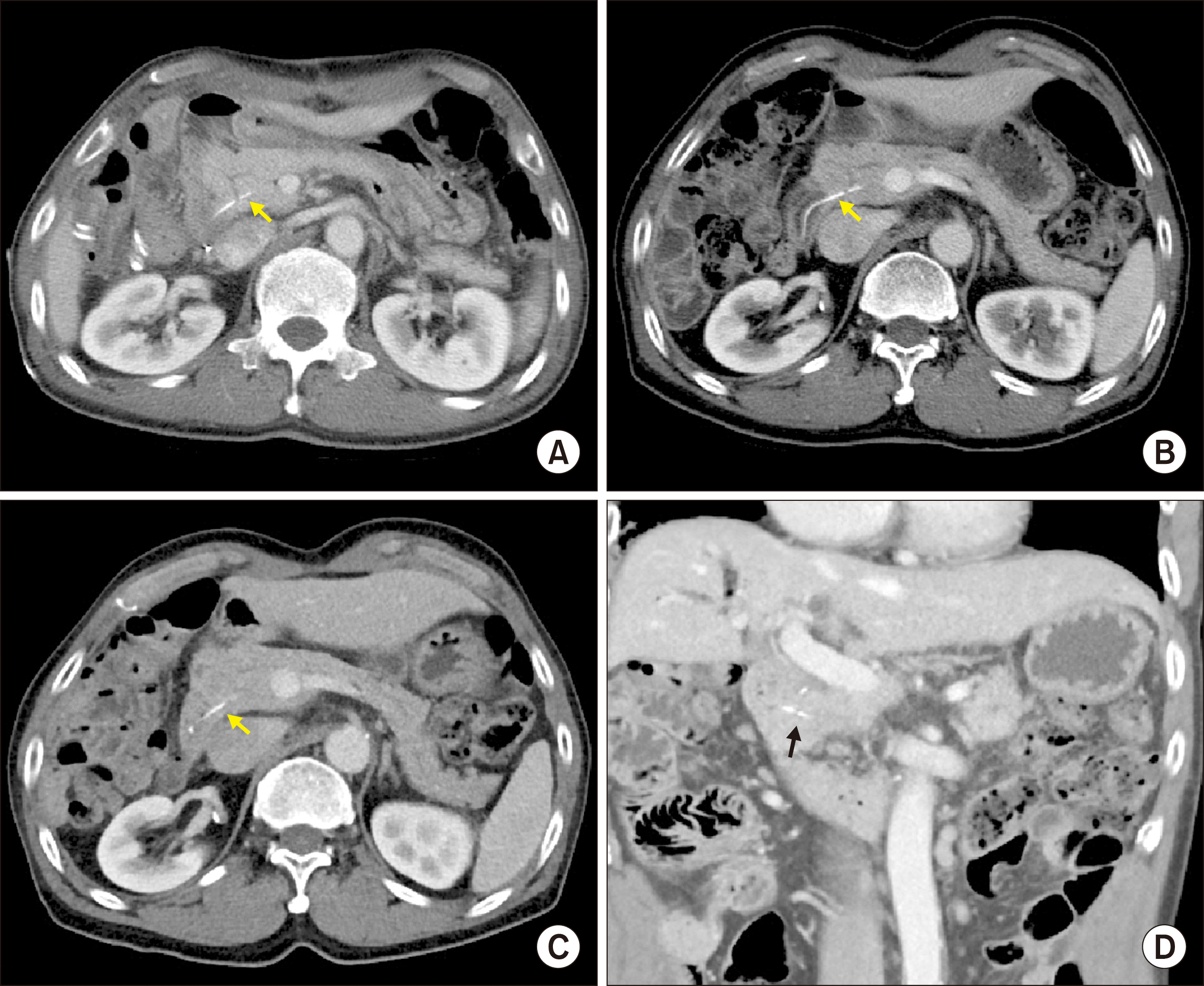
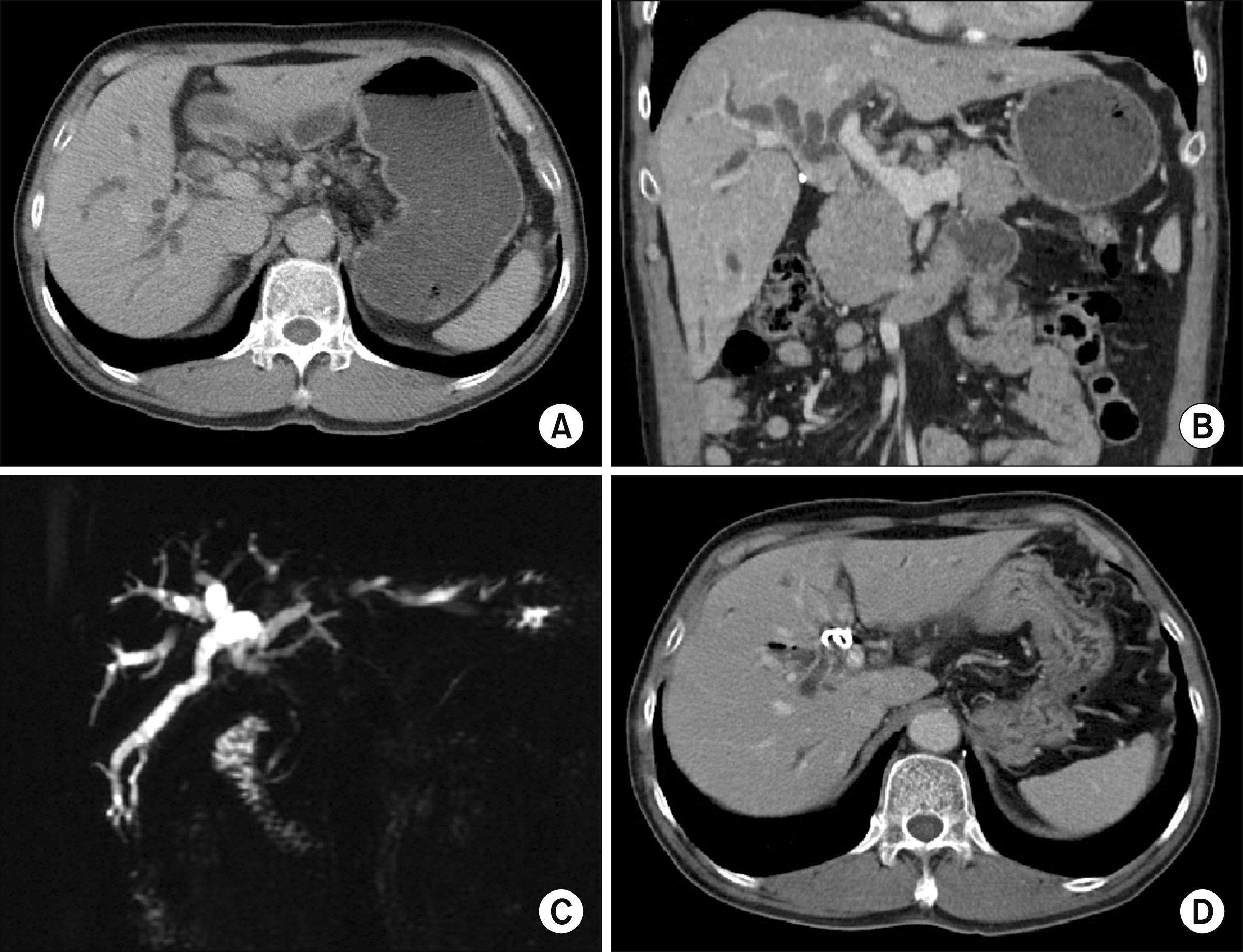
A 70-year-old male patient who experienced jaundice was admitted under the impression of distal bile duct cancer. Computed tomography (CT) scan showed multiple intraductal papillary masses in the extrahepatic bile duct and right intrahepatic duct with diffuse bile duct dilatation, suggesting intraductal papillary neoplasm of the bile duct (IPNB) (Fig. 1A, B). Magnetic resonance cholangiopancreatography (MRCP) showed intrahepatic duct stones and multiple intraductal lesions, suggesting cholangiocarcinoma with intraductal papillary growth (Fig. 1C). Fluorodeoxyglucose-positron emission tomography (FDG-PET) scan showed hypermetabolic lesions in the right liver, common hepatic duct and CBD, suggesting multiple intraductal papillary carcinomas. Endoscopic retrograde cholangiopancreatography was performed for biliary drainage (Fig. 1D), and well-differentiated adenocarcinoma was diagnosed by endoscopic biopsy.
 | Fig. 1Initial preoperative radiologic findings of Case 1. (A and B) Computed tomography scan shows multiple intraductal papillary masses in the extrahepatic bile duct and right intrahepatic duct with diffuse bile duct dilatation. (C) Magnetic resonance cholangiopancreatography shows intrahepatic duct stones and multiple intraductal lesions. (D) Endoscopic retrograde cholangiopancreatography with biopsy was performed.
|
The preplanned extent of resection was extended BDR with/without parenchyma-preserving hepatectomy or pylorus-preserving PD, depending on the intraoperative biopsy findings of the bile duct resection margins. After dissection of the CBD, the hilar bile duct was transected and intrahepatic duct stones were removed. The resection margins of the separated right and left hepatic ducts were tumor-negative on frozen-section biopsies. Dissection of the distal CBD continued into the pancreatic parenchyma, and the distal bile duct was transected at a distance of 1 cm from its end (Fig. 2A). This location was estimated by direct probing with a coronary dilator. As this resection margin was tumor-positive, we dissected the intrapancreatic CBD further beneath the duodenum, and at that time, the ampulla of Vater finally bulged out from the duodenal wall, making a 1 cm-sized round defect (Fig. 2A). The margin of the main pancreatic duct was trimmed to make a cuff measuring 5 mm in diameter. A silastic stent was inserted into the main pancreatic duct and transfixed with 5-0 Prolene. Subsequently, the pancreatic duct cuff was circumferentially sutured to the duodenal wall defect, which was corresponded to the ampulla of Vater (Fig. 2B). The dissected duodenal wall margins were gently sutured to the corresponding pancreatic parenchyma margins. Roux-en-Y hepaticojejunostomy was separately performed on the right and left lateral hepatic ducts. Two sets of suction-type cigarette Penrose drain were inserted around the pancreatic dissection area to cope with potential pancreatic leak.13 The patient recovered uneventfully (Fig. 3A) and was discharged 3 weeks after the operation.
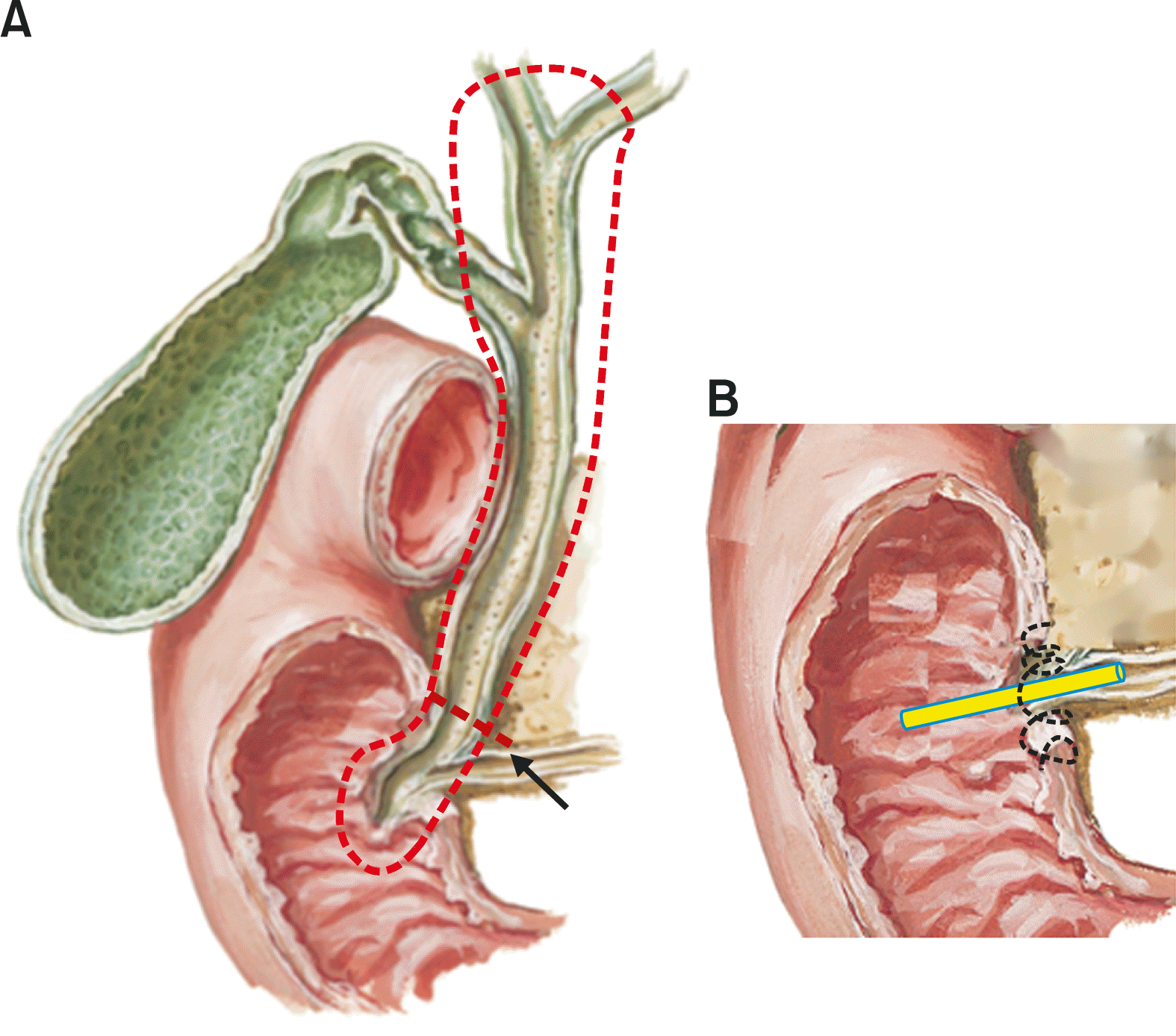 | Fig. 2Schematic illustration of the retroduodenal resection of the whole extrahepatic bile duct (A) and in situ re-implantation of the main pancreatic duct (B) in Case 1. The dotted line and arrow denote the extent of bile duct resection and initial transection line, respectively (A). The tube indicates the internal pancreatic stent (B).
|
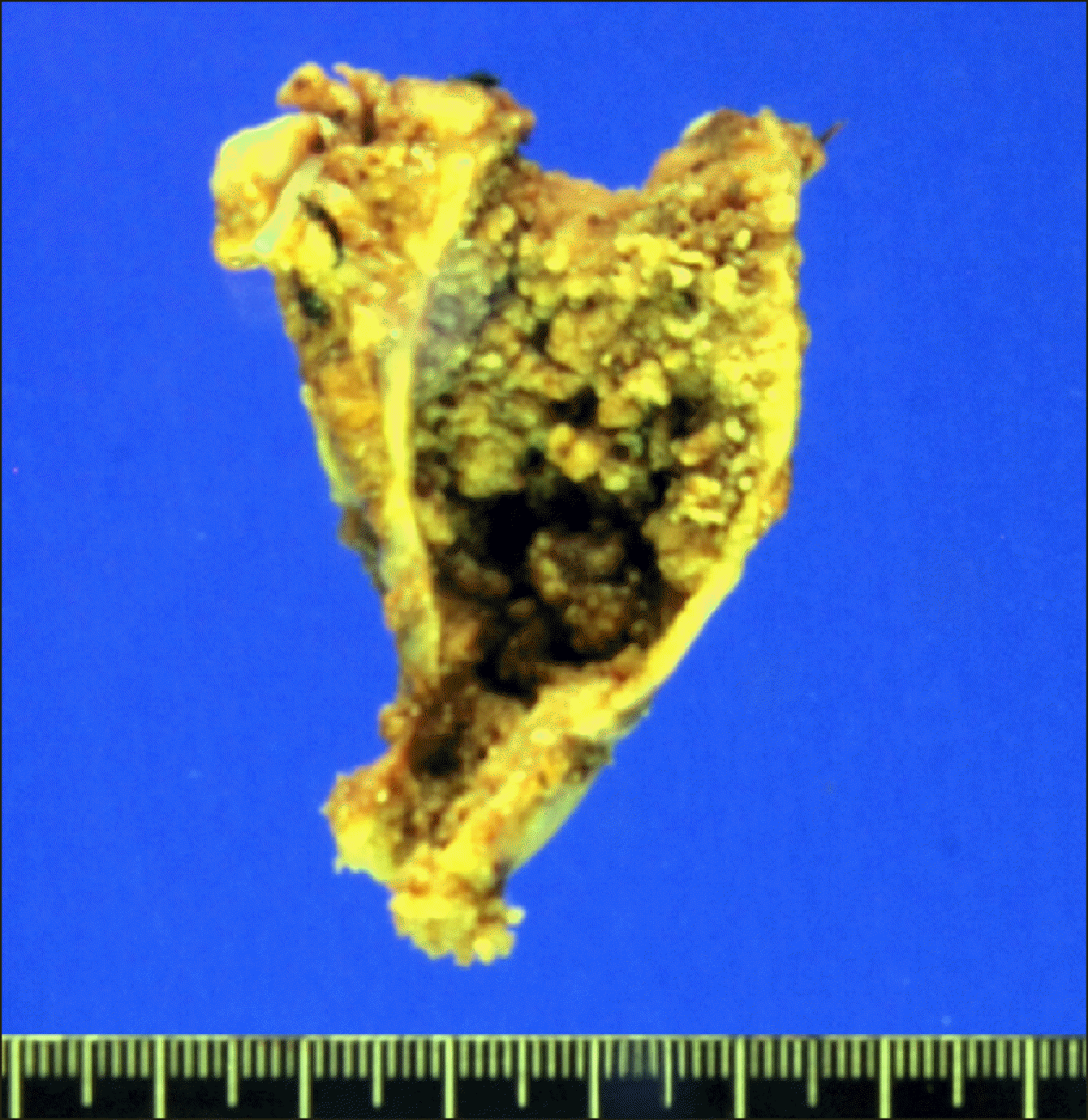
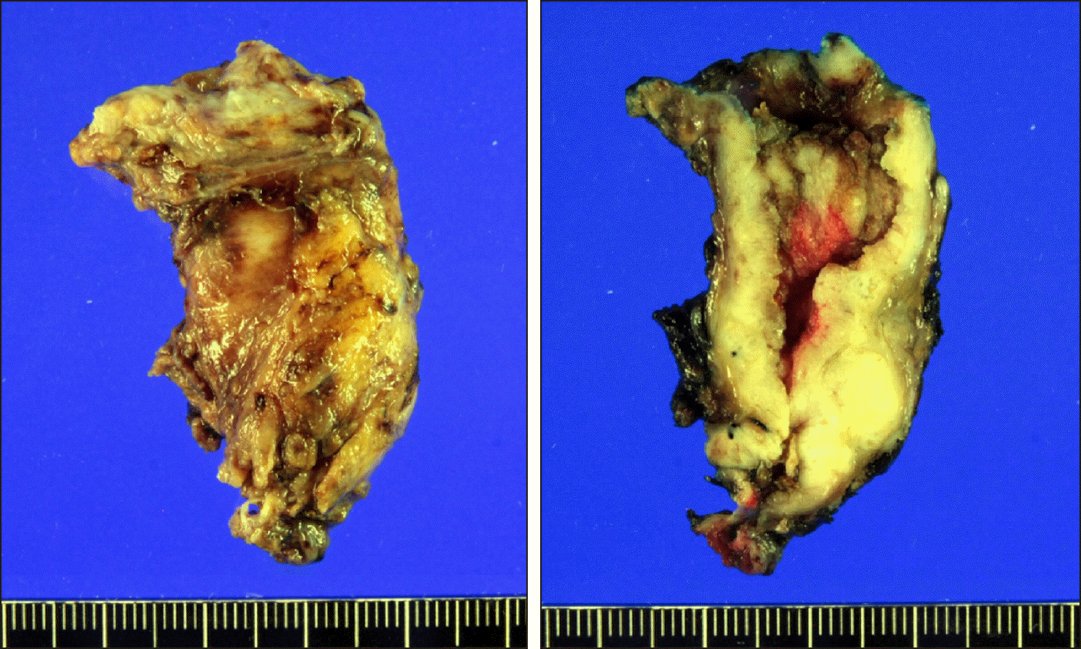
The pathology report showed that the tumor was intraductal papillary neoplasm with invasive cholangiocarcinoma, measuring 6.2 cm in length (Fig. 4). It was extended to the perifibromuscular tissue without lymphovascular or perineural invasion. No metastasis was observed in the 4 lymph nodes. We considered this extent of resection as compatible with R0 resection, thus additional anti-tumor treatment was not provided.
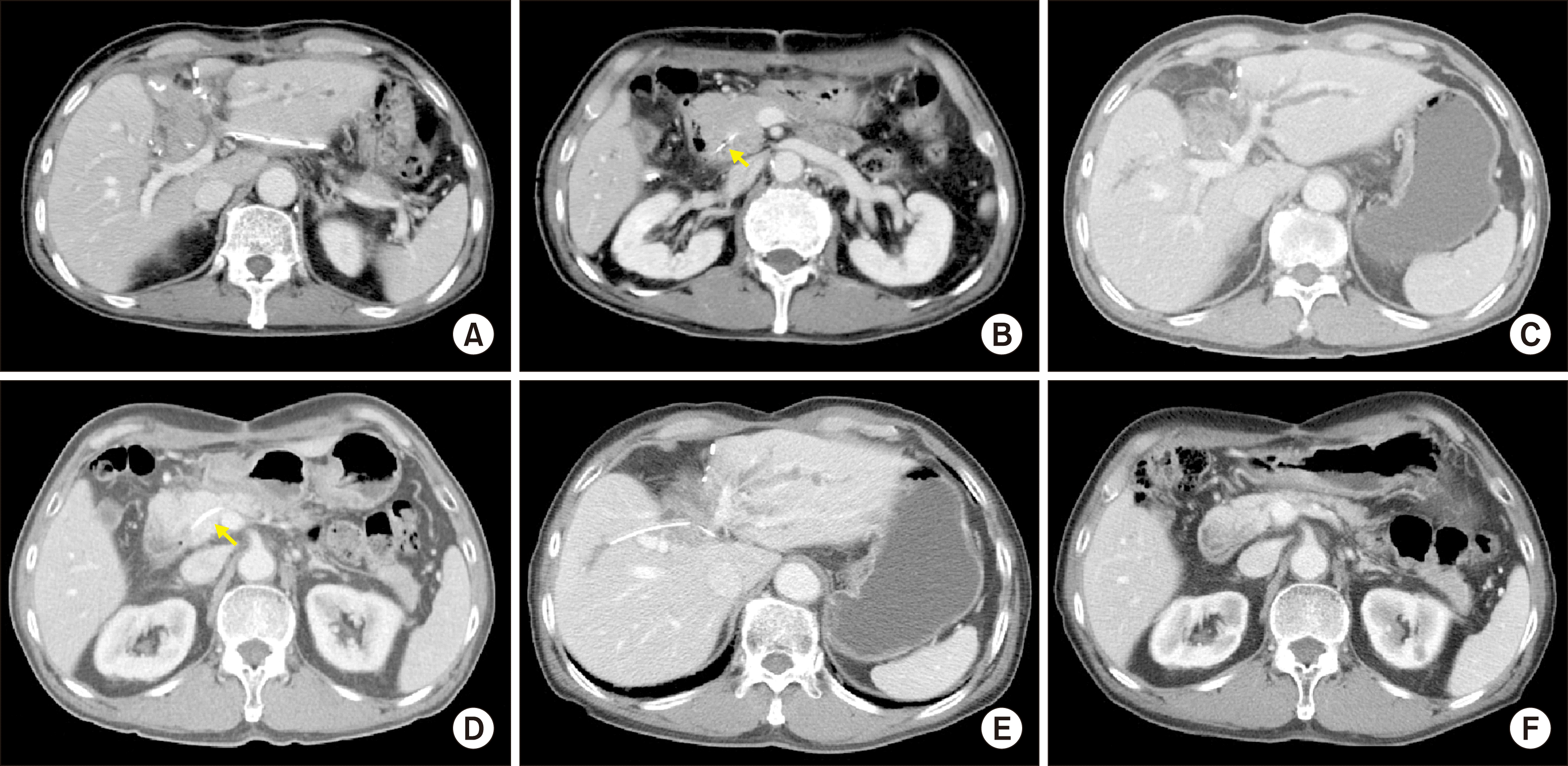
The patient had been regularly followed up every 3-4 months with imaging studies and blood tests for the first 5 years (Fig. 3B-D). Thereafter, he underwent annual follow-ups, showing no evidence of tumor recurrence. However, 8 years after surgery, he experienced cholangitis. A thorough examination with imaging studies revealed occurrence of tumor recurrence at the previous hepaticojejunostomy site with an intact pancreatic duct implantation site. Percutaneous transhepatic biliary drainage was performed to control cholangitis (Fig. 5). This patient is currently undergoing chemoradiation therapy for the treatment of recurrent lesions.
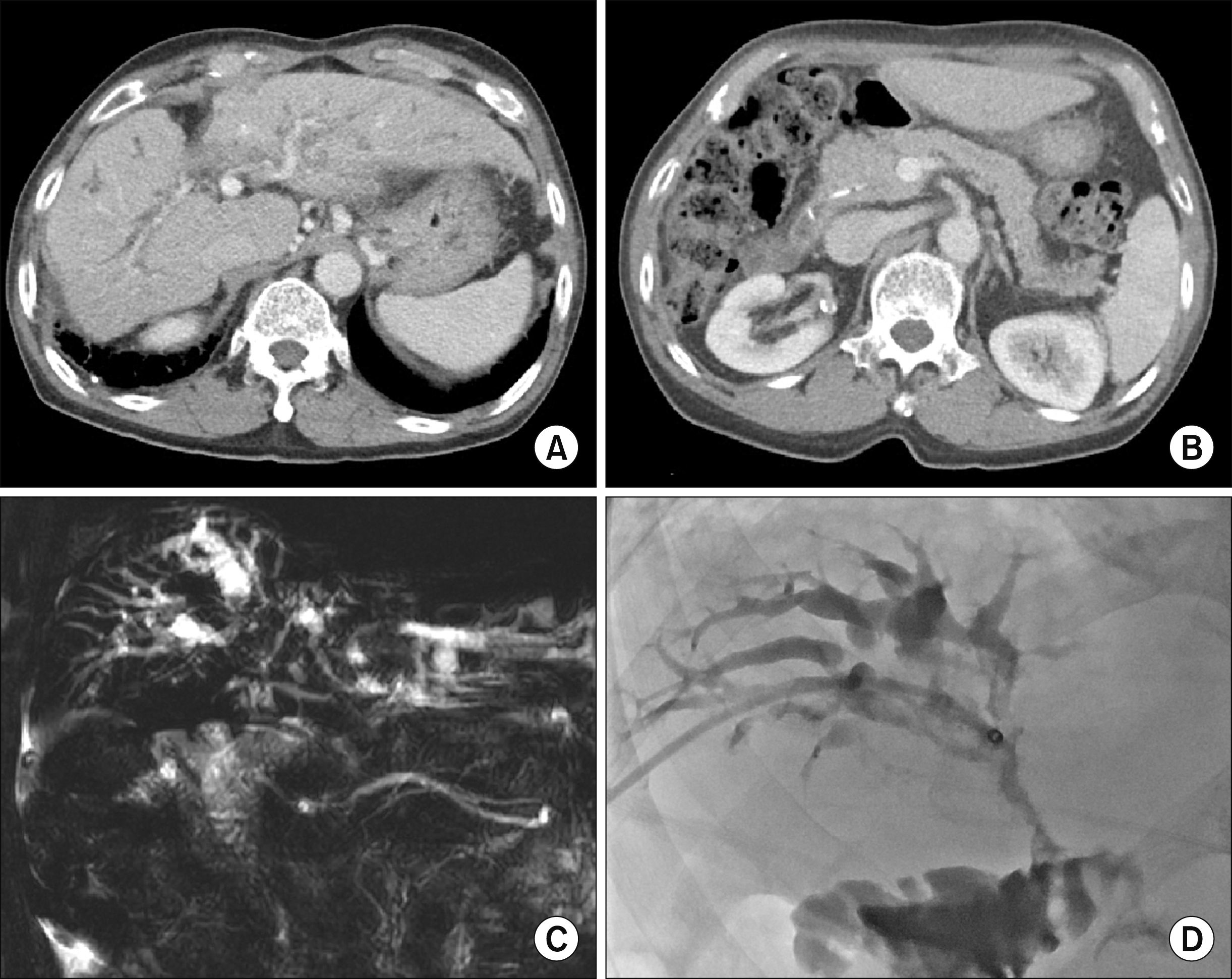 | Fig. 5Imaging study findings of Case 1 showing tumor recurrence at 8 years after the operation. A recurrent tumor is visible at the hepaticojejunostomy site (A), and the pancreatic duct implantation site is intact (B). Magnetic resonance cholangiopancreatography shows intraluminal narrowing (C), thus percutaneous transhepatic biliary drainage was performed for biliary drainage (D).
|
Case 2
A 71-year-old male patient with jaundice was referred to our institution under the impression of proximal bile duct cancer. Three years back, he had undergone laparoscopic cholecystectomy for gallstone. CT scan and MRCP showed that the tumor was compatible with perihilar cholangiocarcinoma of Bismuth-Corlette type I. Biliary decompression was performed sequentially with endoscopic nasobiliary drainage and endoscopic retrograde biliary drainage (Fig. 6).
 | Fig. 6Initial preoperative radiologic findings of Case 2. (A and B) Computed tomography scan shows perihilar cholangiocarcinoma of Bismuth-Corlette type I. (C) Magnetic resonance cholangiopancreatography shows mass within the bile duct. (D) Endoscopic retrograde biliary drainage is performed for biliary drainage.
|
The preoperative plan for the extent of resection was parenchyma-preserving hepatectomy with extended BDR because the general conditions of the patient was poor. After dissection of the hepatoduodenal ligament, the distal intrapancreatic CBD was transected close to its end as a part of extended BDR. However, the frozen-section biopsy showed thar the distal bile duct resection margin was tumor-positive. Consequently, we decided whether to perform PD or not after consideration of the proximal bile duct resection margins. The parenchyma of segment IV was resected. The bile duct resection margin at the left lateral segment was found to be tumor-negative on the frozen-section biopsy. However, the bile duct resection margin of the right liver was tumor-positive, thus the right hepatic duct stump was further resected to obtain tumor-free bile duct resection margin. As the right hepatic duct resection margin was very close to the main mass, we thought that this surgery would be potentially R1 resection. Thus, we decided to remove the remaining ampullary area instead of performing PD. We dissected the distal CBD further beneath the duodenum and the ampulla of Vater was removed from the duodenal wall, as shown in Case 1. The duodenal wall margins of the main pancreatic duct were trimmed to make a cuff measuring 5 mm in diameter. A silastic stent was inserted into the main pancreatic duct and transfixed with 5-0 Prolene, and the pancreatic duct cuff was circumferentially sutured to the duodenal wall defect. Other surgical procedures were similar to those mentioned in Case 1. The patient recovered uneventfully (Fig. 7A, B) and was discharged 3 weeks after the operation.
The pathology report showed that the tumor was a 5.3 cm-sized moderately differentiated adenocarcinoma centered at the common hepatic duct (Fig. 8). It was extended beyond the wall of the bile duct without lymphovascular or perineural invasion. There was no metastasis in the two lymph nodes. We considered this extent of resection as compatible with R1 resection; thus, postoperative chemoradiation therapy was performed.
The patient had been regularly followed up every 3 months with imaging studies and tumor marker tests (Fig. 7C-F). At 3 years after surgery, the segment II duct was dilated with stricture of the left hepaticojejunostomy (Fig. 9A, B); consequently, PTBD was performed. Percutaneous transhepatic cholangioscopy was performed to remove the stones at the left lateral segment (Fig. 9C). However, well-differentiated adenocarcinoma was identified at left hepaticojejunostomy and we planned to perform complete left lateral sectionectomy. To decrease the risk of operative complication, percutaneous left portal vein embolization was performed (Fig. 9D). Meanwhile, cholangiohepatitis with abscess formation developed in the right liver; thus, photodynamic therapy was performed instead of redo hepatectomy (Fig. 9E). One year after the photodynamic therapy, multiple intrahepatic recurrence occurred (Fig. 9F) and the patient finally passed away 4 years and 6 months after surgery.
 | Fig. 9Imaging study findings of Case 2 showing tumor recurrence at 3-4 years after the operation. A recurrent tumor is visible at the left hepaticojejunostomy site (A and B). Percutaneous transhepatic cholangioscopy was performed to remove the intrahepatic stones from the left lateral segment (C). Percutaneous left portal vein embolization was performed (D). Cholangiohepatitis with abscess formation developed in the right liver (E). Finally, multiple intrahepatic recurrences occurred in the liver (F).
|
Go to :
DISCUSSION
PD is a standard surgical procedure for resection of distal cholangiocarcinoma.7 However, PD is associated with a high morbidity rate, despite recent advancements in surgical procedures and perioperative management.9,10 Such a high operative risk is acceptable only when the oncological curativeness of PD is guaranteed. If an invasive surgery has marginal curativeness, a balance between the risk and benefit should be pursued. Extended BDR is a typical example of limited resection to avoid PD,11,12 but this procedure is valid when the distal end of the CBD is not invaded by the tumor.
There are a few case reports on pancreas‐preserving resection of cholangiocarcinoma located at the lower biliary tract. Kolb et al.14 reported an organ‐preserving procedure for resection of an intrapancreatic neuroendocrine tumor. A duodenotomy was carried out and a probe was inserted into the pancreatic duct to avoid inadvertent injury. Subsequently, the bile duct was divided proximal to the lesion and dissected towards the ampulla. Pancreatic parenchyma was dissected dorsally and closed using absorbable interrupted sutures. The duodenal incision was closed, and reconstruction was performed by hepaticojejunostomy. Previously, we reported a case of retroduodenal resection of an ampullary carcinoid tumor because the patient was not indicated for PD due to the cavernous transformation of the main portal vein.15 The duodenal wall defect was primarily repaired, and the pancreatic and bile ducts were separately reconstructed using Roux-en-Y pancreaticojejunostomy and choledochojejunostomy. The surgical techniques employed for retroduodenal dissection for distal CBD removal used in our previous study were similarly applied to our present cases. Nishida et al.16 also reported a case of coring‐out technique for pancreas‐preserving resection of lower biliary tract adenocarcinoma. The bile duct was dissected from the pancreatic parenchyma without pancreatic resection, and duodenotomy was done opposite the ampulla of Vater. The bile duct was completely separated from the pancreatic parenchyma and the lower biliary tract was cored out, and the pancreatic duct was re‐implanted into the duodenal wall. Kim et al.17 reported a case of left hemihepatectomy, caudate lobectomy, and complete extrahepatic bile duct resection using a transduodenal approach in a patient with hilar cholangiocarcinoma and extrahepatic biliary papillomatosis. They approximated the defect around the excised intrapancreatic portion of the CBD through the transduodenal approach. Except for our previous case report,15 a transduodenal approach through an incision of the duodenum wall was employed in the abovementioned three case reports.14,16,17
The unique technical point of our present cases is a retroduodenal approach to expose the running course of the distal CBD within the pancreas head and to remove the ampullary portion and in situ re-implantation of the main pancreatic duct. Kim et al.17 presented complete extrahepatic bile duct resection using a transduodenal approach, in which the insertion site of the pancreatic duct remained untouched probably because of no tumor involvement. On the contrary, if the distal end of CBD or ampulla of Vater is involved, the ampullary portion should be resected and the pancreatic duct should be re-implanted into the duodenal wall. Based on our experience of the previous case report15 and ampullectomy cases,18 in situ re-implantation of the main pancreatic duct into the duodenal wall is considered more technically feasible through a retroduodenal approach than through the usual transduodenal approach, because the duodenal wall used for pancreatic duct re-implantation is already dissected and mobilized in the former method. Placement of an internal stent into the main pancreatic duct is an important point to prevent anastomotic leak. No significant surgical complications occurred in our two cases. Considering that no tumor recurrence occurred at the intrapancreatic area in our two cases, the surgical curability of our method is comparable with that of PD.
In conclusion, complete resection of the extrahepatic CBD through a retroduodenal approach with in situ re-implantation of the main pancreatic duct is feasible and less invasive than PD. Therefore, this less-invasive approach is proposed as an alternative procedure in selected patients requiring complete resection of the distal CBD.
Go to :
 XML Download