

 PDF
PDF Citation
 Print
Print


INTRODUCTION
Resection of choledochal cyst (CC) with an anomalous union of pancreatobiliary duct (AUPBD) is almost always indicated because of its malignant potential. It is well known that persistent reflux of bile juice into the biliary tract by means of AUPBD causes recurrent inflammation of the bile duct, leading to hyperplasia and metaplasia of the epithelium, which predisposes to malignant transformation.1,2 Therefore, resection of the CC theoretically eliminates risk of malignant transformation, since the remnant bile duct is no longer exposed to enzymatic insult from activated pancreatic juice.
However, malignant changes of the remnant CC portion have been sporadically reported worldwide.3-9 Repeated episodes of inflammation at the remnant CC portion for a long period seem to be associated with such malignant transformation. We previously reported a case of adenocarcinoma that arose from the remnant CC that was located deep in the pancreas 16 years after resection.10 We herein present a case of intrahepatic cholangiocarcinoma that arose from the remnant CC portion that was located within the liver ten years after resection.
Go to : 
CASE
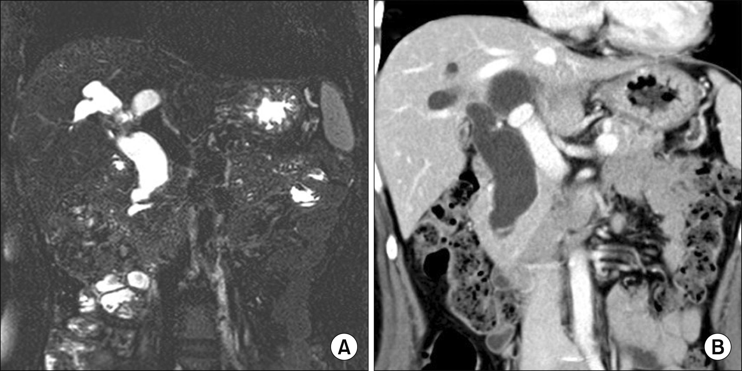
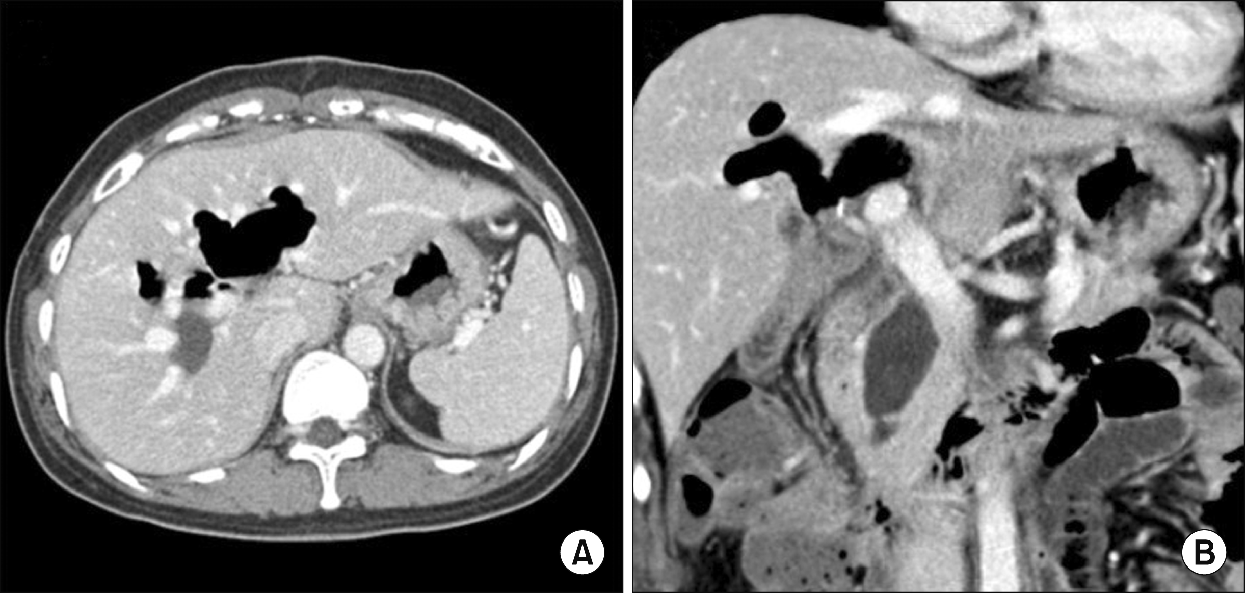
A 59-year-old female patient who had undergone surgical resection of Todani type IV CC with AUPBD 10 years ago at outside hospital (Fig. 1) was transferred for surgical treatment for intrahepatic cholangiocarcinoma. The first operation was resection of the extrahepatic extrapancreatic CC, so the intrahepatic and intrapancreatic portions of the CC were not resected (Fig. 2).
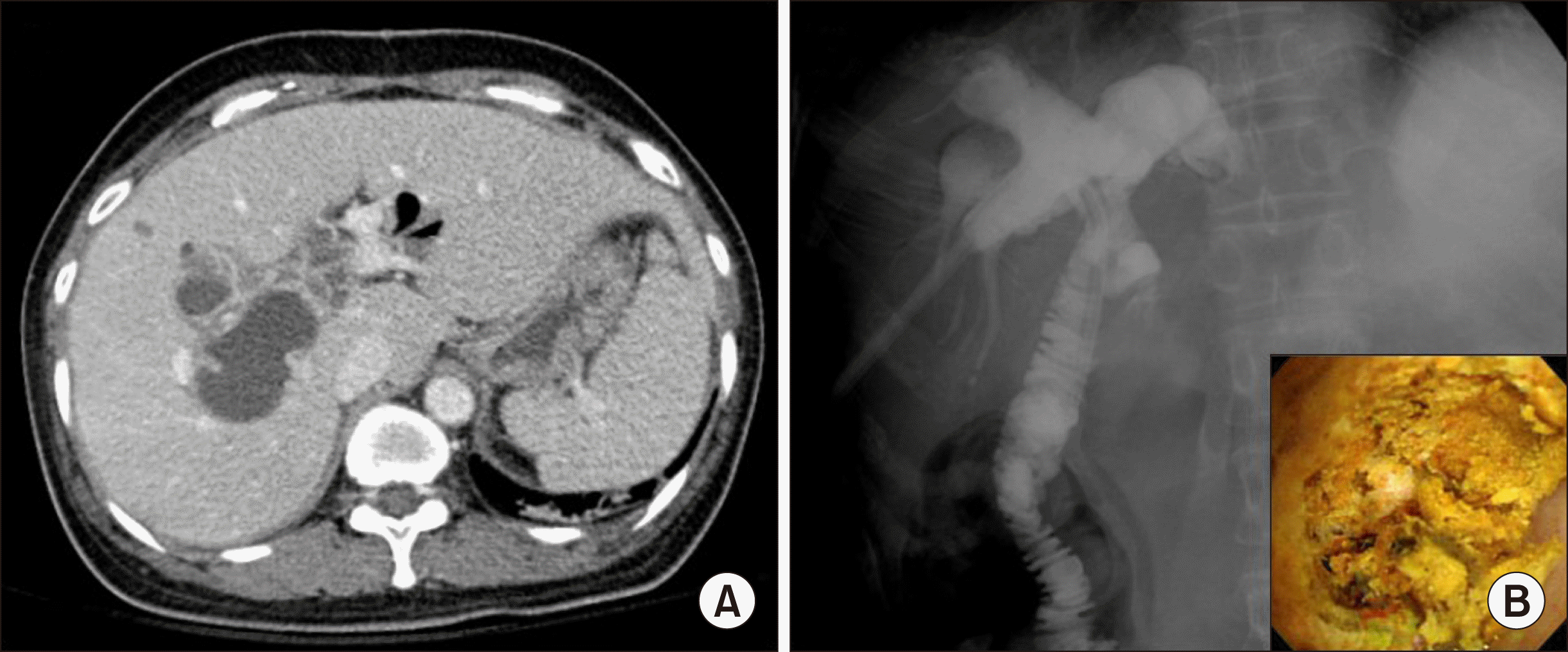
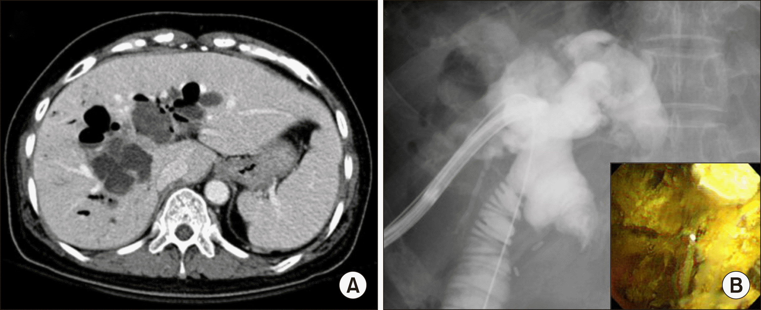
Two years after the operation, a follow-up computed tomography (CT) scan revealed development of intrahepatic stones and formation of liver abscesses. The intrahepatic stones were removed through percutaneous transhepatic cholangioscopy (PTCS) (Fig. 3). Six years later after the operation, intrahepatic duct stones developed again, and the intrahepatic remnant portion of the type IV CC was further dilated, so stone removal by means of PTCS was repeatedly performed (Fig. 4). Thereafter, the patient did not undergo regular follow-up with imaging studies.
Ten years later after the operation, a follow-up CT scan revealed development of intrahepatic stones and formation of a new mass (Fig. 5A, B). We performed PTCS to remove the intrahepatic stones again and performed direct biopsy of the mass developed within the intrahepatic CC portion (Fig. 5C). The tissue biopsy finding was a poorly differentiated adenocarcinoma. Magnetic resonance cholangiopancreatography at one-month intervals showed slow progression of the intrahepatic mass, and 18F-fludeoxyglucose positron emission tomography showed a hypermetabolic intrahepatic cholangiocarcinoma involving the left hepatic duct and hepatic hilum, with metastatic lymph nodes in the periportal and common hepatic artery areas (Fig. 5D). The right liver appeared to be rather shrunken, with noticeable hypertrophy of the left liver, but CT volumetry showed that the proportion of the future remnant right liver volume was 56% of the whole liver volume.
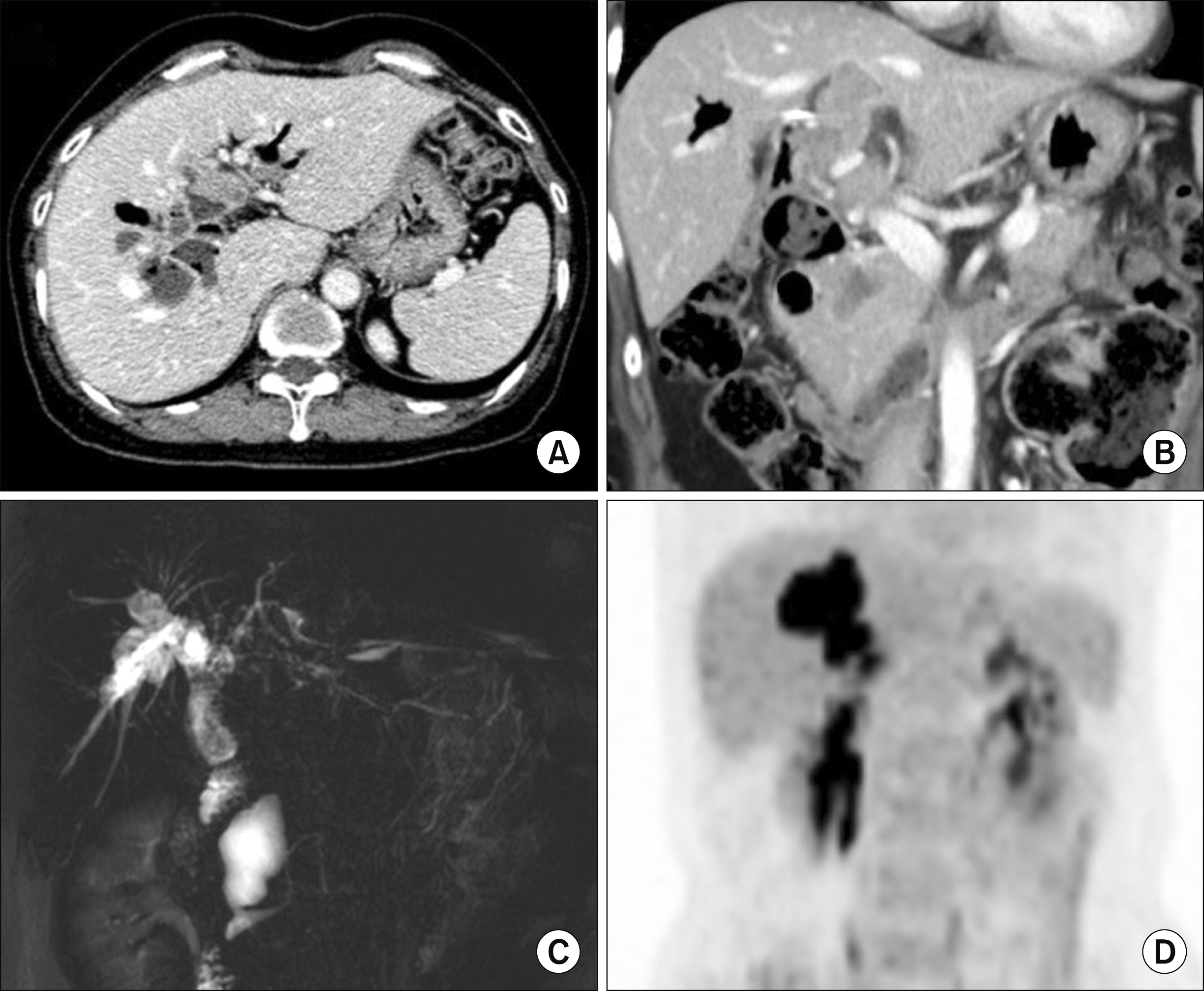 | Fig. 5Imaging study findings taken ten years after the first operation. A computed tomography scan (A and B) and magnetic resonance cholangiopancreatography (C) show development of intrahepatic stones and formation of a new mass. Fludeoxyglucose positron emission tomography shows hypermetabolic intrahepatic cholangiocarcinoma at the remnant cyst portion (D).
|
Because the liver mass appeared to be resectable, we planned to perform left hepatectomy with redo hepaticojejunostomy. First, the hepatic hilum was meticulously dissected, and the hepaticojejunostomy was isolated and transected (Fig. 6A). We then dissected the enlarged lymph nodes around the common hepatic artery and celiac axis. Thereafter, the right hepatic artery and main portal vein were isolated. After clamping of the left hepatic artery and left portal vein, we assessed the extent of liver resection by means of the discoloration of the liver surface. We performed hepatic parenchymal transection under left hemi-hepatic inflow block without Pringle maneuver. The surface of the dilated intrahepatic CC portion was exposed (Fig. 6B). At this step, we opened the right edges of the exposed CC portion with electrocautery to see the luminal structures (Fig. 6C). After thorough inspection of the intraluminal mass within the CC portion, we identified the proper transection plane of the right-sided CC and then transected the CC portion along the resection design. The edges of the right-side CC wall were sampled for frozen-section biopsy. Four samples of the resection margins of the CC wall were tumor-negative. Hepatic transection was continued to remove the left liver with cutting of the left hepatic artery and portal vein (Fig. 6E). The Spigelian lobe was preserved because its bile duct was not involved (Fig. 6F).
 | Fig. 6Intraoperative photographs of left hepatectomy. (A) The hepatic hilum was meticulously dissected, and the hepaticojejunostomy was isolated and transected. (B) We performed hepatic parenchymal transection under left hemi-hepatic inflow block. (C) The right edges of the exposed cyst portion were opened. (D) We identified the transection plane of the right-side cyst. (E) Hepatic transection continued to remove the left liver after cutting of the left hepatic artery and portal vein. (F) The Spigelian lobe was preserved.
|
The Roux-en-Y jejunal limb, which was made at the first operation, was reused for redo hepaticojejunostomy. Because the wall of the right-side residual CC wall was markedly thickened, we applied multiple fixation sutures along its circumferential edges to facilitate anastomosis and to prevent bleeding at the anastomotic site (Fig. 7A, B). We performed a large hepaticojejunostomy with multiple running sutures of the posterior wall anastomosis and multiple interrupted sutures of the anterior wall anastomosis (Fig. 7C, D). The intrapancreatic remnant CC portion was not resected, because it was not possible to completely remove the remnant intrahepatic CC portion within the right liver.
 | Fig. 7Intraoperative photographs of redo hepaticojejunostomy. (A and B) Multiple fixation sutures were applied to the circumferential edges of the remnant cyst wall. (C and D) We performed large hepaticojejunostomy with multiple running sutures of the posterior wall and multiple interrupted sutures of the anterior wall.
|
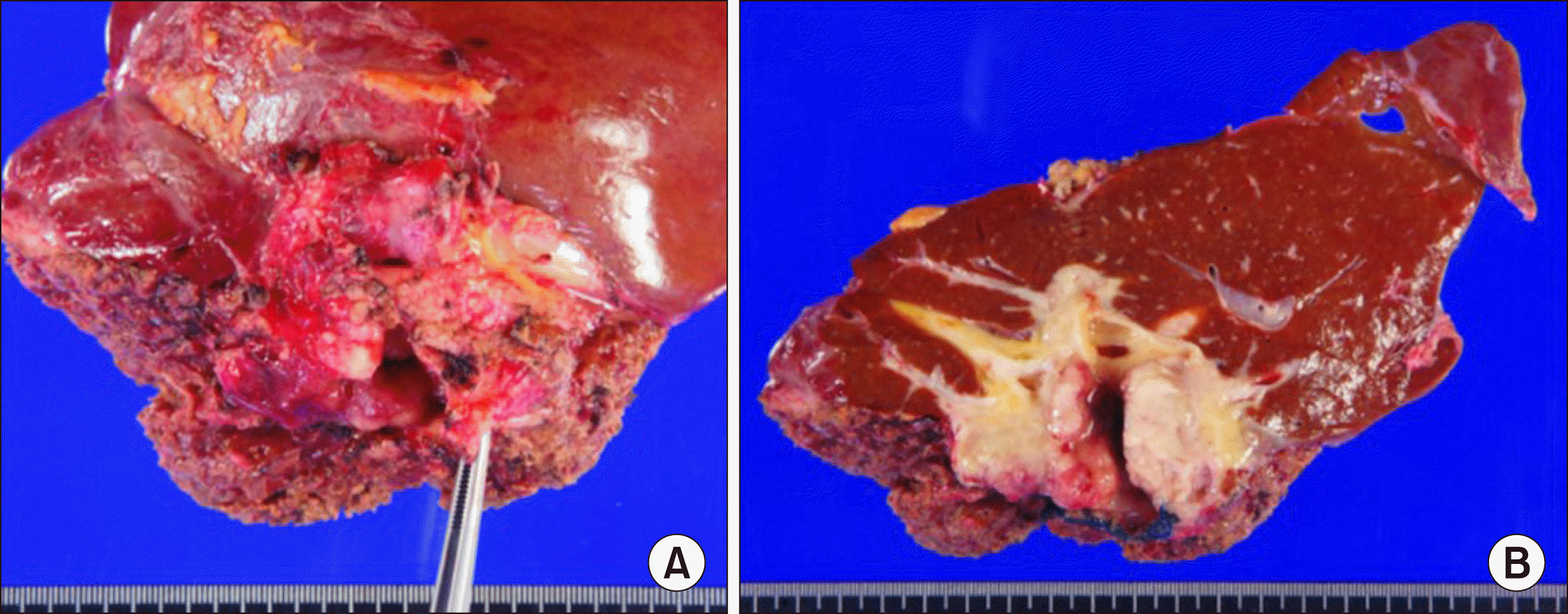
The pathology report revealed that the mass was a 4.5-cm-sized papillary cholangiocarcinoma with moderate differentiation, which was located at the left hepatic duct (Fig. 8). The tumor extended beyond the bile duct with 7-mm-thick wall invasion from the surface epithelia, but the hepatic parenchyma was not involved. Lymphovascular invasion was present, but perineural invasion was not identified. There was lymph node metastasis in 4 of 15 lymph nodes. The patient recovered uneventfully from this second operation (Fig. 9) and is currently undergoing adjuvant chemotherapy for three months.
Considering the clinical sequences of this patient, the reasonable extent of resection seemed to be complete resection of the extrahepatic CC including the intrapancreatic portion combined with proximal partial excision of the intrahepatic CC portion to widen the hepaticojejunostomy opening as much as possible (Fig. 10).
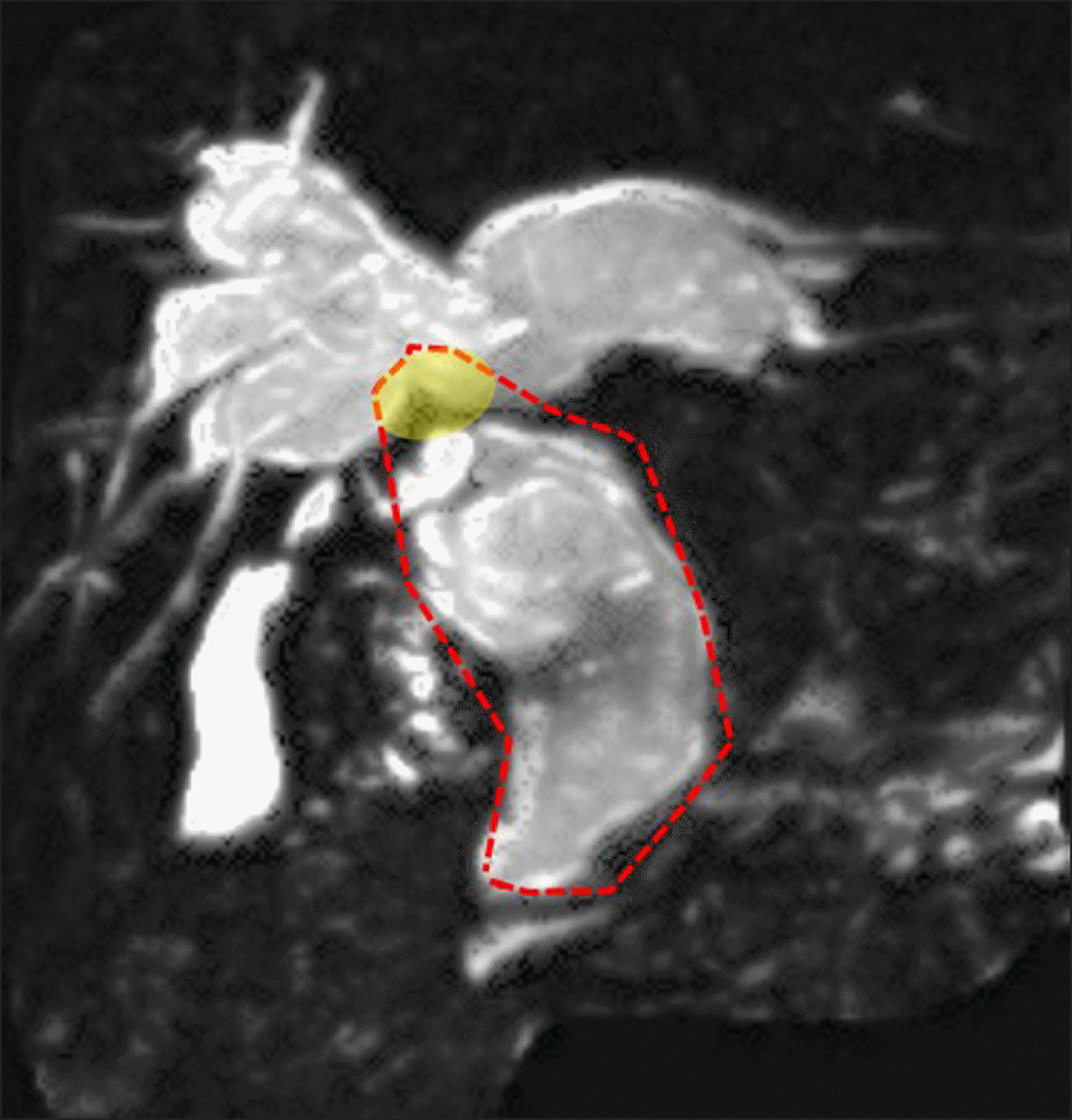 | Fig. 10Initial magnetic resonance cholangiopancreatography before the first operation. The dotted line indicates the reasonable extent of resection, including complete resection of the extrahepatic choledochal cyst including the intrapancreatic portion, combined with proximal partial excision of the intrahepatic cyst portion (yellow shade) to widen the hepaticojejunostomy opening as much as possible.
|
Go to :
DISCUSSION
Complete excision of the CC with biliary reconstruction has been the mainstay in treatment of CC. However, there are some issues regarding the intrahepatic and distal-end part of the CC. Although radical cyst excision is well known to be the treatment of choice, because of the morbidity of porta hepatis dissection and postoperative complications, such as pancreatic fistula and pancreatitis, surgeons are occasionally reluctant to perform aggressive complete excision.11,12 In reality, complete resection of the intrapancreatic CC portion rarely requires pancreatoduodenectomy, but complete resection of the intrahepatic CC is not technically possible in patients with CC of Todani type IV.
It is constantly reported that the remnant CC portion can undergo malignant transformation, which indicates the need for life-long follow-up after the surgery. In a Chinese study that included 78 patients with partial resection of the CC, the patients developed associated symptoms, including new cysts, calculus of the bile duct (65.4%), and carcinogenesis (14.1%) in the residual intrapancreatic bile duct. The authors concluded that surgical re-excision should be considered for patients with a residual intrapancreatic portion of the CC because of prior incomplete surgery, regardless of clinical symptoms.1 We also reported a case of adenocarcinoma that arose from the remnant CC that was located deep in the pancreas 16 years after resection of CC.10
Development of cholangiocarcinoma more than ten years after excision of CC has been rarely reported, with less than 21 cases reported in the literature from 1972 to 2014, with a median period of recurrence at 6 years (range 2-34 years).3-21
Various theories have been proposed to explain development of malignancy in patients with a previously resected CC. First, it was suggested that the epithelium of the remnant bile duct wall is already at a precancerous stage at the time of surgery, hence development of cholangiocarcinoma is merely a result of carcinogenesis during the postoperative period.4 Second, the existence of stenosis at the anastomosis or in the intrahepatic bile duct may induce carcinogenesis. Moreover, some have postulated that carcinogenesis is caused by repeated damage of the biliary epithelium by bile fluid as well as by bacterial contamination, leading to mucosal metaplasia.5 Last, cholangiocarcinoma can develop spontaneously in the general population, which may explain the many different intervals of presentation of cholangiocarcinoma in these patients.
In the present case, we presume that long-term chronic inflammation associated with intracystic stone and abscess formation might be closely related to malignant transformation of the intrahepatic CC. Considering the clinical sequences of this patient, the reasonable extent of resection at the first surgery seems to be complete resection of the extrahepatic CC including the intrapancreatic portion and proximal partial excision of the intrahepatic CC portion to widen the hepaticojejunostomy opening as much as possible to minimize the risk of bile stasis-associated cholangitis.
Although the risk of interval malignancy is well known, there are no practical guidelines for the duration of follow-up and the type of investigations that patients should undergo after initial surgery for CC. Most patients diagnosed with cholangiocarcinoma long after resection of CC were not followed up routinely with radiological imaging studies.3 The common finding between our previous and present cases who experienced malignant transformation at the remnant CC portion was repeated episodes of inflammation with stone formation.10 Therefore, we highly recommend that long-term regular follow-up with imaging studies is important in CC patients who experienced repeated episodes of cholangitis.
Our present case indicates that remnant intrahepatic CC can undergo malignant transformation ten years after resection of the type IV CC. Since the intrahepatic CC portion in type IV CC is usually unresectable, wide hepaticojejunostomy and life-long observation with regular imaging study follow-up are highly recommended for prevention and early detection of malignant transformation.
Go to :
 XML Download