

 PDF
PDF Citation
 Print
Print


INTRODUCTION
Laparoscopic hepatectomy (LH) was introduced in 1996; since then, laparoscopic technology has evolved rapidly around the world. However, due to the associated technical complexity, the adoption of LH has been relatively slow, and many liver surgeons are still reluctant to perform it. Nevertheless, it has several advantages over open hepatectomy, which have led to an increase in the number of LHs performed worldwide.1-3
The previous studies shown that LH has significantly better results in terms of reduced blood loss, reduced postoperative hospitalization, improved cosmesis, better wound recovery, and early return to normal activity.4,5 These result were due to an improvement of surgical procedure and the development of device for LH. However, despite new tools developed for parenchymal transection, the main concern remains the bleeding control during hepatic resection.6-8 Furthermore, blood transfusion has been demonstrated to be a predictor of poor surgical results in liver resection with adverse effects on perioperative morbidity and mortality.9,10
Inflow blocking is one of the most effective methods to control bleeding during parenchymal transection. The Pringle maneuver (PM), first described in 1908 by Pringle,11 is a simple and easily reproducible method for inflow occlusion.
There are several reports on the use of the PM during LH. Here, we describe an easy, safe and unique way of performing totally intra-corporeal PM. More specifically, we describe a simple method for laparoscopic PM with Penrose drain tube which have been using at our center in most laparoscopic liver resections from 2018. This retrospective study was conducted to review our experience with a easily obtainable penrose drain tube for laparoscopic PM and to investigate the safety of intermittent clamping during LH. We report the surgical results and improvements to the totally intra-corporeal PM using penrose drain tube. We also observed that the effective hepatic artery and portal blood flow blocking occurs while using this procedure.
MATERIALS AND METHODS
Patient population and selection
Between 2016 and 2019, we performed LH on 75 consecutive patients at Kosin University Gospel Hospital in Busan, Korea. Laparoscopic left-sided hepatectomy is the main criterion for this study (37 cases).
Our study population was divided into two groups: the No PM group with 12 patients and the PM with Penrose drain tube group with 25 patients.
Surgical technique
In this series, all patients underwent potentially curative hepatic resection with removal of gross pathologies with negative macroscopic margins. All procedures were performed by one experienced hepatobiliary surgeons throughout the study period.
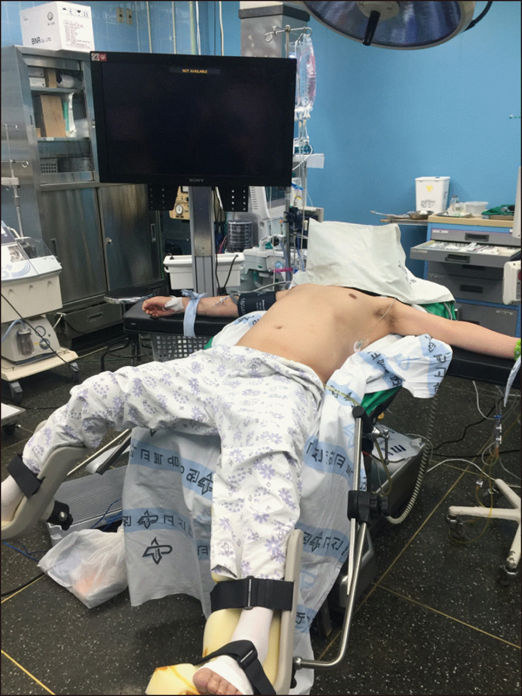
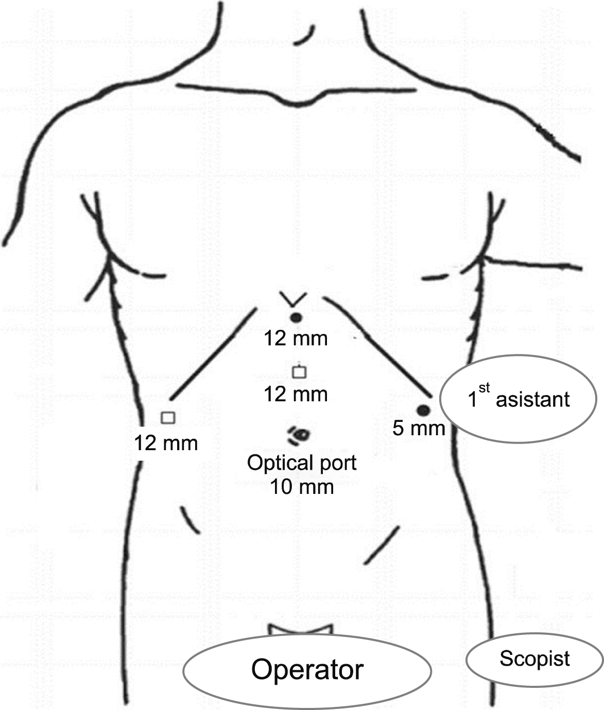
The patients were placed in a supine and low lithotomy position, French position, with the surgeon standing between the patient’s legs (Fig. 1). Five trocars were usually used (Fig. 2). After the introduction of a 12-mm umbilical port using an open technique, continuous carbon dioxide pneumoperitoneum was induced at a pressure limit of 12 mmHg and flow of 6 L/minute to decrease the risk of gas embolism. The laparoscope was inserted through the supraumbilical 12 mm trocar site located 1 to 2 cm above the umbilicus. Five 5- to 12-mm trocars and a flexible, 3D laparoscope (ENDOEYE FLEX 3D, Olympus, Tokyo) were used. The liver was evaluated in all cases using intraoperative laparoscopic ultrasonography (Aloka Medical, Ltd.) to identify the hepatic veins and intrahepatic pathologies.
Subsequently, mobilization of the liver was initiated with the falciform ligament; the left lateral hepatic attachment and the triangular ligament were divided using an energy device (Harmonic scalpel HD 1000i, Ethicon EndoSurgery, Cincinnati, USA) after the round and falciform ligaments were dissected.
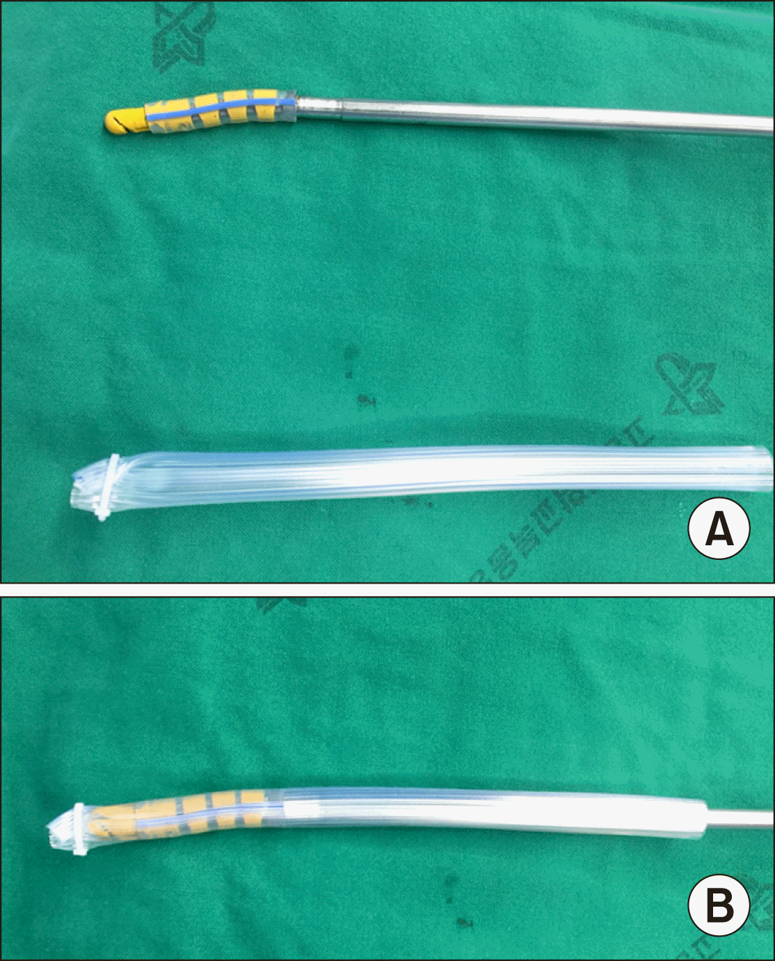
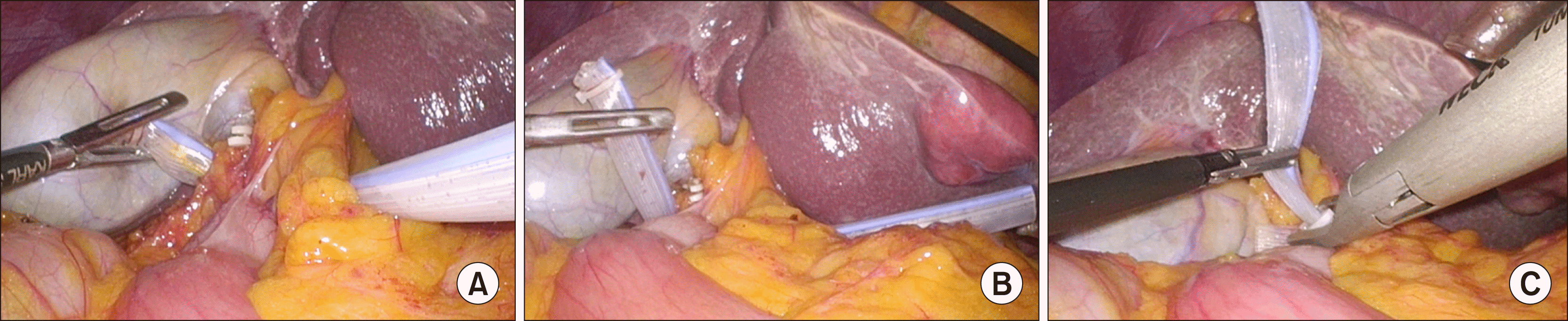
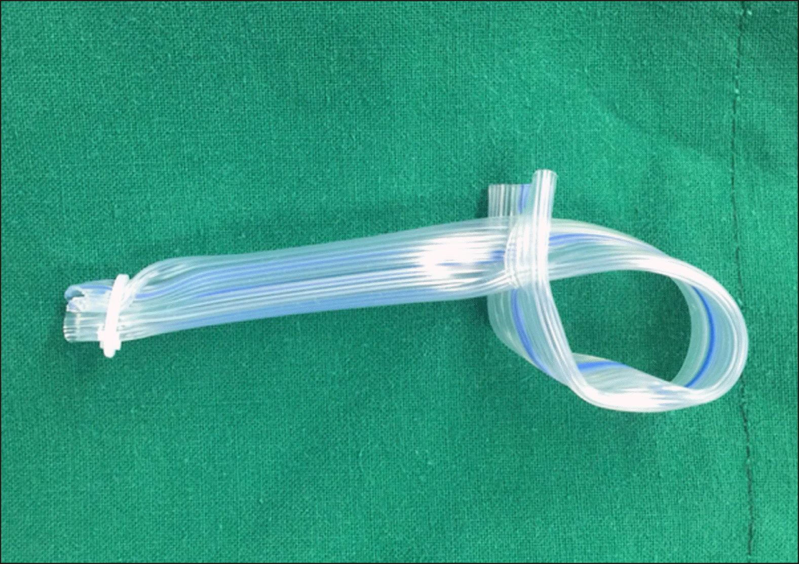
Next, the avascular lesser omentum was divided with electrocautery. From the port that was inserted at the upper mid line, Gold finger dissector (Blunt dissectors, Johnson & Johnson, USA) with 15 cm sized Silicone Penrose drain tube (Sewoonmedical, Korea) was inserted from opened lesser omentum to pass behind the hepatoduodenal ligament (Fig. 3). The forcep was inserted until the Penrose drain tube reached the orifice that was in the Winslow’s foramen (Fig. 4A). The edge of the drain tube was held with a forceps and was pulled out through the side hole of another edge of penrose drain tube for taping of the hepatoduodenal ligament; and finally, the penrose drain tube was pulled out of and lock with the Hemo-o-Lok clip (Weck Closure System, Research Triangle Park, NC, USA) for use as a tourniquet to enable performance of a PM (Fig. 4B, C). Blood flow was occluded by clamping the Penrose drain tube with the Hemo-lock clip in order to perform a totally intracorporeal PM. Intermittent clamping was applied, with 15-minute clamping and 5-minute release periods.
Parenchymal transection was achieved using an energy device and Cavitron ultrasonic surgical aspirator (CUSA EXcel; Valleylab, Boulder, Colorado, USA) under totally intra-corporeal PM. Small vessels were ligated with clip or coagulated using an energy device. Intraparenchymal control of the major vessels was achieved with metal clips or Hemo-o-Lock clips, whereas major glissonean pedicle and major hepatic vein division was obtained with stapling devices. The resected specimen was placed in a plastic retrieval bag and removed through the slightly extended periumbilical incision.
Statistical analysis
Student’s t-test, χ2 test, Fisher’s exact probability test, and the Mann-Whitney U test were used for analysis of parametric and non-parametric data, as appropriate. Statistical analyses were performed using SPSS ver. 25.0 (IBM, New York, NY, USA). Differences of p<0.05 were considered significant.
RESULTS
From 2016 to 2019, a total of 75 patients underwent laparoscopic liver resection. Among these 75 patients, laparoscopic left-sided hepatectomy was performed in 37 (49.3%) patients. There were 12 cases in the no PM group and 25 cases in the PM group. During the study period, there was no morbidity associated with placement of the penrose drain tube and no operative mortality.
The clinical characteristics of patients are shown in Table 1. The median patient age was 63 years (range 36-76), and 20 (54.0%) patients were men. Patients demographics including sex, age, body mass index (BMI), and American Society for Anesthesiologists (ASA) physical status were similar between two groups.
Table 2 summarize the perioperative outcomes between no PM group and PM group. In the no PM group, 12 patients underwent laparoscopic left side liver resection, including 6 left hemihepatectomy, 5 left lateral sectionectomy. In the PM group, 25 patients underwent laparoscopic left side liver resection, including 15 left hemihepatectomy, 10 left lateral sectionectomy. The mean operative time was 156 min in no PM group and 174 min in PM group. The estimated blood loss was higher in no PM group (341 (20-1100) ml vs. 165 (20-700) ml, p= 0.20), but there were not significantly different between groups. There was no blood transfusion during surgery in both group. The mean hospital stay was 8.6 day in no PM group and 10.9 day in PM group. There was one procedure that require surgical exploration after surgery in no PM group, the case was for anastomotic leakage from colorectal anastomosis site. There was no open conversion and no mortality in both group.
DISCUSSION
LH is a safe and effective treatment that is associated with improved short-term outcomes compared to the open technique.12-15 Additionally, LH is comparable to open hepatectomy regarding long-term oncological benefits.15,16 Therefore, progression has been made from simple hepatectomy to major hepatectomy and even laparoscopic donor hepatectomy.
Nevertheless, control of bleeding remains the major concern during parenchymal transection. PM is a traditional and the most commonly used method to decrease blood loss during open liver resection its effectiveness and simplicity. In laparoscopic surgery, various methods for applying PM have been published.7,11,17-20
Some authors have introduced their extra-corporeal PM technique during LH and Robotic hepatectomy. They suggested that compared with the intra-corporeal PM technique, the extra-corporeal PM technique confers the following advantages: it combines all the well-known advantages of the technique used in open operations (ie it is safe, quickly usable in case of bleeding, and achieves complete inflow occlusion). This seems advantageous in that the absence of tactile feedback can lead to serious vascular structural damage during inflow occlusion due to the clamp force applied to the pedicle. However, due to the additional port for PM, it is likely to interfere with the docking and undocking of the robot arm during robot liver surgery, and it is also interfere with the instrumental forcep movement during LH. We assume the advantages and drawbacks of each technique are experience-based and not evidence-based. One of the potential drawbacks of the use of an additional trocar for extra-corporeal PM (compared with intra-corporeal PM) is the risk of gas leakage from the abdominal cavity.21-23
Totally intra-corporeal PM technique also has several methods.24,25 This report demonstrates an easy way to perform a new totally intra-corporeal PM during LH using simple and readily available materials without tourniquet and additional port. Our approach to hepatectomy includes low central venous pressure (CVP) anesthesia and liberal use of PM when bleeding, even minor, occurs during transection. For this reason, before we begin hepatic parenchymal transection, penrose drain tube is passed routinely around the hepatoduodenal ligament, except when taping is impossible due to adhesion. The pedicle clamping is used on demand according to surgeon’s preference and local conditions. This technique is easy and dose not require any special instruments. Our policy of intermittent 15 min clamping, 5 min unclamping is based on practical experience. We have found that the association of PM with low CVP and pneumoperitoneum pressure achieves a bloodless field in most instances.
Our technique has several advantages. It is simple, reliable and reproducible. Due to the fact that it is fully intracorporeal, our technique does not require a working port or placement of an additional port. Its intra-corporeal position is flexible and therefore it does not interfere with the surgeons’ view and actions. Clamping and unclamping can be performed in few seconds, including in emergency situations. It is cost-effective since it does not require any expensive components. Komeda et al.26 suggested Endo intestinal clips for intra-corporeal PM during LH. The benefits of using an Endo intestinal clip are that additional port for PM dose not need to be occupied, and gripping force can be maintained at a steady level. However, with this method, complete occlusion of the inflow through thick and fatty hepatoduodenal ligament is difficult to achieve. A further advantage of our method is the of achieving complete occlusion, by completely surrounding the hepatoduodenal ligament, especially when the porta hepatis is thick or fatty (Fig. 5).
A potential limitation of our technique is that there should be no adhesions to allow the Penrose drain tube to pass through the Winslow foramen between the hepatoduodenal ligament and the inferior vena cava. However, such adhesions are rare in the absence of previous surgery, but not uncommon in repeat hepatectomy or upper abdominal surgery, such as right colectomy or even a simple cholecystectomy.
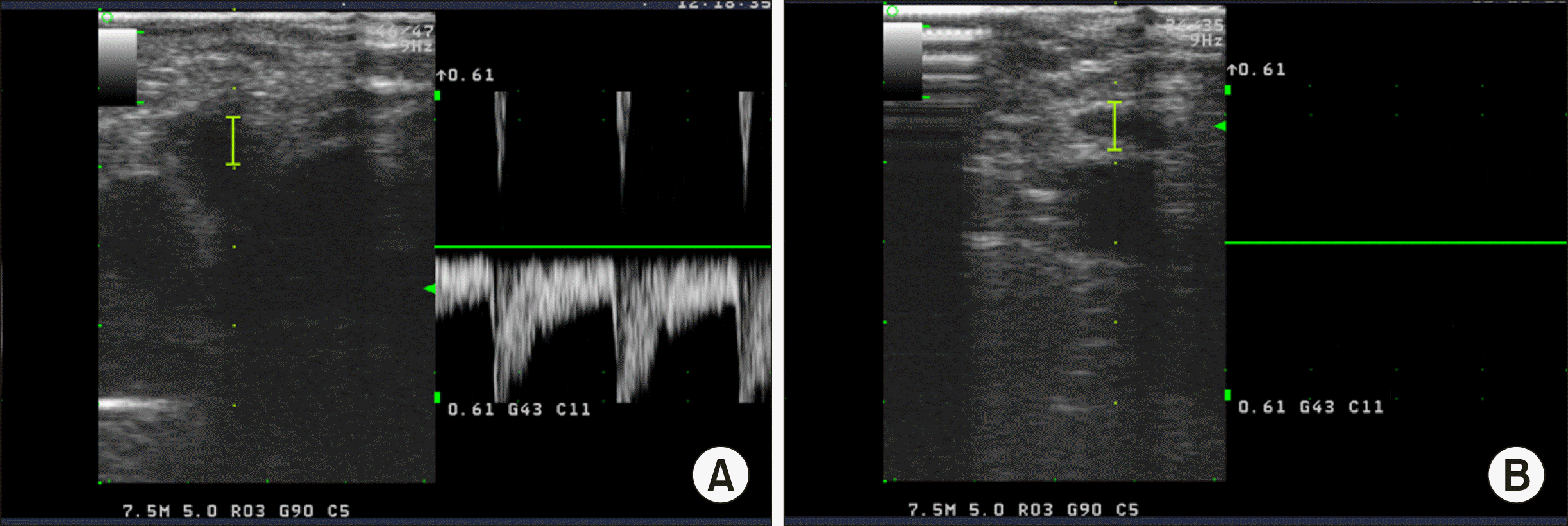
Our study is limited by the small number of patients. Additionally, no objective measurements were performed to assess the degree of clamping by intraoperative Doppler ultrasonography or pressure measurements of the Penrose drain. Although not applicable to all patients during LH, the same PM was applied during open and laparoscopic liver surgery. Although it is difficult to measure the exact quantitative pressure of our PM, Doppler ultrasonography allowed us to observe that the blood flow to the liver disappeared completely after our PM (Fig. 6). It seems to have a study on the minimum compression pressure to laparoscopic PM tool for achieving complete flow blocking effect.
In coclusion, the totally intracorporeal PM with Penrose drain tube may be effective in reducing intraoperative blood loss during laparoscopic left-sided hepatectomy, although the difference is not statistically significant. Moreover, our method is easy, reproducible, effective, safe, can be implemented quickly, and allows intermittent clamping during LH. The current experience-based analysis remains to be confirmed with prospective randomized studies.
 XML Download