Journal List > Korean Circ J > v.50(10) > 1144978

Beom, Choi, Boo, Lee, Kim, and Joo: A Case of Anomalous Right Coronary Artery Infarction Mimicking ST-Segment Elevation Myocardial Infarction at the Left Anterior Descending Artery
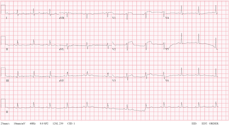
A 48-year-old male visited to the emergency room with chest pain. Electrocardiography showed ST elevations in the precordial leads (Figure 1), and ST-segment elevation myocardial infarction in the left anterior descending (LAD) artery territory was diagnosed. Coronary angiography (CAG) showed significant lesions of the mid-LAD artery and septal branch, and percutaneous coronary intervention (PCI) for the mid-LAD artery lesion was performed (Supplementary Videos 1 and 2). The right coronary artery (RCA) could not be identified despite using pigtail catheter (Supplementary Video 3). The procedure was finished after planning computed tomography coronary angiography (CTCA) to confirm the RCA anatomy.
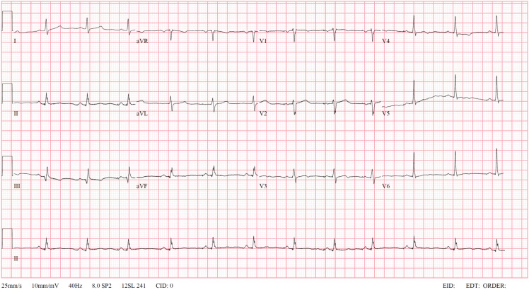
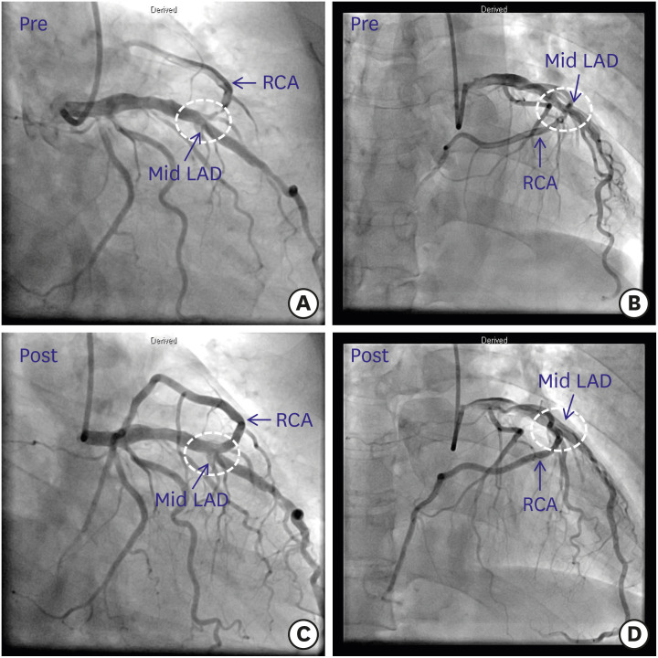
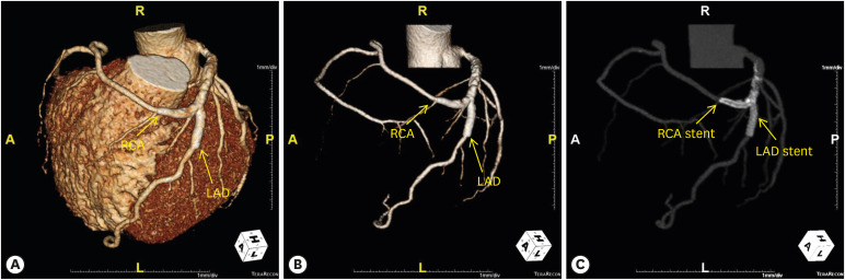
After the procedure, he no longer complained of chest pain, and electrocardiography showed improvement in ST elevations in the precordial leads (Figure 2). Transthoracic echocardiography showed a diastolic D-shape of left ventricle, suggesting right ventricular infarction (Supplementary Video 4). Transradial CAG on the next day revealed an anomalous origin of the RCA arising from the mid-LAD artery with significant stenosis of the RCA ostium. This bifurcation site was treated using T stenting technique (Figure 3). CTCA also confirmed an anomalous course of the RCA and showed the patency of the stents (Figure 4).
A single coronary artery is an extremely rare congenital anomaly of the coronary system in structurally normal heart.1)2)3) To our knowledge, only one patient has been reported to undergo PCI for a true LAD artery/RCA bifurcation lesion treated using two-stent technique.4) We emphasize the importance of clinical effort to evaluate all coronary arteries in patients with coronary artery anomalies.

Notes

Conflict of Interest: The authors have no financial conflicts of interest.

Author Contributions:

  • Supervision: Choi JH.

  • Writing - original draft: Beom JW, Boo KY, Lee JG.

  • Writing - review & editing: Beom JW, Kim SY, Joo SJ.

References

1. Topaz O, DeMarchena EJ, Perin E, Sommer LS, Mallon SM, Chahine RA. Anomalous coronary arteries: angiographic findings in 80 patients. Int J Cardiol. 1992; 34:129–138. PMID: 1737663.
crossref
2. Barriales Villa R, Morís C, López Muñiz A, et al. Adult congenital anomalies of the coronary arteries described over 31 years of angiographic studies in the Asturias Principality: main angiographic and clinical characteristics. Rev Esp Cardiol. 2001; 54:269–281. PMID: 11262367.
3. Yamanaka O, Hobbs RE. Coronary artery anomalies in 126,595 patients undergoing coronary arteriography. Cathet Cardiovasc Diagn. 1990; 21:28–40. PMID: 2208265.
crossref
4. Khan UA, Sharma D, McGlinchey P, Peace A. Percutaneous coronary intervention to left anterior descending artery/right coronary artery bifurcation: this is not a typo! A case report. Eur Heart J Case Rep. 2019; 3:ytz137. PMID: 31425574.
crossref

SUPPLEMENTARY MATERIALS

Supplementary Video 1

Left coronary angiogram showing approximately 80% segmental stenosis of the mid-LAD artery and total occlusion of the septal branch of the LAD artery. LAD = left anterior descending.

Supplementary Video 2

Left coronary angiogram showing successful revascularization of the LAD artery.

Supplementary Video 3

Coronary angiogram via a pigtail catheter. The right coronary artery was not visualized.

Supplementary Video 4

A diastolic D-shape of left ventricle was observed in the parasternal short axis view of echocardiography, suggesting right ventricular infarction.
Figure 1

Twelve-lead electrocardiogram showing ST-segment elevations in leads V1-3 without reciprocal change.

kcj-50-951-g001
Figure 2

Electrocardiogram after percutaneous coronary intervention to the mid-LAD artery lesion showed improvement in ST-segment elevations in the precordial leads.

LAD = left anterior descending.
kcj-50-951-g002
Figure 3

Left coronary angiograms showing an anomalous origin of the RCA from the mid-LAD artery with a significant stenosis of the RCA ostium (A, B). Left coronary angiograms showing successful revascularization at the bifurcation site between the LAD artery and the RCA (C, D).

LAD = left anterior descending; RCA = right coronary artery.
kcj-50-951-g003
Figure 4

Three-dimensional computed tomography coronary angiographic images revealing an anomalous origin of the RCA from the LAD artery (A, B), and showing patent stents at the bifurcation site between the LAD artery and the RCA (C).

LAD = left anterior descending; RCA = right coronary artery.
kcj-50-951-g004
TOOLS
Similar articles