

 PDF
PDF Citation
 Print
Print


INTRODUCTION
Stent apposition refers to the proximity of struts to the vascular wall.1)2)3) Good stent apposition is sufficiently close contact to preclude blood flow between any strut and the underlying artery2) while stent malapposition is separation of any strut from the intimal surface of the arterial wall that is not overlapping a side branch.2) The frequency of stent-vessel wall malapposition after percutaneous coronary intervention varies with the clinical scenario, lesion morphology, and the type of stent implanted. Unfortunately, there is a lack of agreement as to its importance and clinical impact.1) Early short-term intravascular ultrasound (IVUS) studies in small numbers of patients have given way to both long-term IVUS studies in larger numbers of patients as well as to detailed, higher resolution optical coherence tomography (OCT) studies. This review provides a comprehensive summary of the relationship between coronary stent malapposition and its long-term clinical outcomes based on both IVUS and OCT studies.
Go to : 
ACUTE STENT MALAPPOSITION
Definition, prevalence, and mechanisms
The prevalence of acute stent malapposition (ASM), occurring immediately after implantation, is higher when assessed using OCT than with IVUS. On IVUS, ASM has been reported from 7.3% to 38.5% (averaging approximately 13%); on OCT, the prevalence varies from 39.1% to 72.3% (averaging approximately 51%) (Table 1).
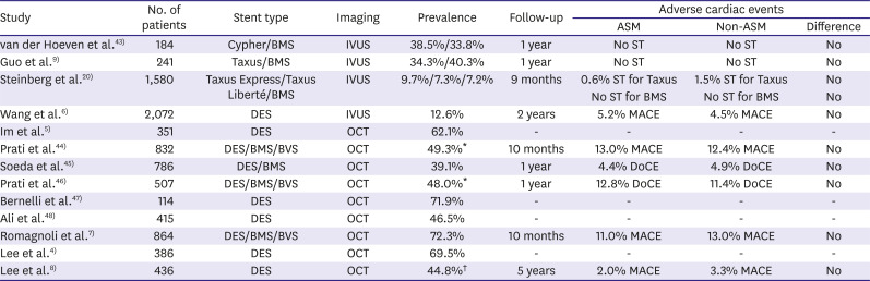
Table 1
Studies investigating ASM
| Study | No. of patients | Stent type | Imaging | Prevalence | Follow-up | Adverse cardiac events | ||
|---|---|---|---|---|---|---|---|---|
| ASM | Non-ASM | Difference | ||||||
| van der Hoeven et al.43) | 184 | Cypher/BMS | IVUS | 38.5%/33.8% | 1 year | No ST | No ST | No |
| Guo et al.9) | 241 | Taxus/BMS | IVUS | 34.3%/40.3% | 1 year | No ST | No ST | No |
| Steinberg et al.20) | 1,580 | Taxus Express/Taxus Liberté/BMS | IVUS | 9.7%/7.3%/7.2% | 9 months | 0.6% ST for Taxus | 1.5% ST for Taxus | No |
| No ST for BMS | No ST for BMS | No | ||||||
| Wang et al.6) | 2,072 | DES | IVUS | 12.6% | 2 years | 5.2% MACE | 4.5% MACE | No |
| Im et al.5) | 351 | DES | OCT | 62.1% | - | - | - | - |
| Prati et al.44) | 832 | DES/BMS/BVS | OCT | 49.3%* | 10 months | 13.0% MACE | 12.4% MACE | No |
| Soeda et al.45) | 786 | DES/BMS | OCT | 39.1% | 1 year | 4.4% DoCE | 4.9% DoCE | No |
| Prati et al.46) | 507 | DES/BMS/BVS | OCT | 48.0%* | 1 year | 12.8% DoCE | 11.4% DoCE | No |
| Bernelli et al.47) | 114 | DES | OCT | 71.9% | - | - | - | - |
| Ali et al.48) | 415 | DES | OCT | 46.5% | - | - | - | - |
| Romagnoli et al.7) | 864 | DES/BMS/BVS | OCT | 72.3% | 10 months | 11.0% MACE | 13.0% MACE | No |
| Lee et al.4) | 386 | DES | OCT | 69.5% | - | - | - | - |
| Lee et al.8) | 436 | DES | OCT | 44.8%† | 5 years | 2.0% MACE | 3.3% MACE | No |
ASM = acute stent malapposition; BMS = bare metal stent; BVS = bioresorbable vascular scaffold; DES = drug-eluting stent; DoCE = device-oriented clinical end point; IVUS = intravascular ultrasound; MACE = major adverse cardiac events; OCT = optical coherence tomography; ST = stent thrombosis.
*Lesions with >200 μm of maximum malapposed distance; †Lesions with ≥400 μm of maximum malapposed distance or ≥1 mm of maximum malapposed length.
![]()
There are 2 main mechanisms of ASM.1)4)5) The most common is a stent whose cross-sectional area is smaller than that of the surrounding lumen due to an undersized stent or an intra-stent aneurysmal/ectatic segment or at the proximal edge because of lumen tapering (for example, proximal to a side branch when treating a bifurcation lesion). The lesser cause of ASM is the transition from non-calcified to calcified plaque (especially a calcified nodule) because the expanded stent cannot conform to abrupt changes in lumen geometry.
Clinical outcomes
Table 1 summarizes the clinical outcomes of ASM. To date there has been no dedicated, prospective study; and most studies are based on retrospective or sub-group analyses. Nevertheless, despite the heterogeneity of study design, patient characteristics, stent types, and imaging modalities, studies consistently show that adverse cardiac events do not differ between patients with vs without ASM.
The IVUS study by Wang et al.6) included 2,072 patients with 2,446 lesions from ADAPT-DES (Assessment of Dual Antiplatelet Therapy With Drug-Eluting Stents) study which was a prospective, multicenter registry designed to assess the relationship between platelet reactivity and other clinical and procedural variables vs subsequent stent thrombosis (ST) and adverse clinical events in patients successfully treated with drug-eluting stent (DES). At 2-year follow-up, there was no significant difference in the incidence of cardiac death; myocardial infarction; early, late, or very late ST; or clinically driven target lesion revascularization in patients with ASM vs those without ASM.6) In fact, the largest areas of ASM were not associated with events. These results were consistent even when ASM was forced into the multivariable model.6)
The study by Romagnoli et al.7) was the most detailed quantitative OCT analysis to date. It analyzed post-procedural OCT findings in 864 patients undergoing percutaneous coronary intervention, assessing prevalence and magnitude of ASM and exploring correlation with clinical outcomes.7) At a median follow-up of 302 days, ASM did not affect risk of major adverse cardiac events (MACE) regardless of its size or length; residual ASM was comparable in terms of size (median 210 μm vs 200 μm distance) and length (2.0 mm vs 2.2 mm) in patients with vs without MACE.7)
The OCT study by Lee et al.8) was a pooled analysis from 6 small randomized trials and included 436 patients with 444 non-complex lesions treated with DES. Adverse cardiac events at 5-year follow-up were compared according to the severity, not simply the presence of ASM. The rate of MACE was 3.3% in patients with ASM ≥400 μm of maximum malapposed strut distance vs 3.1% in those with no ASM or ASM <400 μm (p=0.89) and 1.2% in patients with ASM ≥1mm of maximum malapposed strut length vs 4.6% in those with no ASM or ASM <1mm of maximum malapposed strut length (p=0.06).8)
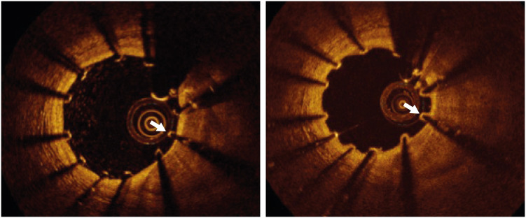
Approximately half of ASM resolve at follow-up (Figure 1). Guo et al.9) reported that 40% of ASM identified by IVUS resolved at follow-up. Im et al.5) similarly reported that 69% of OCT-defined ASM resolved spontaneously. Resolution of ASM depends on its size, especially when the distance from the stent to the vessel wall is less than 350–400 μm.5)10)11) Conversely, Kolandaivelu et al.12) examined the thrombogenicity of malapposed struts via in vitro experiments. In single-strut 2-dimensional simulations with various detachment distances, stent-wall recirculation zones, highly thrombogenic areas around struts, first grew in size, shifted down-stream, and then lost stent communication.12) However, when the distance between stent strut and wall was greater than 320 μm, the recirculation zones became smaller and eventually disappeared.12) Thus, smaller ASM resolve while larger ASM may not affect the blood flow adjacent to arterial wall and, instead, just float in the lumen like struts across the ostium of a side branch.13)
Go to :
LATE STENT MALAPPOSITION
Definition, prevalence, and mechanisms
Late stent malapposition (LSM) refers to stent malapposition that is identified at follow-up using IVUS or OCT. LSM can be further classified into late-persistent stent malapposition (LPSM) or late-acquired stent malapposition (LASM). LPSM is ASM that remains visible at follow-up while LASM is newly developed stent malapposition at follow-up that was not present immediately after stent implantation.1)2)
Table 2 summarizes the prevalence of LSM (combining LPSM and LASM) or just LASM or LPSM in IVUS or OCT studies: 8.3% to 31.8% for LSM and 2.7% to 30.8% for LASM. Importantly, LSM or LASM was higher in first-generation DES compared with bare metal stents (BMSs). The differentiation between LASM and LPSM requires IVUS or OCT at baseline and at follow-up. However, coronary artery aneurysms can be observed at follow-up angiography.14) Because late aneurysm formation represents a type of large-sized LASM, a comparison of angiography between post-intervention and follow-up may be sometimes useful to discriminate between LPSM and LASM in patients who did not have serial intravascular imaging.
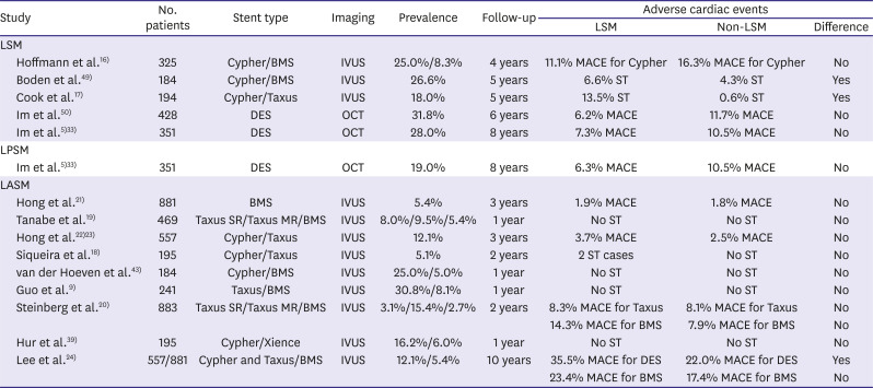
Table 2
Studies investigating clinical outcomes of LSM
| Study | No. patients | Stent type | Imaging | Prevalence | Follow-up | Adverse cardiac events | |||
|---|---|---|---|---|---|---|---|---|---|
| LSM | Non-LSM | Difference | |||||||
| LSM | |||||||||
| Hoffmann et al.16) | 325 | Cypher/BMS | IVUS | 25.0%/8.3% | 4 years | 11.1% MACE for Cypher | 16.3% MACE for Cypher | No | |
| Boden et al.49) | 184 | Cypher/BMS | IVUS | 26.6% | 5 years | 6.6% ST | 4.3% ST | Yes | |
| Cook et al.17) | 194 | Cypher/Taxus | IVUS | 18.0% | 5 years | 13.5% ST | 0.6% ST | Yes | |
| Im et al.50) | 428 | DES | OCT | 31.8% | 6 years | 6.2% MACE | 11.7% MACE | No | |
| Im et al.5)33) | 351 | DES | OCT | 28.0% | 8 years | 7.3% MACE | 10.5% MACE | No | |
| LPSM | |||||||||
| Im et al.5)33) | 351 | DES | OCT | 19.0% | 8 years | 6.3% MACE | 10.5% MACE | No | |
| LASM | |||||||||
| Hong et al.21) | 881 | BMS | IVUS | 5.4% | 3 years | 1.9% MACE | 1.8% MACE | No | |
| Tanabe et al.19) | 469 | Taxus SR/Taxus MR/BMS | IVUS | 8.0%/9.5%/5.4% | 1 year | No ST | No ST | No | |
| Hong et al.22)23) | 557 | Cypher/Taxus | IVUS | 12.1% | 3 years | 3.7% MACE | 2.5% MACE | No | |
| Siqueira et al.18) | 195 | Cypher/Taxus | IVUS | 5.1% | 2 years | 2 ST cases | No ST | No | |
| van der Hoeven et al.43) | 184 | Cypher/BMS | IVUS | 25.0%/5.0% | 1 year | No ST | No ST | No | |
| Guo et al.9) | 241 | Taxus/BMS | IVUS | 30.8%/8.1% | 1 year | No ST | No ST | No | |
| Steinberg et al.20) | 883 | Taxus SR/Taxus MR/BMS | IVUS | 3.1%/15.4%/2.7% | 2 years | 8.3% MACE for Taxus | 8.1% MACE for Taxus | No | |
| 14.3% MACE for BMS | 7.9% MACE for BMS | No | |||||||
| Hur et al.39) | 195 | Cypher/Xience | IVUS | 16.2%/6.0% | 1 year | No ST | No ST | No | |
| Lee et al.24) | 557/881 | Cypher and Taxus/BMS | IVUS | 12.1%/5.4% | 10 years | 35.5% MACE for DES | 22.0% MACE for DES | Yes | |
| 23.4% MACE for BMS | 17.4% MACE for BMS | No | |||||||
BMS = bare metal stent; DES = drug-eluting stent; IVUS = intravascular ultrasound; LASM = late-acquired stent malapposition; LPSM = late-persistent stent malapposition; LSM = late stent malapposition; MACE = major adverse cardiac events; MR = moderate release; OCT = optical coherence tomography; SR = slow release; ST = stent thrombosis.
![]()
Positive vessel remodeling is the leading mechanism for LASM. In the IVUS report by Mintz et al.,15) there was an increase in external elastic membrane radius within the region of LASM, but no change in plaque mass. Besides regional positive vascular remodeling, abluminal thrombus dissolution can contribute to LASM, especially in acute coronary syndromes.1) Post-intervention and follow-up IVUS or OCT is required to differentiate LASM caused by positive remodeling vs LASM caused by thrombus dissolution.
Clinical outcomes
Table 2 summarizes the clinical outcomes of LSM, LASM, or LPSM. Similar to ASM, a dedicated prospective study is absent; and most studies have been based on retrospective or sub-group analyses from randomized trials or registries. Unlike studies of ASM, results from studies of LSM are not consistent.
The study by Hoffmann et al.16) was a pooled analysis from the RAVEL (RAndomised study with the sirolimus-eluting Bx VELocity-stent), E-SIRIUS (European, multicentre, randomised, double-blind trial of the SIRolImUS- coated Bx VELOCITY stent in the treatment of patients with de novo coronary artery lesions), and SIRIUS (SIRolImUS-eluting stent in de novo coronary lesions) comparing sirolimus-eluting stents vs bare metal stents) studies. It included a total of 325 patients that had follow-up IVUS at 6 to 8 months after stent implantation after which 4-year clinical follow-up was available in all included patients.16) The frequency of MACE was 11.1% for patients with LSM vs 16.3% for those without LSM (p=0.48).16) In contrast to this study, Cook et al.17) reported a sub-group analysis of SIRTAX (Sirolimus-Eluting Versus Paclitaxel-Eluting Stents for Coronary Revascularization) showing that very late ST out to 4 years occurred more frequently among patients with vs without incidentally-detected IVUS LSM at 8 months (13.5% vs 0.6%, respectively, p<0.001). However, the type of LSM (LPSM vs LASM) could not be differentiated in the previous 2 studies because of the lack of post-intervention IVUS.16)17)
One registry with a small number of patients with LASM (n=10 patients) raised the possibility of a link between LASM and poor clinical outcomes during median 24.3 months follow-up.18) Conversely, an IVUS study from TAXUS II showed that no ST occurred in patients with LASM over a period of 12 months.19) Furthermore, an integrated IVUS analysis of the TAXUS IV, V, and VI and TAXUS ATLAS Workhorse, Long Lesion, and Direct Stent studies reported LASM in 7 BMS patients, 17 patients with slow-release TAXUS, and 12 patients with moderate-release TAXUS.20) Over the 2 ensuing years, MACE rates were similar in patients with vs without LASM for each of these 3 stent types.20) Two studies by Hong et al.21)22)23) using a large retrospective IVUS registry initially reported that incidentally detected LASM after implantation of first-generation DES or BMSs was not associated with MACE during the first 3-year follow-up period. However, with extended follow-up out to 10 years, LASM was related to a greater risk of MACE (hazard ratio [HR], 1.67; 95% confidence interval [CI], 1.04–2.67; p=0.03) and very late ST (HR, 3.53; 95% CI, 1.15–10.80; p=0.03) vs non-LASM in patients treated with first-generation DESs, but not in patients treated with bare-metal stents.24)
Virmani et al.25) reported the first case of fatal acute myocardial infarction and cardiac rupture as a result of late thrombosis of a first-generation sirolimus-eluting stent deployed 18 months previously. This patient underwent IVUS immediately after stent implantation and at 8-month follow-up when angiographic and IVUS demonstrated enlargement of the stented arterial segments with LASM, indicating positive vessel remodeling.25) Autopsy showed aneurysmal dilatation of these segments with severe localized hypersensitivity consisting predominantly of T-lymphocytes and eosinophils.25) Cook et al.26) analyzed thrombus aspirates in patients presenting with very late DES thrombosis and demonstrated that eosinophils were more common in thrombi harvested from very late DES thrombosis vs aspirates from spontaneous acute myocardial infarction, early bare-metal ST, early DES thrombosis, and late bare-metal ST. The eosinophil counts also correlated with extent of LSM.26) Thus, chronic inflammation and hypersensitivity can weaken the vascular wall, lead to LASM, and induce local stasis of blood flow within the positively remodeled segments.27) These inflammatory responses have been observed most frequently in the first-generation sirolimus-eluting stent among all coronary stents, including BMSs, other first-generation DES, new-generation DES, and polymer-free DESs, highlighting the clinical significance of polymers and drugs in DES system design.28)29)30)31)32)
Go to :
CLINICAL APPROACHES TO DRUG-ELUTING STENTS MALAPPOSITION
Clinical approach to acute stent malapposition
The recent consensus paper from the European Association of Percutaneous Cardiovascular Interventions recommended treating ASM having an axial distance ≥400 μm or longitudinal length ≥1 mm.34) This recommendation was based on OCT studies showing that ASM ≥400 μm usually persists and 3 OCT-based ST registry studies showing that stent malapposition was one of leading mechanisms of (very) late ST.35)36)37) However, these registries did not have control groups, and OCT was performed only at the time of ST. ASM can usually be corrected using a balloon sized to the diameter of the arterial lumen at the site of malapposition inflated to nominal (not high) pressures. For percutaneous coronary intervention of bifurcation lesions, the recent consensus document from the European Bifurcation Club suggested that a stent optimization (the so-called POT technique) should be performed routinely during the bifurcation procedure before side branch rewiring as it facilitated access towards the side branch and reduced the possibility that the wire might cross into the side branch behind the main-branch stent along with main branch stent crush.38)
Clinical approach to late stent malapposition
There is no recommendation or consensus on proper managements of patients with incidentally detected LSM. However, fortunately, rapid advances of DES technology have made stent struts thinner and have applied biocompatible or biodegradable polymers to metallic struts. This progress has reduced the thrombogenicity of new-generation DES. From the autopsy study by Otsuka et al.,31) second-generation cobalt-chromium everolimus-eluting stents (Xience®; Abbott Vascular, Santa Clara, CA, USA or Promus®; Boston Scientific, Marlborough, MA, USA) demonstrated greater strut coverage with less inflammation, less fibrin deposition, and less late and very late ST compared with first-generation DES. Clinically, these everolimus-eluting stents showed a lower frequency of LASM than first-generation sirolimus-eluting stents at 8-month follow-up IVUS39) and were associated with a significant reduction of ST compared with first-generation DES.40) When the type of LSM is discriminated as LASM rather than LPSM, the long-term clinical outcomes appear to be worse. Thus, prolonged dual antiplatelet or anticoagulation therapy may be considered in patients having LASM. The study from Doi et al.41) showed that patients with coronary artery ectasia receiving optimal anticoagulation therapy did not experience the occurrence of MACE. Surgical resection or percutaneous angioplasty with covered stents may be considered for multiple or giant coronary artery aneurysms.42) In contrast to LASM, the studies by Im et al.5)33) suggest that treatments may not be necessary for patients with LPSM.
Go to :
CONCLUSION
The effects of DES malapposition on long-term clinical outcomes have been controversial. Regardless of its severity, ASM has not been associated with adverse cardiac events across all IVUS and OCT studies. In contrast to ASM, cumulative evidences have demonstrated that LASM of first-generation DES is likely to cause (very) late ST through chronic inflammatory reactions. This association is not clear in LPSM and may be attenuated in recent DESs which have thin struts and biocompatible/biodegradable polymers. Figure 2 provides the illustrative overview about the association between DES malapposition and long-term clinical outcomes. Although prolonged anti-thrombotic therapy and/or percutaneous intervention using large-sized balloon or covered stent may be considered in patients with LASM, the treatments should be made considering the clinical situations of individual patient due to lack of firm evidences.
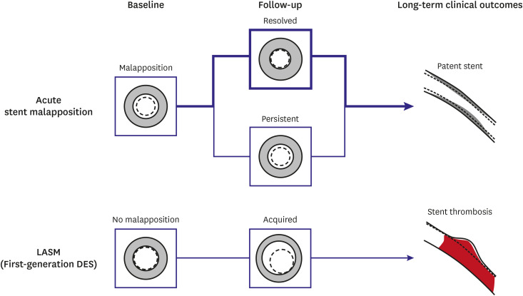 | Figure 2Overview of stent malapposition assessed by intravascular imaging modality. Even though malapposed struts immediately after stent implantation remain occasionally during follow-up, they usually do not link to ST. However, LASM increases the risk of ST through chronic inflammatory reactions especially in first-generation DESs.DES = drug-eluting stent; LASM = late-acquired stent malapposition; ST = stent thrombosis.
|
Go to :
 XML Download