

 PDF
PDF Citation
 Print
Print


The patient was an 81-year-old female with medical treatment for old myocardial infarction which occurred in the middle left anterior descending artery in 1989. She had multiple admission for heart failure, with the most recent admission being associated with aggravated symptoms by low left ventricular (LV) ejection fraction with severe tricuspid regurgitation (TR) despite optimal medical therapy. Chest X-ray showed cardiomegaly with bilateral pleural effusion. As shown in Figures 1 and 2, echocardiography demonstrated a degenerative mitral valve with a prolaptic motion of anterior mitral leaflet with tethering (Supplementary Videos 1 and 2). There was incomplete coaptation of A2-P2. This resulted in severe mitral regurgitation (MR) with an effective regurgitant orifice area (EROA) of 0.68 cm2, the regurgitant volume of 73 mL. She had enlarged LV dimensions and LV ejection fraction of 42% due to old myocardial infarction. The Society of Thoracic Surgeons score for mortality was 12.3%. After a Heart Team discussion, the patient was offered the transcatheter mitral-valve repair according to recent European Society of Cardiology guidelines (Class IIb).1)2)3) The first grasp was performed centrally with careful attention to grasp both leaflets (A2-P2) adequately. With a single clip, the MR was reduced from IV to III. Thus, the second grasp was performed at the lateral side parallel to the first clip. After the second grasp (Supplementary Videos 3), MR was moderate in severity with EROA of 0.21 cm2 and regurgitant volume of 36 mL (Supplementary Videos 4 and 5). Additionally, TR was reduced from IV to III. She was discharged with improved functional status from New York Heart Association Classification IV to II.
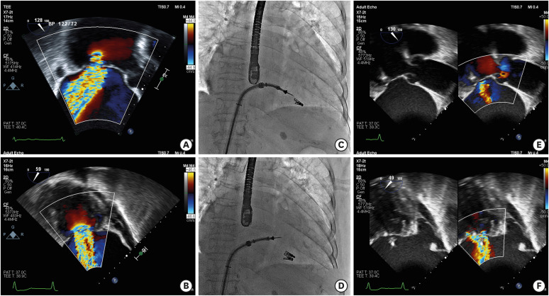 | Figure 1Severe MR demonstrated on (A, B) Pre-procedural transesophageal echocardiographic left ventricualar outflow tract and intercommissural view. (C) First grasp. (D) Second grasp. Reduced MR reduced after clipping demonstrated on (E,F) Post-procedural transesophageal echocardiographic left ventricualar outflow tract and intercommissural view.MR = mitral regurgitation.
|
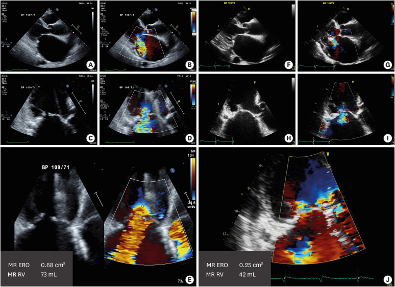 | Figure 2(A, B) Transthoracic echocardiographic parasternal long axis and (C,D) apical 3-chamber views showing degenerative mitral valve with severe MR. (E) pre-procedural EROA and regurgitant volume is 0.68 cm2 and 73 mL, respectively. After the MitraClip, (F, G) transthoracic echocardiographic parasternal long axis and (H, I) apical 3-chamber views showing degenerative mitral valve with severe MR. (J) post-procedural EROA and regurgitant volume is 0.21 cm2 and 36 mL, respectively.MR = mitral regurgitation, EROA = effective regurgitant orifice area.
|

 XML Download