

 PDF
PDF Citation
 Print
Print


INTRODUCTION
Transcatheter aortic valve replacement (TAVR) has been used as an alternative to surgical aortic valve replacement (SAVR) in severe aortic stenosis patients with successful mid-term outcomes.1)2)3)4)5) Although it is less invasive than SAVR, TAVR also has risks of peri-procedural or post-procedural complications, including paravalvular leak, cerebrovascular events, vascular injury, arrhythmia, and acute kidney injury.6) Stroke is a particularly devastating complication of TAVR associated with severe disability and a high incidence of mortality.7) In the Placement of AoRTic TraNscathetER Valve (PARTNER) trial, detection rates of high signal intensities on diffusion-weighted magnetic resonance imaging (MRI) after TAVR were higher than after SAVR.8) Occurrence of post-TAVR stroke was associated with increased 30-day mortality, and the causes of stroke during or after TAVR should thereby be studied in greater depth.
Recent articles about transcatheter heart valve (THV) thrombosis have focused on leaflet thrombosis, and alterations of hemodynamics in sinuses of Valsalva are a significant cause of THV thrombosis.9)10) However, sinus of Valsalva thrombosis after TAVR has only been reported in a single case study by Tsunaki et al.11) Sinus of Valsalva thrombosis after TAVR should be studied as a potential target of anticoagulation treatment to prevent systemic embolization or coronary obstruction. This study describes the clinical and computed tomography (CT) characteristics of patients with sinus of Valsalva thrombosis via a retrospective review of post-TAVR CT scans. We performed subgroup analysis for the association between sinus of Valsalva thrombosis and embolic stroke in patients with eligible brain MRI after TAVR.
METHODS
Patients
This study was approved by the Institutional Review Board of Asan Medical Center (approval number: 2019-0312). The requirement for informed consent was exempted due to its retrospective nature. In our hospital, TAVR has been performed since 2011. Total 665 patients underwent TAVR procedures between March 2011 and August 2019, and 192 patients underwent cardiac CT after TAVR during this period to evaluate device location, valve function, and potential device thrombosis based on the established guidelines for the appropriate use of cardiac CT.12)13) Indications for CT scan after TAVR were determined by the patient's clinician, and the reasons for each CT scan are as follows: for evaluation of paravalvular leakage (n=78), for research purpose (n=54), for evaluation of stent position (n=31), for routine follow-up (n=10), because of increased transaortic pressure gradient detected on echocardiography (n=6), because of patients' symptoms such as chest pain (n=5) and dyspnea (n=5), concomitant mitral valve disease (n=2), or for left ventricular (LV) wall motion abnormality (n=1).
Echocardiography
Transthoracic echocardiography with 3–5 MHz real-time transducers (iE33, EPIC; Philips Medical Systems, Andover, MA, USA; Vivid 7, E9, General Electric Healthcare, Waukesha, WI, USA) was performed by 4 cardiologists with at least 8 years of clinical experience. Two-dimensional and Doppler images were obtained in accordance with the American Society of Echocardiography recommendations.14) End-systolic volume, end-diastolic volume, and LV ejection fraction were obtained with the biplane Simpson method. Maximal aortic jet velocity and degree of paravalvular leak were recorded, and maximal and minimal pressure gradients across the aortic valve (AV) were estimated using a modified Bernoulli equation. LV mass and LV mass index were calculated using the LV cavity dimension and LV wall thickness at end-diastole.
Computed tomography protocols
Cardiac CT scans were obtained using dual-source CT scanners (Somatom Force or Somatom Definition Flash; Siemens Medical Solutions, Forchheim, Germany). If the initial heart rate was higher than 75 beats/min measured 1 hour before the CT examination, patients were given 2.5 mg oral bisoprolol (Concor; Merck, Darmstadt, Germany) as long as beta-blockers were not contraindicated. A bolus of 60–70 mL of contrast material (Iopamidol, iodine 370 mg/mL; Patheon Italia S.p.A., Monza, Italy) was injected using a power injector at a rate of 3.5–5 mL/s (Stellant D; Medrad, Indianola, PA, USA), followed by 40 mL of saline chaser. The threshold of the bolus-tracking method was 100 HU with region of interest in the ascending aorta. Electrocardiogram-gated retrospective spiral scans were performed with tube current modulation (dose pulsing window of 0–90% of the R-R interval). Tube voltage and tube current/exposure time product were adjusted in accordance with patient's body size as follows: tube voltage, 100–120 kV; tube current/exposure time product, 185–380 mAs; collimation, 2 × 192 × 0.6 mm or 2 × 128 × 0.6 mm; gantry rotation time, 250 ms or 280 ms. Iterative reconstruction at 10% intervals was performed via a 0.6 mm slice and 0.4 mm interval. CT image analysis was performed by the consensus of 2 expert cardiovascular radiologists blinded to all clinical information, including the results of post-TAVR echocardiography. Multiphase images of CT scans were transported into commercially available software (Aquarius iNtuition Edition ver. 4.4.12; TeraRecon, Foster City, CA, USA) for post-processing.
Computed tomography image analysis
End-systolic phase (20–30% R-R interval) data was selected to generate the AV en-face view with a 1 mm thickness.15) Aortic annulus maximal and minimal diameters, aortic annulus area, sinus of Valsalva diameter, and sinotubular junction diameter were measured on the AV en-face view. The diameters of sinus of Valsalva, TAVR stent, and residual aortic cusp excluded from the stent on post-TAVR CT images were also measured (Supplementary Figure 1). Sinus of Valsalva thrombosis was defined as low-attenuation soft tissue within the sinuses of Valsalva, detected on the en-face view and oblique coronal or sagittal views of the TAVR device. CT attenuation values were obtained at 3 different points of the lesions, and their medians were recorded to minimize the effects of motion or beam hardening artifacts. Hypoattenuating leaflet thickening (HALT) indicating leaflet thrombosis was visible in the diastolic phase and detected in 2 or more phases using the multiplanar reconstruction method.16) The multiplanar reconstruction images were obtained in the optimal phase and view (either en-face or oblique sagittal plane) to visualize HALT with the fewest artifacts.
Brain magnetic resonance imaging
Because this study is a retrospective design, not all patients underwent brain MRI for evaluation of embolic stroke. To exclude periprocedural embolic infarction during the TAVR, we only included patients with brain MRI acquired at least a month after the TAVR. Also, we included brain MRI underwent within a month from the date of post-TAVR cardiac CT for a connection between the 2 imaging studies. Embolic infarction was defined as a new high signal intensity in the cerebral cortex or subcortical white matter on diffusion weighted image or fluid attenuated inversion recovery (FLAIR) image compared to the initial brain MRI after TAVR. Finally, we found 64 patients with eligible brain MRI. The reasons for brain MRI were for the research purpose (n=54), perioperative work-up (n=4), mental change (n=2), headache (n=1), recurrent transient ischemic attack (n=1), left side weakness (n=1), and syncope (n=1).
Statistical analysis
Clinical information and CT characteristics of the patients were shown in a descriptive manner. CT findings of the patient with and without sinus of Valsalva thrombosis or HALT were compared using Student t-test, χ2 test, and Fisher's exact test. We used median values of three sinuses of Valsalva for per-patient analysis, and used values of each sinus of Valsalva for per-sinus analysis. Subgroup analysis of the patients who have eligible brain MRI was performed. Univariate logistic regression analysis of the CT findings associated with the occurrence of embolic stroke was also performed.
RESULTS
Patients
Among the 192 patients (male:female=100:92) that underwent post-TAVR CT scan, 117 Edward Sapien (Sapien, n=3; Sapien XT, n=53; Sapien 3, n=61), 64 CoreValve, and 11 Evolut R valves were implanted. Eight patients receiving CoreValve (n=8) underwent immediate valve-in-valve procedures with a CoreValve (n=7) or Evolut R (n=1) due to inappropriate implantation of the first THV. One patient who received TAVR using Sapien valve at 2011 underwent valve-in-valve TAVR using Evolut R valve after 8 years from initial TAVR. The second inserted valve type was recorded as the final valve type for evaluation of post-TAVR CT findings in these patients. Among all patients, sinus of Valsalva thrombosis presented in 9 patients (4.7%). In these patients, 7 CoreValve (including valve-in-valve), 3 Edwards Sapien (Sapien XT n = 1; and Sapien 3, n = 2) valves, and 1 Evolute R valve were used.
Clinical characteristics of patients with sinus of Valsalva thrombosis are shown in Table 1. All patients were elderly, with a median age of 82 years (interquartile range [IQR], 77–85), and 7 were female. All patients were taking aspirin and clopidogrel. None received anticoagulation therapy to prevent post-TAVR thrombosis.
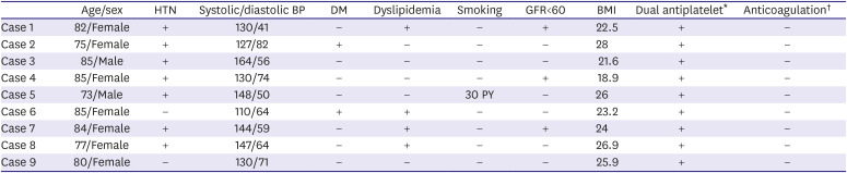
Table 1
Clinical characteristics of the patients with sinus of Valsalva thrombosis after TAVR
BMI = body mass index; BP = blood pressure; DM = diabetes mellitus; GFR = glomerular filtration rate; HTN = hypertension; PY = pack-year; TAVR = transcatheter aortic valve replacement.
*Aspirin and clopidogrel administration; †Anticoagulation to prevent valve thrombosis.
![]()
Echocardiography
The echocardiographic findings of the 9 patients with sinus of Valsalva thrombosis after TAVR are summarized in Table 2. All patients had severe aortic stenosis before TAVR, and most of them showed preserved cardiac function. Four patients showed a mild paravalvular leak on immediate post-TAVR echocardiography. All patients demonstrated a significant drop in transaortic pressure gradient and maximal transaortic jet velocity after TAVR. However, one patient presenting with concurrent HALT with severe leaflet opening reduction (case 9) demonstrated high transaortic peak velocity (3.9 m/s) and mean pressure gradient (33 mmHg) on an echocardiogram obtained 772 days after TAVR.
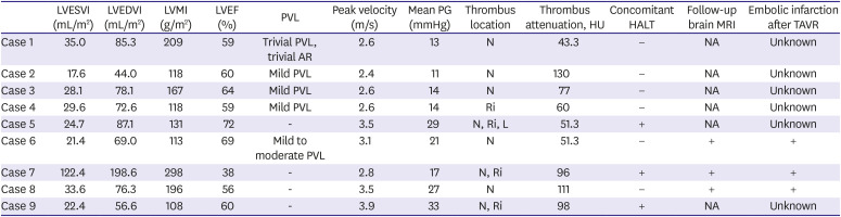
Table 2
Echocardiography, cardiac CT, and brain MRI findings of patients with sinus of Valsalva thrombosis after TAVR
AR = aortic regurgitation; CT = computed tomography; HALT = hypo-attenuating leaflet thickening; HU = Hounsfield unit; L = left sinus of Valsalva; LVEF = left ventricular ejection fraction; LVEDVI = left ventricular end-diastolic volume index; LVESVI = left ventricular end-systolic volume index; LVMI = left ventricular mass index; MRI = magnetic resonance imaging; N = non-coronary sinus of Valsalva; NA = not available; PG = pressure gradient; PVL = paravalvular leak; POD = post-operative day; Ri = right sinus of Valsalva; TAVR = transcatheter aortic valve replacement.
![]()
Computed tomography findings
CT findings of the patients with and without sinus of Valsalva thrombosis are noted in Table 3. HALT was common in patients with sinus thrombosis, but not statistically significant (15.8% vs. 33.3%, p=0.17). Embolic infarction was more common in patients with sinus thrombosis, but showed marginal trend (16.4% vs. 66.7%, p=0.09). CT parameters measured on both pre- and post-TAVR CT scans were not statistically different between the patients with and without sinus thrombosis. Even in per-sinus comparison on post-TAVR CT images, no definite statistical difference was observed between the 2 groups (Supplementary Table 1).
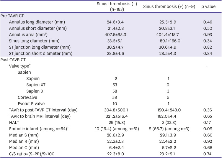
Table 3
Comparison of the patients with and without sinus of Valsalva thrombosis
Data are shown as mean±standard deviation or number (%).
C =cusp diameter; CT = computed tomography; HALT = hypo-attenuating leaflet thickening; MRI = magnetic resonance imaging; R = stent diameter at sinus level; S = sinus diameter; TAVR = transcatheter aortic valve replacement.
*In case of valve-in-valve, second implanted valve was counted; †Only 64 patients have eligible brain MRI.
![]()
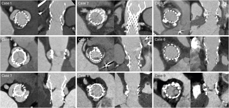
Multiplanar reconstructed CT images of sinus of Valsalva thrombosis in 9 patients are shown in Figure 1. The detection rates of sinus thrombosis were different by valve types: Sapien, 3/117 (2.6%); CoreValve, 5/64 (7.8%); and Evolute R, 1/11 (9.1%). After combining the CoreValve group and Evolute R valve, there was no statistical difference by the χ2 test (p=0.16). Two patients showed sinus of Valsalva thrombosis (18.2%) among the eleven patients who had undergone valve-in-valve procedures. There was no statistically significant association between valve-in-vale procedures and sinus of Valsalva thrombosis by the Chi-square test (p=0.59). For the 9 patients with sinus of Valsalva thrombosis on post-TAVR CT scan, the median interval between TAVR and CT scan was 12 days (IQR, 6–184). Eight patients (89%) presented with thrombus in the non-coronary sinus of Valsalva. In all patients, thrombi presented at the base of the sinus and extended towards the sinotubular junction, but not yet obstructed the coronary artery ostium. Three patients had concomitant HALT. The median attenuation value of sinus thrombosis was 77 (IQR, 51.3–98).
Aortic annulus long diameter on pre-TAVR CT was shorter in patients with HALT (23.1 mm vs. 25.0 mm, p=0.002) (Supplementary Table 2). The interval from the TAVR to post-TAVR CT scan was longer in patients with HALT (601 days vs. 236 days, p=0.004). Embolic infarction was common in patients with HALT, but not statistically significant (16.7% vs. 30.0%, p=0.38).
Brain magnetic resonance imaging
Subgroup analysis of the patients (n=64) with eligible brain MRI after TAVR was performed (Supplementary Tables 3 and 4). Univariate logistic regression analysis showed that sinus thrombosis increased the risk of embolic stroke by 10.2 times with marginal significance (p=0.068).
Among the 9 patients with sinus of Valsalva thrombosis, 3 underwent brain MRI scans on the same date of their post-TAVR CT scans. These patients presented with newly developed subcortical high signal intensity foci on FLAIR images when compared to brain MRIs taken immediately after TAVR, indicating new embolic infarction (Figures 2 and 3). Three patients were provided anticoagulation treatment, including vitamin K antagonist or direct factor Xa inhibitor, for THV thrombosis suspected on post-TAVR CT scan or echocardiography. All patients underwent one post-TAVR CT scan and none of them received an additional follow-up scan thereafter. The effect of anticoagulants could not be evaluated as these patients did not undergo follow-up CT scans to assess resolution of thrombosis. Last follow-up date from TAVR procedure ranged from 2 weeks to 6 years, and 3 patients transferred to other geriatric hospitals and did not receive follow-ups at our institution. One of the three patients (case 2) died 1.5 years after the post-TAVR CT scan, but cause of death is unknown.
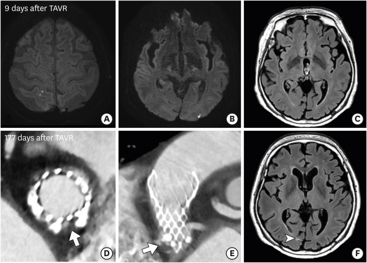
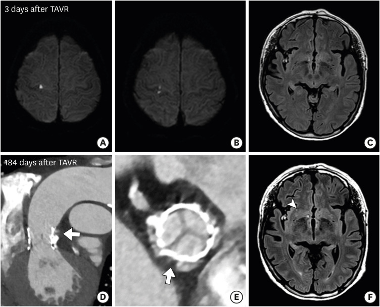
Figure 2
An 85-year-old woman diagnosed with severe aortic stenosis. (A, B) Diffusion-weighted magnetic resonance images and (C) FLAIR image obtained 9 days after TAVR. Hyperintense acute infarction lesions can be noted in right parietal and left occipital lobes. (D, E) Cardiac gated CT obtained 177 days after TAVR shows low attenuation thrombus in the non-coronary sinus of Valsalva, and (F) FLAIR image acquired at the same time shows new cortical/subcortical hyperintensity consistent with prior embolic infarction.
CT = computed tomography; FLAIR = fluid attenuated inversion recovery; TAVR = transcatheter aortic valve replacement.
![]()
Figure 3
A 79-year-old woman who underwent TAVR. (A, B) Diffusion-weighted magnetic resonance images and (C) FLAIR image obtained 3 days after TAVR. Hyperintense acute infarction lesions were noted in the right frontal lobe. (D, E) Cardiac gated CT obtained 184 days after TAVR shows low attenuation thrombus in the non-coronary sinus of Valsalva, and (F) FLAIR image acquired at the same time shows new subcortical white signal intensity consistent with prior embolic infarction.
CT = computed tomography; FLAIR = fluid attenuated inversion recovery; TAVR = transcatheter aortic valve replacement.
![]()
DISCUSSION
Among the 192 patients that underwent post-TAVR CT scan, 9 (4.7%) presented with sinus of Valsalva thrombosis. Of them, 3 patients had concomitant HALT. Three patients underwent brain MRIs on the same day with their post-TAVR CT scans, and showed newly developed subcortical high signal intensities on FLAIR images, indicating embolic stroke.
It is unclear why sinus of Valsalva thrombosis is rarely reported, but it may be due to under-evaluation on follow-up CT or overall low incidence of sinus of Valsalva thrombosis. A retrospective cohort study on a large population from 521 US hospitals (n=101,430) found that rates of stroke and transient ischemic attacks were 2.3% and 0.4%, respectively, at 30 days after TAVR.17) The PARTNER trial for high-risk patients demonstrated that the detection rates of high signal intensities on diffusion-weighted brain MRI after TAVR were higher than after SAVR, at 86% for the Edwards Sapien, 80% for the CoreValve, and 48% for SAVR.18) Rates of clinical stroke with neurologic event after TAVR and SAVR were reported to be 5.5% vs. 2.4%, respectively, at 30 days, and 8.3% vs. 4.3% at 1 year.4) Risk factors for stroke after TAVR vary depending on the timing of stroke onset.19) Early stroke, within the first week after surgery, is considered related to the embolization of tissue fragments or thrombi during the procedure.20) Late onset stroke might be induced by HALT or other thrombosis associated with the TAVR device. The altered flow dynamics in sinuses of Valsalva combined with patient-specific blood chemistry and interaction with foreign THV materials can result in THV thrombosis.21)
In the present study, 8 patients (89%) had thrombus in the non-coronary sinus of Valsalva. The thrombus was located at the bottom of the sinuses and extended upward towards the sinotubular junction. These results, along with those from previous studies, indicated that the surface-averaged length of blood residence time on the non-coronary leaflet was longer than on the coronary leaflet in an intra-annular, valve-in-valve setting,22) and that blood flow stagnation was observed at the bottom of the Valsalva sinuses during the entire heart beat cycle.9)
We found that the detection rates of sinus thrombosis were different by valve types. Moreover, valve-in-valve procedure also showed high rates of sinus thrombosis (18.2%) even though there was no statistical significance. These results are limited due to small number of patients, and further studies are needed. Preventive measures should be considered when performing a valve-in-valve procedure or delayed post-dilatation in the event of suboptimal prosthesis implantation.
Sinus of Valsalva thrombosis has been reported not only after TAVR but also after SAVR, and even in native AVs with or without Valsalva sinus aneurysm. Protein C or S deficiency, oral contraceptives, LV hypokinesia, atheromatous changes, and aortic stenosis have been implicated in native sinus of Valsalva thrombosis.23)24)25) Several studies demonstrated that native sinus of Valsalva thrombosis resulted in acute myocardial infarction, renal infarction, or arterial thromboembolism in the lower legs, as well as ischemic stroke.26)27)28)29) Even though there were only 3 cases of silent embolic stroke detected on brain MRI in these studies, sinus of Valsalva thrombosis in post-TAVR patients also have potential for coronary arterial occlusion and cerebral or peripheral arterial embolization. As such, assessing potential sinus of Valsalva thrombosis should be an essential component of post-TAVR CT scans.
Dual antiplatelet therapy after TAVR might not be enough to prevent THV thrombosis, including sinus of Valsalva thrombosis and leaflet thrombosis. Although the administration of unfractionated heparin is recommended for stroke prophylaxis during the TAVR, a consensus about optimal antithrombotic therapy after the procedure has yet to be established.19) European guidelines (European Society of Cardiology/European Association for Cardio-Thoracic Surgery) recommend dual antiplatelet therapy for 3–6 months followed by single antiplatelet therapy, whereas the American Association (American Heart Association/American College of Cardiology) recommends oral anticoagulation for the first 3 months in patients with low bleeding risk (class IIb, level of evidence C).30) Additional prospective studies to determine optimal anticoagulation and/or antiplatelet therapy after TAVR could provide valuable information for setting overall guidelines.
There are several limitations in our study. First, selection bias is an inherent part of all retrospective observational study. In our study, not all patients received a post-TAVR CT scan and/or brain MRI, and total incidences of sinus of Valsalva thrombosis and concurrent silent embolic stroke could not be determined. Second, follow-up CT scans to check if the sinus of Valsalva thrombosis persisted or had resolved were not available. We recommend additional follow-up CT examinations in the patients who show sinus of Valsalva thrombosis to evaluate the effect of anticoagulants for lysis of thrombi. Finally, because of the range of time intervals from TAVR to CT scan, we were unable to pinpoint the moment when sinus of Valsalva thrombosis developed.
In conclusion, sinus of Valsalva thrombosis is uncommon but can be detected on post-TAVR CT scans. Silent embolic stroke could be a comorbidity despite dual antiplatelet therapy. Therefore, cardiac CT to detect sinus of Valsalva thrombosis could guide optimal treatment in the future.
 XML Download